


STUDY OF EFFECT OF STERCULIA FOETIDA SEEDS ON INSULIN RESISTANCE IN TYPE II DIABETES MELLITUS MODEL IN RATS
HTML Full TextSTUDY OF EFFECT OF STERCULIA FOETIDA SEEDS ON INSULIN RESISTANCE IN TYPE II DIABETES MELLITUS MODEL IN RATS
Sneha Bhadoriya *, S. C. Mehta and Madhu Rajput
Shri Ramnath Singh Institute of Pharmaceutical Sciences & Technology, Sitholi, Gwalior, Madhya Pradesh, India.
ABSTRACT: Type 2 Diabetes Mellitus (T2DM) is characterized by insulin resistance and impaired glucose metabolism. With the rising interest in plant-based interventions, Sterculia foetida seeds have attracted attention for their potential antidiabetic properties. This study aimed to evaluate the effects of Sterculia foetida seed extract on insulin resistance in a rat model of T2DM. Male Wistar rats were divided into normal, diabetic control, and treatment groups. T2DM was induced using a combination of high-fat diet and low-dose streptozotocin (STZ). After confirmation of diabetes, rats in the treatment group received Sterculia foetida seed extract orally for a specified duration. Fasting blood glucose, serum insulin levels, and insulin resistance (assessed by HOMA-IR) were measured. Additional biochemical parameters and histopathological examination of pancreatic tissues were conducted. Treatment with Sterculia foetida significantly reduced fasting blood glucose and HOMA-IR values compared to the diabetic control group. Serum insulin levels improved, indicating enhanced insulin sensitivity. Histological analysis supported these findings, showing improved islet morphology in treated rats. Sterculia foetida seed extract demonstrated promising effects in reducing insulin resistance and improving glycemic control in T2DM rats. Further studies are needed to isolate active compounds and validate its therapeutic potential in humans.
Keywords: Sterculia foetida, Diabetes, STZ-High fat diet
INTRODUCTION: Diabetes is a chronic metabolic disorder characterized by hyperglycemia, a high level of glucose in the blood. Hyperglycemia is a common effect of uncontrolled diabetes and it could lead to long-term damage and functional disorders of various tissues and organs such as eyes, kidneys, nerves, heart, and blood vessels 1.
Diabetes significantly affects the number of microvascular and macrovascular diabetic complications 2. Heart attack, stroke, and peripheral vascular diseases are the macrovascular complication of diabetes, while retinopathy, neuropathy, and nephropathy are the microvascular complication of the disease 3.
Both complications could reduce the quality of life and increase mortality 2. Insulin resistance is the most common cause of chronic hyperglycemia in T2DM. It activates numerous factors and pathways linked to decreased insulin sensitivity and dysfunction of β-cells. Healthcare practitioners frequently lack the skills to begin, intensify, or de-escalate therapy when required. This perplexity frequently results in underachievement of the anticipated goal and the induction of life-threatening hypoglycemia. As a result, there is a need to investigate treatment drugs that are both efficacious and have a high safety profile 3. Herbal medications are widely employed in most countries’ health care. As per the World Health Organization (WHO), in underdeveloped countries such as Africa and Asia, over 80% of the population uses this form of medicine for primary care 4, 5.
Several medicinal plants are used as antidiabetic agents, and some of them have shown their efficacy through pharmacological evaluation. The World Health Organization (WHO) strongly supports researches related to the discovery and development of new drugs, especially the medicinal plants that have been traditionally used by the community and proven to be able to control and cure the diseases 6. Sterculia foetida, commonly known as the Wild Almond or Java Olive, is a tropical plant traditionally used in various cultures for its medicinal properties. Preliminary phytochemical studies have shown that the seeds of Sterculia foetida contain bioactive compounds such as flavonoids, sterols, and fatty acids, which are known to possess antioxidant, anti-inflammatory, and lipid-lowering effects 7. These pharmacological activities suggest potential benefits in managing metabolic syndromes including insulin resistance.
Although there is some ethnomedicinal evidence supporting the use of Sterculia foetida seeds in metabolic disorders, there is limited scientific data evaluating their specific role in insulin sensitivity and glucose regulation. Therefore, investigating the effect of Sterculia foetida seed extract on insulin resistance using a validated T2DM animal model (such as high-fat diet or streptozotocin-induced diabetic rats) could provide valuable insights into its therapeutic potential 8, 9. This study aims to bridge the gap in current knowledge by exploring the anti-diabetic properties of Sterculia foetida seeds, focusing specifically on their impact on insulin resistance. The outcomes of this research could contribute to the development of novel, plant-based treatments for T2DM and offer a scientific basis for traditional uses of this plant.
MATERIALS & METHODS:
Plant Material: The plant (Sterculia foetida) seed were collected locally from herbal store and botanical garden of the garden, Gwalior District. The plant was identified and authenticated by comparison with herbarium specimens. The seeds of Sterculia foetida were authenticated by comparison with herbarium specimens and authentification. The weighed coarse powder was used for the extraction by successive solvent extraction by Soxhlet apparatus using various solvents 10.
Animals: Wistar Albino rats (150 – 250 g) used for the study were obtained from the animal house of the Department of Pharmacology, Shri Ramnath Singh Institute Of Pharmaceutical Sciences & Technology, Sitholi, Gwalior (M.P.). The animals are randomly selected, marked to permit individual identification, and kept in their cages for at least 5 days prior to dosing to allow for acclimatisation to the laboratory conditions. The animals were housed three per cage in a polypropylene cage and maintained in standard laboratory conditions with free access to food and water ad libitum. All animal experiments were conducted incompliance with (Organization for Economic Cooperation and Development) OECD Guideline and approved by the Institutional Animal Ethics 11, 12.
Extraction: The seeds of Sterculia foetida, dried under shade are carefully removed and grinded using a blender. The coarse power so obtained was used for the extraction by successive solvent extraction by Soxhlet apparatus using various solvents. The assembly of Soxhlet apparatus is as shown in the figure 13. Marc obtained from the above extract was dried and extracted with 2.5litres of ethanol (90%) in Soxhlet apparatus for 36 hours. Then the extract obtained were collected and concentrated by vaccum distillation. The concentrated extract were then dried by in a vaccum desciccator.
Antioxidant Content & Activity:
Determination of Total Flavonoid Content: The determination of the total flavonoid content the samples used then method of Chang and Pharmacopoeia Herbs. Preparation of standard parent solution of quercetin. The Quercetin weighed 10 mg, dissolve in 96% ethanol to obtain 10 ml (1000 μg / ml) solution. Created n dilution 50, 40, 30, 20, and 10 μg / ml. From the concentrations taken consecutively 0.5; 0.4; 0.3; 0.2; and 0.1 ml of solution. Add 3 ml of 96%n ethanol, 0.2 ml AlCl3 10%, 0.2 ml Na-acetate 1 M, and distilled up to 10 ml volume. The solution mixture was incubated for 30 min at room temperature. Measure absorbance at 437 nm wavelength using UV-Visspectrophotometer 14. Calculate the linear regression equation between the concentration and absorbance relationship. The standard curve equation is obtained from the linear regression between quercetin (x) and absorbance (y).
DPPH Scavenging Ability Assay: Antioxidant activity of methanol extract was evaluated using DPPH scavenging ability assay, which was conducted in a 96-well plate according to previously used method with slight modification. Samples in different concentrations (100, 250, 500, 1000, 1500, 2000 ppm) and 0.114 mM DPPH solution 180 µL in methanol were added to each well. The absorbance was read at 517 nm after 30 min of reaction in dark with a micro-plate reader 15. The scavenging ability (%) was calculated as follows:
Scavenging (%) = Ac – As / Ac × 100%
AC was the absorbance of control (without sample), AS is the absorbance of sample. Ascorbic acid was used as positive standard. All tests were performed in triplicate. Concentration of samples resulting in 50% n inhibition on DPPH (IC50 value) were calculated.
Acute Toxicity Study: The Acute Oral Toxicity - OECD Guideline 423 is a widely accepted method used to assess the potential toxicity of substances when administered orally. It's known as the Acute Toxic Class Method 16. The study will be conducted using female Wistar albino rats, with a total of 12 animals divided into four groups, each comprising three rats. An initial dose of 5 mg/kg of the test substance will be administered orally. Following administration, the animals will be kept under close observation for 3 hours to monitor any immediate effects, and then further observed for 14 days for any delayed reactions. Throughout the study period, various parameters will be assessed, including body weight, mortality status, and clinical signs. Special attention will be given to any changes in the central nervous system (CNS), autonomic nervous system (ANS), and general behavioral patterns to detect signs of toxicity or adverse effects.
TABLE 1: EXPERIMENTAL DESIGN OF ACUTE TOXICITY STUDY
| Group | Number of Animals | Dose (mg/kg) |
| Group A | 3 | 5 |
| Group B | 3 | 50 |
| Group C | 3 | 300 |
| Group D | 3 | 2000 |
Pharmacological Studies: The animals were separated into six groups (n = 6 in each).
- Group 1 (normal control; 10 mL/kg normal saline)
- Group 2 (diabetic control; DC)
- Group 3 (standard drug; metformin)
- Group 4 (negative control; 200 mg/kg EESF i.p.)
- Group 5 (DC + EESF 200 mg/kg i.p.)
- Group 6 (DC + EESF 400 mg/kg i.p.) 17
Induction of Diabetes in Experimental Animals: The animals in groups 1 and 2 were fed a conventional, normal pellet diet. Whereas animals in groups 3 to 6 were initially fed a high-fat diet (HFD: 66.5% standard pellet feed, 13.5% lard, and 20% sugar) for 2 weeks, 40 mg/kg streptozotocin (STZ) made from 0.5% normal saline was injected intraperitoneally (i.p.) to induce type-2 diabetes. To avoid hypoglycemia, the animals were given a 5% glucose solution for the first 12 h. As previously stated, the HFD diet was prepared 18.
Assessment of Diabetes: Diabetes was confirmed after 48 hr of streptozotocin injection, the blood samples were collected through tail vein and plasma glucose levels were estimated by glucose oxidase method (accu check active glucometer). The rats having fasting plasma glucose levels more than 200mg/dL were selected and used for the present study 19.
Glucose Tolerance Test: Fasting blood glucose (FBG) levels were assessed 3 days after STZ injection using a digital glucometer with test strips (Contour, Ascensia Diabetes Care Holdings AG, M, Switzerland), and animals were considered diabetic if FBG levels were >250 mg/dl, and those animals were included in the study 20.
Body Weight, Blood Glucose, and Insulin Measurement: The animals used in the study were weighed on days 0, 7, and 14 to monitor changes in body weight over the experimental period. This routine measurement served as a general indicator of health and response to the test substance. All rats were observed daily for clinical signs, and any abnormal findings were recorded. Regular weight tracking is also essential to assess the potential toxicity or therapeutic effects of a compound in preclinical studies. In addition to glucose levels, fasting serum insulin concentrations were measured on day 14 using the radioimmunoassay method with a commercial insulin assay kit (Cisbio International, France) 21. The insulin resistance index (HOMA-IR) was calculated to assess insulin sensitivity using the formula:
HOMA-IR = serum insulin (mmol/L) × (blood glucose (mmol/L) / 22.5
This index is widely used in metabolic research to estimate insulin resistance, which plays a critical role in the development of type 2 diabetes and other metabolic disorders.
Intraperitoneal Glucose and Insulin Tolerance Test: At the end of the 14-day treatment period, an intraperitoneal glucose tolerance test (IPGTT) was performed to evaluate the glucose handling capacity of the animals. Prior to the test, all animals were fasted overnight to ensure a consistent baseline for glucose measurement. Each rat received an intraperitoneal injection of glucose at a dose of 2 g/kg body weight, and blood glucose levels were measured at 0 (baseline), 30, 60, and 120 minutes post-injection. These time points were selected to capture both the initial glucose spike and subsequent clearance, allowing for an accurate assessment of glucose tolerance 22.
The ITT provides a complementary perspective to the IPGTT, offering a direct measure of peripheral insulin sensitivity. A normal insulin response would be reflected by a rapid decrease in blood glucose levels after insulin administration.
In contrast, a blunted or delayed response may suggest insulin resistance, a key marker in the pathophysiology of type 2 diabetes and other metabolic disorders 23.
To quantify the overall responses from both tests, the area under the curve (AUC) was calculated for each animal using the trapezoidal rule. The AUC serves as a comprehensive summary measure, integrating the glucose levels over time to reflect the magnitude and duration of the glycemic response. Comparisons of AUC values between control and treatment groups provided a robust statistical basis to evaluate the efficacy or toxicity of the tested compound in modulating glucose and insulin tolerance 24.
Determination of Biochemical Parameters: On day 16 of the study, the animals were subjected to anesthesia following a 12-hour fasting period to facilitate the collection of pancreatic tissue. Anesthesia was induced via intraperitoneal injection of ketamine hydrochloride (90 mg/kg) in combination with xylazine hydrochloride (10 mg/kg). These agents were selected for their synergistic effect in providing sufficient depth of anesthesia with minimal stress to the animals. Once a surgical plane of anesthesia was confirmed, the rats were ethically sacrificed in accordance with institutional animal care guidelines 25.
Statistical Analysis: GraphPad Prism version 6 (San Diego, USA) was used to analyze the data. The study results are presented as mean± SD. Tukey’s test was used after a one-way analysis of variance to calculate the differences between the different study groups 26.
RESULTS AND DISCUSSION:
Extraction: The percentage yield of the ethanol extract was found to be approximately 8.66% w/w, indicating efficient solubility of active constituents in ethanol. The ethanol extract exhibited a distinctive aromatic odor and a slightly bitter taste, with good miscibility in polar solvents such as methanol and ethanol. The consistency of the extract suggests the presence of a complex mixture of phytoconstituents, likely including fixed oils, flavonoids, alkaloids, saponins, and sterols—compounds commonly reported in Sterculia species.
Antioxidant Content and Activity:
Flavonoid Total: The total flavonoid content of the ethanol extract of Sterculia foetida was found to be 44.653 mg/g of extract, with a standard deviation (SD) of ±1.0752. This indicates a high concentration of flavonoid compounds in the extract. Flavonoids are a major class of polyphenolic compounds known for their strong antioxidant activity, primarily due to their ability to scavenge free radicals, chelate metal ions, and modulate enzymatic activity. The high flavonoid content in the ethanol extract may play a significant role in the antioxidant activity observed in the DPPH assay.
Antioxidant Activity: The antioxidant activity of the ethanol extract of Sterculia foetida was evaluated using different concentrations (25–200 ppm). The antioxidant activity increased in a dose-dependent manner, with the percentage of radical scavenging rising from 6.534% at 25 ppm to 76.6444% at 200 ppm. This suggests that the extract has a strong potential to donate electrons or hydrogen atoms to neutralize free radicals. The IC₅₀ value, which indicates the concentration required to inhibit 50% of free radicals, was calculated to be 87.214 ppm for the ethanol extract. This relatively low IC₅₀ value demonstrates a considerable antioxidant potential. In comparison, the standard antioxidant (Vitamin C) exhibited an IC₅₀ of 6.78 ppm, indicating that while Sterculia foetida extract is less potent than Vitamin C, it still possesses significant antioxidant activity.
TABLE 2: ANTIOXIDANT ACTIVITY OF ETHANOL EXTRACT OF STERCULIA FOETIDA
| Concentration (ppm) | Antioxidant Activity (%) | IC50 (ppm) |
| 25 | 6.534 | |
| 50 | 17.673 | |
| 100 | 30.521 | 87.214 |
| 150 | 44.298 | |
| 200 | 76.6444 | |
| Vit C | 2.23 |
Acute Toxicity Study: An acute oral toxicity study of the ethanol extract of Sterculia foetida was conducted using Wistar rats, following OECD Guideline 423. The extract was administered at a limit dose of 2000 mg/kg body weight. Throughout the 14-day observation period, no mortality or significant signs of toxicity were observed in any of the 12 treated rats. All animals survived and remained healthy for the duration of the study. Observations were conducted intensively during the first 2 hours post-administration, followed by intermittent monitoring up to 6 hours, and then daily thereafter. No abnormalities were detected in any of these parameters. Additionally, there were no signs of tremors, convulsions, salivation, diarrhea, lethargy, sleep disturbances, or coma, which are commonly monitored indicators of acute systemic toxicity. These findings suggest that the ethanol extract of Sterculia foetida is non-toxic at a dose of 2000 mg/kg, indicating a high margin of safety. According to the Globally Harmonized System (GHS) of Classification and Labelling of Chemicals, substances with LD₅₀ values greater than 2000 mg/kg are classified as Category 5 or unclassified (low toxicity).
Pharmacological Studies:
Effect of EESF on Body Weight: Body weight is a key general health indicator in experimental models, especially in metabolic disorders such as diabetes. In this study, changes in bodyweight were observed over a 14-day period in different experimental groups.
In the normal control group, a consistent increase in body weight was observed over the 14 days, from 190 ± 5.63 g on day 0 to 215.12 ± 9.12 g on day 14, indicating normal growth and metabolic function. In contrast, the diabetic control group showed a significant reduction in body weight, from 278.64 ± 6.43 g at baseline to 209.12 ± 5.19 g by day 14 (p < 0.001). This decline reflects the catabolic effects of uncontrolled diabetes, including increased muscle wasting and fat breakdown due to insulin deficiency or resistance. Rats treated with metformin (standard drug) showed a minor, non-significant reduction in body weight from 269.12 ± 9.34 g to 257.12 ± 9.64 g, indicating its role in controlling hyperglycemia without promoting further weight loss. The non-diabetic group treated with EESF (200 mg/kg) displayed a normal increase in body weight, similar to the normal control, suggesting that the extract did not exert toxic or adverse effects on normal metabolism.
Importantly, diabetic rats treated with EESF showed a dose-dependent improvement in body weight:
- At 200 mg/kg, body weight increased from 179.534 ± 4.75 g to 210.12 ± 9.54 g by day 14 (p < 0.01).
- At 400 mg/kg, the increase was more pronounced, from 181.23 ± 11.4 g to 239.17 ± 11.79 g (p < 0.05).
These results indicate that EESF not only prevents weight loss in diabetic rats but may also promote recovery of body mass, possibly by improving glucose metabolism and reducing muscle catabolism.
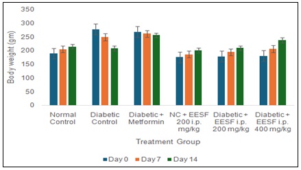
FIG. 1: EFFECT OF EESF ON ALTERATION IN BODY WEIGHT OF RATS. Data are demonstrated as mean ± SD (n = 6) at p < .05. One-way ANOVA followed by Tukey’s t-test to compare means. *p < .05, **p < .01, ***p < .001 compared to day Zero body weight.
Effect of EESF on Glycemic Control in HFD-STZ-induced Diabetic Rats: The hypoglycemic effect of the ethanol extract of Sterculia foetida (EESF) was evaluated in high-fat diet and streptozotocin (HFD-STZ)-induced diabetic rats over a 14-day period. Fasting blood glucose (FBG) levels were measured on days 0, 7, and 14, and the results are summarized in Table 3.
TABLE 3: EFFECT OF EESF ON FBG LEVELS IN HFD-STZ-INDUCED DIABETIC RATS
| Treatment Group | Day 0 (mg/dL) | Day 7 (mg/dL) [Change %] | Day 14 (mg/dL) [Change %] |
| Normal Control | 79.65 ± 4.98 | 85.54 ± 4.90 [-7.54%] | 75.65 ± 7.60 [4.98%] |
| Diabetic Control | 233.54 ± 5.70 | 249.32 ± 6.70 [-6.45%] | 242.34 ± 6.23 [-5.23%] |
| Diabetic + Metformin | 219.23 ± 5.54 | 192.23 ± 4.60* [9.35%] | 135.23 ± 6.40** [35.57%] |
| NC + EESF 200 mg/kg | 82.23 ± 6.65 | 75.23 ± 5.90 [8.23%] | 72.24 ± 5.70 [10.03%] |
| Diabetic + EESF i.p. 200 mg/kg | 191.57 ± 6.35 | 173.23 ± 5.90 [9.02%] | 142.23 ± 2.65** [25.04%] |
| Diabetic + EESF i.p. 400 mg/kg | 227.23 ± 4.97 | 179.23 ± 4.90** [19.23%] | 151.34 ± 5.64** [33.03%] |
Values are expressed as Mean ± SEM. The values of percentage reduction in glycemia are shown in brackets. *p < .05, **p < .001 compared to day zero values of the same group; NC: normal control.
In the normal control group, FBG levels remained within the normal physiological range across the study period, with minor fluctuations (79.65 ± 4.98 mg/dL at day 0 to 75.65 ± 7.6 mg/dL at day 14), indicating stable glucose metabolism. The diabetic control group exhibited persistently elevated FBG levels, rising from 233.54 ± 5.7 mg/dL at baseline to 249.32 ± 6.7 mg/dL on day 7 and 242.34 ± 6.23 mg/dL on day 14, showing no significant self-recovery and confirming the induction of hyperglycemia. The slight reduction by day 14 (-5.23%) was not statistically significant, emphasizing the chronic nature of diabetes in this model. Rats treated with metformin showed a significant and progressive reduction in blood glucose, with levels decreasing from 219.23 ± 5.54 mg/dL at baseline to 135.23 ± 6.4 mg/dL on day 14 (35.57% reduction, p < 0.001), validating the efficacy of metformin as a standard antidiabetic drug. In the non-diabetic group treated with EESF (200 mg/kg), FBG levels slightly decreased from 82.23 ± 6.65 to 72.24 ± 5.7 mg/dL by day 14, reflecting that the extract does not induce hypoglycemia in normal rats, confirming its safety.
Significantly, EESF treatment in diabetic rats led to a dose-dependent decrease in blood glucose:
- At 200 mg/kg, FBG decreased from 191.57 ± 6.35 mg/dL to 142.23 ± 2.65 mg/dL on day 14 (25.04% reduction, p < 0.001).
- At 400 mg/kg, a greater reduction was observed, from 227.23 ± 4.97 mg/dL to 151.34 ± 5.64 mg/dL (33.03% reduction, p < 0.001).
These reductions were statistically significant and comparable to the standard drug metformin, indicating that EESF exhibits strong antidiabetic activity. The results demonstrate that EESF has a potent antihyperglycemic effect in diabetic rats, and its activity appears to be dose-dependent. The mechanism of action may be attributed to the presence of flavonoids, phenolic compounds, and other bioactive constituents that improve insulin sensitivity, enhance glucose uptake, or stimulate pancreatic β-cell function. Furthermore, EESF did not lower blood glucose in normoglycemic rats, which suggests a glucose-normalizing rather than glucose-lowering effect, an important safety characteristic for potential therapeutic agents. Overall, these findings support the potential of EESF as a natural antidiabetic agent. However, mechanistic studies and long-term evaluations are recommended to confirm its efficacy and safety.
Effect of EESF on Serum Metabolic Indexes in HFD-STZ Diabetic Rats: The impact of ethanol extract of Sterculia foetida (EESF) on serum metabolic parameters including fasting serum insulin (FSI), fasting blood glucose (FBG), HOMA-IR, and HOMA-β was evaluated in HFD-STZ-induced diabetic rats. The data are summarized in Table 4.
TABLE 4: EFFECT OF EESF ON SERUM METABOLIC INDEXES IN HFD-STZ DIABETIC RATS
| Group | FSI (mmol/L) | FBG (mmol/L) | HOMA-IR | HOMA-β |
| Normal Control | 18.23 ± 0.34 | 5.45 ± 0.29 | 4.38 ± 0.76 | 252.12 ± 18.32 |
| Diabetic Control | 34.23 ± 1.45 # | 11.34 ± 0.34 # | 16.22 ± 0.98 # | 94.21 ± 9.54 # |
| Diabetic + Metformin | 22.34 ± 3.22 | 8.34 ± 0.76 *** | 8.34 ± 0.97 *** | 106.32 ± 6.22 |
| NC + EESF 200 mg/kg | 15.67 ± 1.20 | 5.33 ± 0.08 | 3.72 ± 0.35 | 170.44 ± 5.10 |
| Diabetic + EESF i.p. 200 mg/kg | 24.22 ± 3.23 ** | 9.34 ± 0.86 * | 9.32 ± 0.76 *** | 100.23 ± 4.23 |
| Diabetic + EESF i.p. 400 mg/kg | 23.23 ± 1.35 *** | 7.56 ± 0.23 ** | 7.54 ± 0.73 *** | 98.12 ± 6.12 |
#p < .001 compared to normal control group; *p < .05, **p < .01, ***p < .001 compared to diabetic control group; NC: normal control.
In the normal control group, FSI and FBG levels remained within the physiological range (18.23 ± 0.34 mmol/L and 5.45 ± 0.29 mmol/L, respectively), with corresponding HOMA-IR and HOMA-β values of 4.38 ± 0.76 and 252.12 ± 18.32, indicating normal insulin sensitivity and β-cell function. The diabetic control group showed a significant disturbance in all metabolic indices. FSI and FBG were markedly elevated (34.23 ± 1.45 mmol/L and 11.34 ± 0.34 mmol/L, respectively; p < 0.001), indicating severe hyperinsulinemia and hyperglycemia. Consequently, HOMA-IR was significantly increased (16.22 ± 0.98, p < 0.001), pointing to pronounced insulin resistance. Meanwhile, HOMA-β was drastically reduced to 94.21 ± 9.54, reflecting β-cell dysfunction due to STZ-induced damage. Treatment with metformin, a standard antidiabetic drug, partially normalized these indices. FBG and HOMA-IR were significantly reduced (p < 0.001) compared to the diabetic group, indicating improved insulin sensitivity. However, HOMA-β (106.32 ± 6.22) remained relatively low, suggesting modest recovery of β-cell function.
Importantly, EESF treatment in diabetic rats improved all metabolic parameters in a dose-dependent manner:
- At 200 mg/kg (i.p.), FBG decreased to 9.34 ± 0.86 mmol/L (p < 0.05) and HOMA-IR to 9.32 ± 0.76 (*p < 0.001), indicating improved glycemic control and insulin sensitivity. HOMA-β showed slight improvement (100.23 ± 4.23).
- At 400 mg/kg (i.p.), FBG was further reduced to 7.56 ± 0.23 mmol/L (p < 0.01) and HOMA-IR to 7.54 ± 0.73 (*p < 0.001), with HOMA-β nearing normal values (98.12 ± 6.12), suggesting improved β-cell function alongside enhanced insulin action.
In the non-diabetic group treated with EESF (200 mg/kg), metabolic markers remained stable and comparable to normal controls, suggesting the extract does not disrupt glucose-insulin homeostasis in healthy animals. These results demonstrate the therapeutic potential of EESF in managing insulin resistance and hyperglycemia in diabetic rats. The significant reduction in HOMA-IR and partial restoration of HOMA-β indicate that the extract improves both insulin sensitivity and β-cell function, which are key targets in diabetes management.
The dose-dependent efficacy suggests that 400 mg/kg of EESF is more effective, achieving improvements comparable to or better than metformin in some parameters. The activity could be attributed to bioactive constituents such as flavonoids, phenolics, and other antioxidants present in Sterculia foetida, which are known to modulate insulin signaling and reduce oxidative stress-induced β-cell damage. Overall, EESF shows promise as a natural antidiabetic agent, but further mechanistic and long-term toxicity studies are warranted to validate its clinical relevance.
Impact of EESF Intervention on IPGTT and ITT in Rats: The intraperitoneal glucose tolerance test (IPGTT) was conducted to evaluate the glucose clearance ability of ethanol extract of Sterculia foetida (EESF) in HFD-STZ-induced diabetic rats. Glucose levels were measured at 0, 30, 60, and 120 minutes after glucose administration. The area under the glucose curve (AUC) was calculated as an index of overall glucose exposure. Results are shown in Table 5
TABLE 5: EFFECT OF EESF ON INTRAPERITONEAL GLUCOSE TOLERANCE TEST IN HFD-STZ DIABETIC RATS
| Group | 0 min | 30 min | 60 min | 120 min | AUC (mg·min/dL) |
| Normal Control | 94.11 ±3.09 | 117.65 ± 7.24 | 104.23 ± 6.4 | 95.34 ± 4.9 | 12432.6± 423.7 |
| Diabetic Control | 207.34 ± 4.09 | 232.45± 4.09 | 220.43 ± 5.11 | 211.65±6.98 | 26211 ± 473.2 # |
| Diabetic + Metformin | 165.23± 5.32 | 183.12 ± 2.24 c | 169.00 ± 3.2 c | 151.33 ± 2.5 c | 19990 ± 233.8 *** |
| NC + EESF 200 mg/kg | 98.17 ± 3.1 | 123.00 ± 3.6 | 123.23 ± 5.24 | 106.12 ± 4.4 | 1375.4 ± 421.3 |
| Diabetic + EESF i.p. 200 mg/kg | 175.23 ± 4.23 | 204.12 ± 7.21c | 199.11 ± 4.76b | 191.23 ± 5.61 a | 23432± 563.2 ** |
| Diabetic + EESF i.p. 400 mg/kg | 171.23 ± 8.12 | 186.11 ± 9.23c | 178.23 ± 7.32 c | 165.23 ± 8.34 c | 21322.5 ± 783.2 *** |
#p < .001; compared to normal control group; *p < .05, **p < .01, ***p < .001 compared to diabetic control group. The letters (a, b, c), represent the significant difference among the groups ap < .05, bp < .01, cp < .001.
In the normal control group, glucose levels peaked at 30 minutes (117.65 ± 7.24 mg/dL) and gradually returned to near baseline by 120 minutes (95.34 ± 4.9 mg/dL), with an AUC of 12432.6 ± 423.7 mg·min/dL, indicating efficient glucose handling and normal insulin sensitivity. The diabetic control group showed markedly elevated glucose levels at all time points, with minimal decline by 120 minutes (211.65 ± 6.98 mg/dL) and a significantly increased AUC of 26211 ± 473.2 mg·min/dL (#p < 0.001 vs. normal control), reflecting impaired glucose tolerance and insulin resistance. Treatment with metformin significantly improved glucose tolerance. Glucose levels decreased consistently over time (165.23 → 151.33 mg/dL) and the AUC was reduced to 19990 ± 233.8 mg·min/dL (*p < 0.001 vs. diabetic control), confirming metformin's efficacy as a positive control. The non-diabetic group treated with EESF (200 mg/kg) maintained normal glucose regulation, with peak glucose at 30–60 minutes and near-basal levels by 120 minutes (106.12 ± 4.4 mg/dL), and an AUC of 1375.4 ± 421.3 mg·min/dL, showing that EESF does not cause glucose intolerance in healthy animals.
In diabetic rats, EESF produced a dose-dependent improvement:
- At 200 mg/kg, glucose levels showed a modest reduction over 2 hours (175.23 → 191.23 mg/dL) with a significantly lower AUC of 23432 ± 563.2 mg·min/dL (p < 0.01 vs. diabetic control).
- At 400 mg/kg, a more pronounced improvement was observed (171.23 → 165.23 mg/dL) with an AUC of 21322.5 ± 783.2 mg·min/dL (*p < 0.001 vs. diabetic control), indicating superior glucose clearance compared to the lower dose.
The IPGTT results confirm that EESF improves glucose tolerance in diabetic rats, with the 400 mg/kg dose demonstrating a strong antihyperglycemic effect comparable to metformin.
The improved glycemic response could be attributed to enhanced insulin sensitivity, glucose uptake, or pancreatic β-cell function mechanisms likely mediated by bioactive compounds such as flavonoids and phenolics in the extract. Furthermore, the lack of hyperglycemia in non-diabetic rats treated with EESF suggests the extract is safe and does not cause hypoglycemia in normoglycemic conditions an important feature for any antidiabetic therapy. These findings, when combined with earlier data on fasting glucose, insulin levels, and HOMA indices, support the therapeutic potential of EESF as a natural antidiabetic agent, particularly at higher doses.
Impact of EESF on the Endogenous In-vivo Antioxidant Levels: Oxidative stress plays a key role in the pathogenesis and complications of diabetes mellitus. In this study, the impact of the ethanol extract of Sterculia foetida (EESF) on endogenous antioxidant enzymes superoxide dismutase (SOD), catalase (CAT), and glutathione (GSH) was evaluated in HFD-STZ-induced diabetic rats. The results are summarized in Table 6.
TABLE 6: IMPACT OF EESF ON THE ENDOGENOUS IN-VIVO ANTIOXIDANT LEVELS IN HFD-STZ DIABETIC RATS
| Treatment Group | Superoxide Dismutase (SOD) (U/mg protein) | Catalase (CAT) (U/mg protein) | Glutathione (GSH) (µmol/mg protein) |
| Normal Control | 8.56 ± 0.32 | 14.21 ± 0.49 | 25.23 ± 1.42 |
| Diabetic Control | 3.76 ± 0.64# | 5.39 ± 0.69 # | 10.07 ± 0.82 # |
| Diabetic + Metformin | 7.87 ± 0.73 | 11.04 ± 0.83 | 19.75 ± 1.62 |
| NC + EESF 200 mg/kg | 7.74 ± 0.53 | 13.43 ± 0.73 | 23.12 ± 0.54 |
| Diabetic + EESF i.p. 200 mg/kg | 7.01 ± 0.53** ** | 9.56 ± 0.79** | 15.66 ± 0.83** |
| Diabetic + EESF i.p. 400 mg/kg | 7.62 ± 0.43*** | 10.03 ± 0.69*** | 16.23± 0.29*** |
Data are indicated as mean ± SD (n = 6) at p < .05. One-way ANOVA thereafter by Tukey’s t-test is performed to compare means. #p < .001, compared to normal control group; *p < .05, **p < .01, ***p < .001 compared to diabetic control group.
The normal control group exhibited robust antioxidant activity, as evidenced by high levels of antioxidant enzymes: superoxide dismutase (SOD) at 8.56 ± 0.32 U/mg, catalase (CAT) at 14.21 ± 0.49 U/mg, and reduced glutathione (GSH) at 25.23 ± 1.42 µmol/mg, indicating a healthy oxidative balance. In contrast, the diabetic control group showed a significant decline in all measured antioxidant parameters, with SOD reduced to 3.76 ± 0.64 U/mg, CAT to 5.39 ± 0.69 U/mg, and GSH to 10.07 ± 0.82 µmol/mg (#p < 0.001), signifying severe oxidative stress and impaired antioxidant defense due to diabetic conditions.
Treatment with metformin significantly restored antioxidant enzyme levels, with SOD at 7.87 ± 0.73 U/mg, CAT at 11.04 ± 0.83 U/mg, and GSH at 19.75 ± 1.62 µmol/mg, demonstrating its effective role in mitigating oxidative stress. Similarly, administration of the ethanolic extract of Sterculia foetida (EESF) also showed notable antioxidant-restoring effects. At a dose of 200 mg/kg (i.p.), EESF significantly increased SOD to 7.01 ± 0.53 U/mg, CAT to 9.56 ± 0.79 U/mg, and GSH to 15.66 ± 0.83 µmol/mg (p < 0.01). A more pronounced effect was observed at the 400 mg/kg (i.p.) dose, where SOD, CAT, and GSH levels were restored to 7.62 ± 0.43 U/mg, 10.03 ± 0.69 U/mg, and 16.23 ± 0.29 µmol/mg, respectively (p < 0.001), further validating the dose-dependent antioxidant potential of EESF in combating oxidative stress associated with Type II diabetes. The non-diabetic rats treated with EESF (200 mg/kg) showed no significant deviations from normal control values, indicating that EESF does not disrupt normal oxidative balance and is safe under physiological conditions. These findings underline the potential of EESF as a promising adjunct therapy in the management of diabetes and its complications.
CONCLUSION: The findings from this study clearly demonstrate that the ethanolic extract of Sterculia foetida (EESF) possesses significant antidiabetic and antioxidant properties. This is evidenced by the restoration of glucose homeostasis and insulin function, improvement in oxidative stress markers such as SOD, CAT, and GSH, and the reversal of diabetes-induced weight loss and pancreatic β-cell damage in treated rats. The therapeutic effects of EESF were found to be dose-dependent, with the 400 mg/kg intraperitoneal dose showing comparable efficacy to metformin across most evaluated parameters. These beneficial actions are likely attributed to the presence of bioactive phytoconstituents, particularly flavonoids, known for their antioxidant and insulin-sensitizing properties. Notably, the extract was observed to be non-toxic, safe, and effective in both normoglycemic and diabetic conditions, further supporting its potential as a promising natural therapeutic agent for managing Type II diabetes and its associated oxidative stress.
ACKNOWLEDGEMENT: Nil
CONFLICT OF INTEREST: Nil
REFERENCES:
- WHO Traditional medicine strategy: 2014-2023. Hong Kong, SAR, China: World Health Organization 2013.
- Global burden of diabetes. International Diabetes federation. Diabetic atlas fifth edition 2011, Brussels. Available at http://www.idf.org/diabetesatlas (Accessed 18th December 2011).
- Chamnan P, Simmons RK, Forouhi NG, Luben R. Khaw Ky and Wareham NJ: Incidence of type 2 diabetes using proposed HbA1c diagnostic criteria in the EPIC-Norflok cohort: Implication for preventive strategies. Available at http://care.diabetesjournal.org (Accessed 19th December 2011).
- Department of Health and Human Services. Centres for Disease Control and Prevention, 2011. National diabetes fact sheet: national estimates and general information on diabetes and prediabetes in the United States, 2011. Available at http://www.cdc.gov/diabetes/pubs/pdf/ndfs_2011.pdf (Accessed December, 20th 2011).
- Si-Yuan Pan, Gerhard Litscher, Si-Hua Gao, Shu- Feng Zhou, Zhi-Ling Yu and Hou-Qi Chen: Historical perspective of traditional indigenous medical practices: the current renaissance and conservation of herbal resources, Evidence-Based Complementary and Alternative Medicine 2014; 1-20.
- Popa IC: The lists of plant synonyms in De materia medica of Dioscorides, Global Journal of Science Frontier Research 2010; 10(3): 46-49.
- Bijauliya RK, Alok S, Chanchal DK, Sabharwal M and Yadav RD: An updated review of pharmacological studies on Azadirachta indica (neem). International Journal of Pharmaceutical Sciences and Research 2018; 9(7): 2645-55.
- Moshi MJ: Current and future prospects of integrating traditional and alternative medicine in the management of diseases in Tanzania, Tanzania Health Research Bulletin 2005; 7(3): 159-166.
- Maria Russo, Carmela Spagnuolo, Idolo Tedesco and Gian Luigi Russo: Phytochemicals in Cancer prevention and therapy: Truth or Dare, Toxins 2010; 2(4): 517-551.
- Agarwal P, Amreen Fatima and Singh PP: Herbal medicine scenario in India and European countries, Journal of Pharmacognosy and Phytochemistry 2012; 1(4): 88-93.
- Azevedo M and Alla S: Diabetes in sub-saharan Africa: kenya, mali, mozambique, Nigeria, South Africa and zambia. Int J Diabetes Dev Ctries 2008; 28(4): 101-108 10.4103/0973-3930.45268
- Zimmet P, Alberti KG and Shaw J: Global and societal implications of the diabetes epidemic. Nature 2001; 414(6865): 782-787 10.1038/414782a [DOI] [PubMed]
- Bijauliya RK, Alok S, Sabharwal M and Chanchal DK: Syzygium cumini (linn.)-an overview on morphology, cultivation, traditional uses and pharmacology. International Journal of Pharmaceutical Sciences and Research 2018; 9(9): 3608-20.
- Wild S, Roglic G, Green A, Sicree R and King H: Global prevalence of diabetes: estimate for the year 2000 and projections for 2030. Diabetes Care 2004; 127(5): 1047-1053. 10.2337/diacare.27.5.1047
- Yach D, Hawkes C, Gould CL and Hofman KJ: The global burden of chronic diseases: overcoming impediments to prevention and control. JAMA 2004; 291(21) :2616-2622 10.1001/jama.291.21.2616 [DOI] [PubMed] [Google Scholar]
- Ripsin CM, Kang H and Urban RJ: Management of blood glucose in type 2 diabetes mellitus. Am Fam Physician 2009; 79(1): 29-36
- Hu FB, Manson JE, Stampfer MJ, Colditz G, Liu S and Solomon CG: Diet, lifestyle, and the risk of type 2 diabetes mellitus in women. N Engl J Med 2001; 345(11): 790-797 10.1056/NEJMoa010492
- Lang IA, Galloway TS, Scarlett A, Henley WE, Depledge M and Wallace RB: Association of urinary bisphenol A concentration with medical disorders and laboratory abnormalities in adults. JAMA 2008; 300(11): 1303-1310 10.1001/jama.300.11.1303 [DOI] [PubMed] [Google Scholar]
- Niran Vipunngeum and Chanida Palanuvej: Fatty acids of Sterculia foetida seed oil, Journal of Health Research 2009; 23(3): 157.
- Raja TAR, Ramanarayana Reddy RV and Uma Meheswara Rao K: Evaluation of Anticonvulsant effect of Sterculia foetida (pinari) in Pentylenetetrazole (PTZ) and Maximal Electrical Shock induced convulsions in albino rats, World Journal of Pharmacy and Pharmaceutical Sciences 2014; 3(3): 1898-1907.
- Shivakumar Singh P and Vidhyasagar GM: In-vitro antidermatophytic activity of low polar petroleum ether and inter polar methanolic extract of Sterculia foetida Linn, International Journal of Pharma and Bio Sciences 2014; 5(2): 872-879.
- Narsing Rao Galla: In-vitro Antioxidants activity of Sterculia foetida Linn., seed methanol extract. AJPTR 2012; 2(6): 572-581.
- Pierangeli G: Antimicrobial activity, cytotoxicity and phytochemical screening of Ficus saptic Burn and Sterculia foetida. J of Med Plant Res 2011; 4(1): 58-63.
- Majumdar AM: Pharmacological studies on Sterculia foetida leaves, Pharmaceutical Biology 2011; 38(1): 13-18.
- Usharani P and Rajasekharreddy P: Toxic and antifeedant activity of Sterculia foetida Linn., seed crude extract against Spodoptera litura F. and Achaea janata L., Journal of Biopesticide 2009; 2(2): 161-164.
- Noamesi BK, Idigo JC, Adeoye AO and Fadiran EO: Preliminary report on the bronchodilator properties of the aqueous stem bark extract of Sterculia foetida, Planta Med 1986; (6): 547.
How to cite this article:
Bhadoriya S, Mehta SC and Rajput M: Study of effect of Sterculia foetida seeds on insulin resistance in type ii diabetes mellitus model in rats. Int J Pharmacognosy 2026; 13(8): 887-97. doi link: http://dx.doi.org/10.13040/IJPSR.0975-8232.IJP.13(8).887-97.
This Journal licensed under a Creative Commons Attribution-Non-commercial-Share Alike 3.0 Unported License.
Article Information
15
887-897
537 KB
103
English
IJP
Sneha Bhadoriya *, S. C. Mehta and Madhu Rajput
Shri Ramnath Singh Institute of Pharmaceutical Sciences & Technology, Sitholi, Gwalior, Madhya Pradesh, India.
snehabhadoriya0610@gmail.com
10 July 2026
22 July 2026
23 July 2026
10.13040/IJPSR.0975-8232.IJP.13(8).887-97
01 August 2026
 Download