


RHEUMATOID ARTHRITIS: HERBAL DRUG APPROACHES
HTML Full TextRHEUMATOID ARTHRITIS: HERBAL DRUG APPROACHES
Malati Salunke * and Shweta Aher
Department of Pharmacognosy, Poona College of Pharmacy, Bharati Vidyapeeth (Deemed to be University), Pune, Maharashtra, India.
ABSTRACT: Rheumatoid arthritis (RA) is a long-term systemic autoimmune disease marked by systemic problems, progressive joint deterioration, and persistent synovial inflammation. Environmental contaminants, asbestos and silica dust from the workplace, smoking, vitamin D insufficiency, and hormonal factors all contribute to oxidative stress and immunological activation, which in turn promotes autoimmunity and further RA. Despite better clinical results from Non-steroidal anti-inflammatory drug (NSAID), Disease-modifying antirheumatic medications (DMARD) and biologics, drawbacks such as side effects and long-term toxicity. As alternative therapy alternatives in the management of RA, herbal and phytopharmaceutical substances with anti-inflammatory, antioxidant, and immunomodulatory qualities have thus drawn more attention. Furthermore, new approaches for the targeted and long-term distribution of synthetic and herbal medicines are provided by recent developments in innovative drug-delivery systems. This review summarizes the major RA pathogenesis, conventional therapies, natural therapies and emerging therapeutic delivery strategies aimed at improving efficacy and safety.
Keywords: Rheumatoid arthritis, Drug delivery system, Herbal plant, Novel drug delivery system
INTRODUCTION: The long-term, systemic autoimmune illness known as rheumatoid arthritis (RA) impacts the synovial joints mainly, resulting in stiffness, pain, swelling, and ultimately joint destruction. Globally, 0.5% to 1% of adults have RA, with a higher prevalence in women than man 1. The illness is so chronic and progressive, it not only lowers quality of life and physical function but also raises morbidity and healthcare costs. Immune cell infiltration into the synovium, the thin membrane lining the joints, is a hallmark of RA.
This eventually causes cartilage and bone degradation, pannus development, and chronic inflammation. Immune dysregulation, environmental stressors, and genetic predisposition are all implicated, albeit the precise cause is still unknown 2.
A number of genes, particularly in younger individuals, HLA-DRB1 alleles has strongly link with increased chance of getting RA. Environmental elements like smoking, air pollution, and an imbalanced gut microbiome and the pathophysiology of RA is also known to be influenced by hormones. These factors can initiate autoimmune responses in genetically predisposed individuals, leading to chronic inflammation and joint damage 3. Symmetrical joint discomfort, morning stiffness lasting more than half an hour, exhaustion, low-grade fever, and in more severe cases, joint deformity are the classic clinical signs of RA. Rheumatoid nodules, interstitial lung disease, vasculitis, and cardiovascular issues are examples of systemic symptoms 4. To avoid irreparable joint injury and systemic consequences, early diagnosis and timely treatment are crucial. Clinical, serological, and radiographic results are used to determine the urgent diagnostic criterion. Imaging modalities like X-ray, ultrasound, and MRI help in assessing joint damage and disease progression 5. Over the past few decades, there has been a major evolution in the therapy of RA. While corticosteroids and non-steroidal anti-inflammatory treatments (NSAIDs) relieve symptoms, disease-modifying anti-rheumatic medications (DMARDs) such methotrexate, sulfasalazine, and leflunomide are the cornerstone of RA treatment, as they reduce synovial Inflammation, supress autoimmune activity and improve long term functional outcomes. By focusing on particular immune pathways, biologic medicines such as TNF inhibitors, IL-6 inhibitors, and β-cell depleting treatments have completely changed how moderate to severe RA is managed 6. However, these medications are expensive and may have negative side effects, so careful patient selection and monitoring are required. Despite the availability of effective pharmacological treatments, there is growing interest in complementary and alternative therapies, including herbal medicines, due to concerns over long-term safety and side effects of conventional drugs. Ayurveda, the traditional Indian system of medicine, offers several herbal formulations and lifestyle interventions aimed at reducing inflammation and restoring immune balance.
Pathophysiology of RA: Rheumatoid arthritis, a complicated inflammatory disease that mostly affects the joints, has an unclear cause. Due to the intricacy of RA, which is based on a poorly known pathophysiological process, effective care requires a multidisciplinary approach. However, further research is needed before RA may be treated 7.
Despite the fact that the pathophysiological mechanisms behind RA remain unclear, a number of theories have been put forth that the immunological processes can take place years before joint inflammatory symptoms appear this is known as the pre-RA period 8.
Various environmental, Lifestyle factors, and immunological parameters contributing to this early disease onset are discussed below, are also show in Fig. 1 as they play a crucial role in triggering and progressing rheumatoid arthritis.
Environmental Factor:
Air Pollution: Rheumatoid arthritis development and progression have lately been linked to air pollution as a significant environmental risk factor. It consists of a complex mixture of gases and different-sized suspended particulate matter (PM). Pollutants are released into the atmosphere by a variety of man-made and natural causes, such as the burning of fossil fuels, industrial emissions, agricultural practices, solvent use, volcanic eruptions, and wind-blown dust. The development and aggravation of RA can be facilitated by these airborne pollutants, which can cause oxidative stress, immunological dysregulation, and systemic inflammation 9.
The pathophysiology of RA can be linked to air pollution, according to three important epidemiological studies. Three significant epidemiological studies carried out in the US, Canada, and Sweden have found a connection between the pathophysiology of RA and air pollution. One study looked at the connections between RA activity and air pollutionthe means of the regression models, Sulphur dioxide and nitrates were found to be significant risk factors for the onset of RA 10.
Occupational Dust: In fact, the epidemiological research that looked into a potential link between the silica breathed and the development of RA found an elevated risk. Evidence of noticeably elevated levels of Interleukin (IL) 1α is part of the hypothesis underlying this relationship. Tumor necrosis factor-alpha (TNF-α), IL-1β, IL-2, IL-4, IL-6, IL-10, and reduced antioxidant state in ceramic workers, indicating an activated immune system and the inflammatory reaction to contact with silica. According to certain research, this correlation appears to grow with the length of time spent exposed to silica 11.
Lifestyle Factors:
Smoking and Development of RA: Primary environmental risk factor for developing RA are exposure to tobacco smoking, which is thought to be the cause of 20% of all RA cases and 35% of people who test positive for the anti-citrulline antibody (ACPA). In 1996, twin research confirmed the long-suspected link between smoking and the onset of RA. One twin from each of the 71 dizygotic and 79 monozygotic twin pairs whose smoking habits were assessed had RA. Smoking raised the likelihood of having RA by a factor of four in twin pairs with inconsistent smoking patterns 12.
Vitamin D: One of the things that leads to the development of autoimmune rheumatic disorders is a lack of vitamin D. Numerous global research have demonstrated that vitamin D insufficiency is seen in RA patients and appears to be negatively correlated with the illness. Because vitamin D acts on monocytes, natural killer cells, and antigen-presenting cells to create a tolerogenic immune phenotype, a lack of it exacerbate autoimmunity 13.
Gut Microbiota: The pathophysiology of rheumatoid arthritis is significantly influenced by the gut microbiota, the most densely populated microbial community in the human body. Because it can activate autoimmune pathways through a variety of mechanisms, such as molecular mimicry, increased intestinal permeability, activation of antigen-presenting cells via toll-like receptors (TLRs), stimulation of T-cell differentiation, and amplification of mucosal inflammation, intestinal dysbiosis has been connected to the aetiology of RA 14.
Role of Dendritic Cells in RA Inflammation: Dendritic cells (DCs) are essential in rheumatoid arthritis (RA) because they stimulate inflammation and present antigens to T lymphocytes. As a result of conventional and plasmacytoid DCs (pDCs) migrating to inflammatory joints through CCR6–CCL20 interactions, their distribution and function are altered, which leads to autoimmune inflammation 15. Pro-inflammatory cytokines including IL-12 and IL-23 are released by mature DCs within the joint, causing Th17 responses and upsetting the Th1/Th2/Th17 balance. Furthermore, CD14⁺ inflammatory DCs and pDCs increase inflammation via TGF-β, IL-1β, IL-6, IL-23, and type I interferons (IFN-α/β) 16. pDCs also stimulate the generation of autoantibodies via BAFF, especially in RA patients who have anti-citrullinated protein antibody (ACPA) positivity 17.
Immune Mediators:
T-Cell: T-cells are essential to the pathophysiology of rheumatoid arthritis because they promote autoimmune reactions and maintain inflammation in the synovium. Antigen-presenting cells in lymphoid organs and joint tissues stimulate autoreactive T cells, which then migrate into the synovium and interact with osteoclasts, macrophages, dendritic cells, and synoviocytes to promote chronic inflammation and joint degeneration 18.
Th1 cells play a role by secreting IL-2, IFN-γ, and TNF-β, which promote inflammation and macrophage activation. By encouraging neutrophil recruitment and osteoclast differentiation, Th17 cells which are triggered by IL-6, IL-1β, and IL-23 further accelerate the course of the disease, especially in early RA 19. Moreover, malfunctioning regulatory T cells (Tregs) and increased cytotoxic CD4⁺CD28⁻ T-cell populations are linked to RA's ongoing inflammation and loss of immunological tolerance 20.
β Cell: β cells contribute to adaptive immune responses and synovial inflammation, which are important aspects of the pathophysiology of rheumatoid arthritis. Under the effect of chemokines, B-lymphocyte stimulatorand APRIL, βcells concentrate into tertiary lymphoid follicles or T-cell–β-cell aggregates in the RA synovium. The therapeutic effectiveness of rituximab, which targets CD20⁺ B cells in RA patients, supports their pathogenic role. In addition to producing autoantibodies, β cells also aid in the advancement of illness by presenting antigens and secreting pro-inflammatory cytokines including lymphotoxin and TNF-α, which increase humoral immunity and synovial inflammation 21.
Macrophages: In RA synovitis, macrophages are important effector cells that perpetuate inflammation and joint destruction. Major cytokines including IL-1β, IL-6, and TNF-α are released by activated macrophages in the pro-inflammatory synovial milieu 22. These cytokines trigger inflammatory cascades and attract neutrophils and other innate immune cells to the synovitis site. Proteases, phospholipases, defensins, myeloperoxidase, and TNF-α are among the oxidants and inflammatory mediators that are released in significant quantities within damaged joints as a result of these cytokines additional activation of neutrophils. This accelerates joint deterioration and increases tissue damage in RA 23.
Fibroblasts: Moreover, RANK-L from cytokine-stimulated fibroblasts and TNF-α and IL-6 from activated immune cells encourage the development of preosteoclasts and macrophages into osteoclasts, which is specialized in breaking down bone. Activated fibroblasts not only directly cause local joint damage by generating RANK-L and MMPs, However, they also go from one joint to another, inflaming additional joints which explains the symmetrical pattern of the disease 24. Therefore, joint inflammation in RA is characterized by a specific tissue response in which local fibroblasts adopt an aggressive pro-inflammatory phenotype with invasive, matrix-regulating, and osteoclast-generating properties, in addition to the activation of resident and invasive immune cells 25.
Cytokines Role in RA Inflammation: In RA, cytokines are important mediators in the development and upkeep of inflammation. Progressive joint deterioration results from a prolonged inflammatory milieu in the synovium caused by an imbalance between pro- and anti-inflammatory cytokines. Tumor necrosis factor-α (TNF-α), interleukins (IL)-1, IL-6, IL-17A, and interferon-γ (IFN-γ) are examples of pro-inflammatory cytokines that coordinate immune cell recruitment, activation, and retention at the site of inflammation 26. These cytokines cause endothelial cells, fibroblast-like synoviocytes, and synovial macrophages to become activated, which prolongs the creation of mediators of inflammation and exacerbates synovitis 27. By stimulating the appearance of several downstream cytokines and chemokines, TNF-α functions as a key regulator of inflammation, encouraging leukocyte infiltration and sustaining the inflammatory cascade in the synovial region. By stimulating the synthesis of cytokines and chemokines, encouraging neutrophil recruitment, and escalating local inflammation, IL-17A amplifies inflammatory responses and accelerates the course of disease 28. By improving antigen presentation and macrophage activity, IFN-γ supports cytokine-driven immune activation within the joint and helps initiate early inflammatory reactions. Furthermore, via influencing immune cell contacts and maintaining chronic inflammation in RA joints, cytokines including RANK-L, IL-33, IL-36, IL-32, and IL-34 contribute to the inflammatory milieu. Overall, cytokines play a major role in the pathophysiology of RA by stimulating immune cell activation, cytokine amplification loops, and persistent inflammatory signalling 29.
Contribution of Autoantibodies to the Pathogenesis of RA: The pathophysiology of rheumatoid arthritis is significantly influenced by autoantibodies produced as a result of abnormal activation of autoreactive β cells. Rheumatoid factor (RF) and anti-citrullinated protein antibodies (ACPAs) are the two main autoantibodies linked to RA, and their presence characterizes seropositive RA 30. Type II collagen, fibrinogen, vimentin, histones, fibronectin, and α-enolase are examples of citrullinated self-proteins that are recognized by ACPAs, which are primarily of the IgG, IgA, and IgM isotypes. These autoantibodies have a high clinical specificity and are seen in 60–80% of RA patients. ACPAs are crucial for early illness development and diagnosis since they can be seen in the bloodstream years before clinical symptoms appear 31. The main way that autoantibodies contribute to RA pathogenesis is by forming immune complexes and then activating complement, which causes inflammation in the synovium. ACPAs boost osteoclast development and bone resorption via stimulating macrophages and boosting the synthesis of pro-inflammatory cytokines such as RANK-L and TNF-α 32.
Furthermore, by binding to citrullinated proteins produced on osteoclast precursor cells and promoting their maturation into bone-resorbing osteoclasts, ACPAs directly contribute to bone erosion. Their harmful involvement is further supported by a substantial correlation between the development of bone erosions and ACPA positive. By encouraging IL-8-dependent osteoclast activation, ACPAs have been linked to joint discomfort in addition to structural damage, suggesting that autoantibodies actively contribute to both the inflammatory and symptomatic features of RA 33.
Role of CD147: The immunoglobulin superfamily includes the CD147 (an extracellular matrix metalloproteinase inducer, or EMMPRIN). The expression of matrix metalloproteinases (MMPs) is induced by CD147. It has been discovered in the synovial membrane of RA patients that Peripheral blood CD14² monocytes have more CD147 receptors, which boosts the cellular response to a molecular stimulation. According to reports, RA patients have higher levels of CD147 expression on monocytes and macrophages, which may be the cause of increased MMP secretion, cyclophilin A-mediated cell migration into the patient's joints, and cell invasion all of which ultimately lead to the patients' loss of bone and cartilage 34.
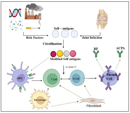
FIG. 1: IMMUNOPATHOGENESIS OF RHEUMATOID ARTHRITIS
Symptoms: Rheumatoid arthritis is a condition that is somewhat common chronic inflammatory disease that mostly affects synovial joints. It results in discomfort, edema, stiffness, inflammation, and ultimately loss of joint function 35. Early-stage RA and poorly treated later stages of the illness have somewhat different clinical presentations. RA in its early stages is distinguished by widespread illness symptoms such fatigue, malaise, flu-like sensation, swollen and aching joints, and morning stiffness. It is associated with higher erythrocyte sedimentation rate (ESR) and C-reactive protein (CRP) levels 35, 36. Patients often experience rigidity from the start of the illness and their inability to move their fingers when they wake up, this is sometimes characterized as having trouble making a fist. Swelling and restricted movement are frequently linked to arthralgia. These signs are probably to manifest in the knees, wrists, elbows, cervical spine, and joints in the fingers and toes (including the metatarsophalangeal, proximal interphalangeal, and metacarpophalangeal joints). However, the early start is rarely found in the distal interphalangeal joints 37.
Common concomitant symptoms include xerostomia from 40% of individuals have sialadenitis, 35% have subcutaneous rheumatoid nodules on the forearm's extensor surface, and 45% have dry eyes linked to keratoconjunctivitis sicca. compressive neuropathy-related numbness in the fingers and feet 25%, and interstitial pneumonia-related dry cough after physical activity 15% 38. Subcutaneous nodules, interstitial pneumonia, pericarditis, conjunctivitis, and hepatosplenomegaly might also accompany the illness.
If the arthritis is not treated in a timely manner, the degradation of bone and cartilage in the joints could cause permanent disability. In order to eradicate symptoms, manage disease activity, reduce development, and prevent joint degeneration, early treatment is crucial 39.
Diagnosis: When treating RA, a precise diagnosis is crucial. Early diagnosis reduces joint degeneration and prevents disability in 90% of patients in addition to slowing the progression of the disease. RA patients have stiffness, discomfort, and inflammation in their joints especially in early days. A doctor's evaluation is typically used to make the diagnosis of RA 40. Due to its lack of sensitivity in early RA, the American College of Rheumatology classification standards (1987) was criticized and replaced by the American College of Rheumatology (ACR, 2010) criteria, which were created by European 41. EULAR stands for League against Rheumatism. RA disease activity is measured using various scoring methods.
Laboratory testing and clinical imaging techniques are used in the diagnosis of RA. Anaemia, the presence of rheumatoid factor, antibodies against cyclic citrullinated peptides, and an increase in the rate of erythrocyte sedimentation are among the laboratory testing techniques. While Long-lasting pain and stiffness in the morning are symptoms that provide some insight into the illness. For the diagnosis and tracking of disease activity in RA patients, both MRI and ultrasound have been suggested. Additionally, bone erosions can be detected by ultrasound. Because of these features, ultrasonography is frequently utilized in both RA diagnosis and disease state monitoring through clinical practice and clinical trials. Despite the fact that magnetic resonance imaging (MRI) is a highly sensitive diagnostic tool that can identify, for instance, synovial hypertrophy or pannus formation before bone erosion occurs, its routine use in the diagnosis of RA is restricted by cost considerations and the limited capacity to image multiple joints in a single measurement 42.
Risk Factor: The development of RA is influenced by a number of risk factors, and the complexity of the condition depends on how the host and risk factors interact to ascertain the total risk of disease severity, persistence, and susceptibility. And various factors are dietary, sex, caffeine, hormonal environmental, genetic, vitamin D deficiency, Microbiota and infectious agents 43 are seen in Fig. 2.
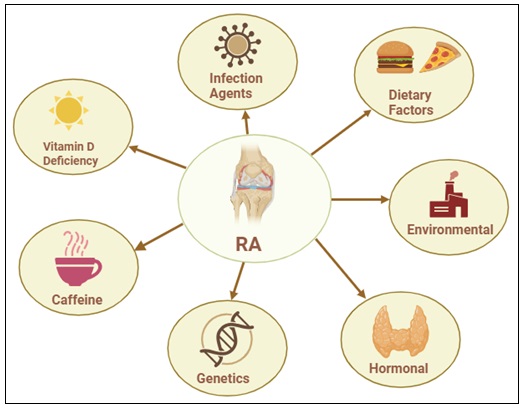
FIG. 2: RISK FACTOR IN RA DISEASE
Dietary Factors: Over time, dietary determinants and consumption patterns have also been assessed. RA is influenced by dietary factors, and research has demonstrated that vegetarian diets and fasting can slow the disease's progression. Additionally, increasing fruit and fatty fish while avoiding red meat intake are linked to a lower incidence of RA. Coffee drinking may increase the risk of RA, this could be due to its role in the creation of RF 44. According to the case research, drinking liquor might possess a positive impact on RA by reducing the chance of getting ACPA-positive RA, however further research is needed to support this theory. As a result, each person should have a customized diet 45.
Microbiota and Infectious Agents: Rheumatoid arthritis has long been thought to have an infectious cause, according to the infectious hypothesis 46. This idea is supported indirectly by the fact that RA incidence has decreased in populations with better health and cleanliness, indicating that microbial exposure may serve as a risk factor for the disease 47. Translational and epidemiological research that has identified particular bacteria, viruses, and other microbes linked to RA lends more credence to this theory 48. These infectious pathogens may contribute to the onset and progression of RA by inducing autoimmune responses through non-specific immune activation, molecular mimicry, and disturbance of host–microbiota equilibrium 49.
Sex: Generally speaking, RA is two to three times more common in women than in men. According to estimates, the cumulative life span risk of developing an adult-onset disease is 1.7% for men and 3.6% for women. The condition is more common in women, which is attributed to the corresponding effects of estrogen on a person's immune system The role of hormones in the genesis of disease, however, is still debatable 50. Nulliparity has been identified as one of the factors that raises the incidence of this illness in females, and pregnancy is also associated with the disease's remission. Even Nevertheless, illness flickers are typical throughout the postpartum phase. The majority of the disease's symptoms in females appear at menopause or middle age. Because they have significantly greater levels of ACPAs and are more likely to have RA, men become ill later than women 51.
Caffeine: There have been reports regarding the impact of caffeine on the onset of RA, but the inconsistent results make a true impact improbable. There was no correlation found in the Iowa Women's Health Study between daily caffeine intake and the quantity of cups of caffeinated coffee intake, while consuming more than four cups of decaffeinated coffee per day was linked to a 2.5-fold higher risk. They also discovered that drinking three cups of tea a day had a protective effect. For people with seropositive disease, these correlations were more pronounced 52.
Hormonal: The anti-inflammatory qualities of progesterone and androgens, which are diminished in RA patients of both sexes, the hormone imbalance typically linked to estrogens is frequently considered as being proinflammatory. But their behavior is much more complicated, and estrogens really have anti-inflammatory qualities in a variety of tissues and cells 53. The overall net effect is likely to be influenced by additional elements include the reproductive stage, the main cell types and estrogen receptors that are involved, and serum and tissue levels 54. Understanding the contradictory results for several hormonal and reproductive factors in the risk of RA requires an understanding of these mechanistic features. Early menarche, age of the first childbirth, parity, breastfeeding, Pregnancy loss and hormone replacement medication have all been linked to a higher, unchanged, chance of developing RA 55, 56. However, circumstances such as the post-menopausal phase and early menopause, puerperium, and anti-estrogen medications, such as aromatase inhibitors and selective estrogen receptor modulators, have increasingly been identified as RA risk factors 57.
RA Risk Factors Related to the Environment: The primary risk factors for the environment that double the chance of getting rheumatoid arthritis are non-genetic variables like smoking, air pollution, silica, and asbestos. Patients with ACPA-positive diseases are the only ones affected 58. In recent times, the field of RA has concentrated more onair pollution, which is a combination of chemicals like carbon monoxide, ozone (O3), sulfur dioxide (SO2), nitrates, and suspended particle matter (PM) of different sizes 59.
These pollutants are mostly caused by fixed fuel burners, heavy traffic, industry, solid fuel combustion, and forest fires. In this sense, the production of pro-inflammatory cytokines in RA patients is regulated by nuclear factor ̙app a B (NF-B), which can be induced by when gaseous pollutants or fine/ultrafine PM are inhaled into the respiratory system, free reactive oxygen species are produced. Auto-antigens are released by resting monocytes when they mature into dendritic cells. After being activated by these cytokines, self-reactive T lymphocytes travel to target tissues, such as synovial joints, and eliminate cells that express autoantigens. Reactive oxygen species can also be made worse by unfavorable environmental risk factors for RA 60.
Genetic: Numerous indications have made it clear that genetics plays a significant role in the onset of RA. The greater prevalence of RA in families as a whole is one of these reasons, estimates of the familial risk contribution range from 40 to 50 percent of seropositive RA, with the biggest risks being borne by first-degree relatives (FDRs) 61. Furthermore, the higher incidence of RA incertain racial groups, including Native Americans, They exhibit prevalence rates of 5–7% 62. Suggests that genetic factors play a role in the disease 63.
Non-genetic familial or cohort factors may also affect family or racial ethnic group risk, even though several unique genetic loci have been discovered to be associated with an elevated risk for RA and in other circumstances a decreased risk 64. The main genetic risk factors are "shared epitopes" (SE), a collection of alleles in the major histocompatibility complex (MHC) that generate amino acid sequences that predict structural similarities in the human leukocyte antigen (HLA) peptide-binding groove. Although some study suggests a lesser percentage, it is believed that SE alleles together account for up to 40% of the hereditary risk for RA. Notably, previous nomenclature has reflected the large number of alleles thought to contain the SE, many of which are located in the HLA DRB1 region 65.
Vitamin D Deficiency: Vitamin D is thought to have immunomodulatory properties. The risk of RA has indirectly related with vitamin D consumption 66. It has been demonstrated that taking vitamin D supplements for five years, with or withoutomega-3 fatty acids, lowers the risk of developing an autoimmune illness 67, 68.
Treatment of RA: The main objectives are to minimize joint discomfort and inflammation, maximize joint function, and prevent joint deformity and destruction and approaches for managing rheumatoid arthritis 69. In the treatment of RA, controlling pain, preventing long-term joint damage, and reducing inflammation are the main challenges. There is currently no long-term treatment for this illness. The goals of treatment are to improve physical function and quality of life, reduce inflammation and discomfort, preserve joint functioning to reduce joint damage and consequences, and decrease the disease's course 70. Treatment available for Rheumatoid arthritis are as follows:
First-Line Management: NSAIDS and Corticosteroids: Relieving pain and reducing inflammation are the main objectives of first-line therapy. Nonsteroidal anti-inflammatory medications, such as acetylsalicylate, naproxen, ibuprofen, and etodolac, are regarded as fast-acting medications. aspirin, it is among the first NSAIDs to be used to treat joint pain 71.
High dosages of aspirin can cause tinnitus, hearing loss, and gastrointestinal intolerance as side effects. In addition to aspirin, there are additional NSAIDs that are equally effective but more recent to the market. These medications also require fewer daily doses. NSAIDs limit the production of prostacyclin, thromboxane, and prostaglandins via blocking cyclo-oxygenase. Gastrointestinal (GI) bleeding, ulcers, nausea, and abdominal pain are typical adverse effects. Glucocorticoids and mineral corticoids are the two categories of corticosteroids. Glucocorticoids have vasoconstrictive, immunosuppressive, and anti-inflammatory qualities. Mineralocorticoids are involved in the regulation of electrolytes and water balance. Patients with RA are frequently prescribed glucocorticoids, such as dexamethasone, methylprednisolone, prednisone, and triamcinolone, due to their potent anti-inflammatory and immunosuppressive properties 72, 73. Because glucocorticoids bind toglucocorticoid receptors and create the receptor-glucocorticoid complex, they have anti-inflammatory properties. This complex changes the transcription of several genes linked to the inflammatory response by binding to DNA regions called glucocorticoid response elements 74. Similar to other medications, glucocorticoids can have a number of side effects, particularly. When taken in large or prolonged doses. These adverse effects include, among others, peptic ulcers, diabetes, osteoporosis, metabolic syndrome, and cataracts 75.
Second-Line Management: Disease-Modifying Antirheumatic Drugs: By reducing the advancement of joint degradation and deformity, second-line therapy aims to induce remission. Because they take weeks or months to start working, medications are referred to as slow-acting. Additionally, disease-modifying antirheumatic medications (DMARDs) can lower the incidence of lymphoma, which is linked to RA 76.
The cornerstone of RA treatment is DMARDs, which include a variety of medications that slow the disease's progression and manage symptoms. Methotrexate (MTX), a traditional synthetic DMARD, has anti-proliferative properties similar to those of folic acid. MTX causes T cell and platelet death, inhibits the synthesis of amino acids and polyamines, and impairs the metabolism of purines and pyrimidines. However, in clinical practice, there have been reports of gastrointestinal, infectious, pulmonary, and hematologic adverse effects, as well as risks of developing skin cancer and bone marrow abnormalities. MTX should be used as a monotherapy for patients with moderate or severe illness. MTX can be used in conjunction with a supplementary medication when the disease does not respond well to it, or it can be completely replaced by another DMARD if side effects are noted 77.
Other synthetic DMARDs include sulfasalazine, leflunomide, Chloroquine and hydroxychloroquine. Hydroxychloroquine can be used as an initial treatment for people with mild illness courses. For the treatment of RA, leflunomide and sulfasalazine are other often prescribed medications, primarily in situations where patients are contraindicated for MTX. MTX, sulfasalazine, and hydroxychloroquine are sometimes used as triple-drug therapy. Notably, due of its affordability and therapeutic effectiveness, MTX is recommended for usage in patients. Nonetheless, it is said that using MTX in conjunction with other medications is a more effective therapeutic approach than using MTX by itself 78.
When reactions to artificial DMARDs are insufficient, biological DMARDs are chosen. In Japan, medications that target TNF (such as infliximab, etanercept, adalimumab, golimumab, and certolizumab), IL-6 and the T cell-selective Abatacept, a co-stimulation modulator, can be given by drip infusion or injection. These medications all have strong and quick clinical effects. In around half of the cases, they can induce remission when used in conjunction with methotrexate. For extended periods of time, biological DMARDs can also stop the deterioration and functioning of joints.
Combinational Therapy: Here, the use of biological agents in conjunction with DMARDs is referred to as combinational therapy in RA. Clinical recommendations suggest using both of them in combination when DMARDs are either not tolerated by RA patients or do not exhibit any reaction and the use of DMARDs alone is ineffective. These recommendations were developed in response to evidence from clinical trials that concurrent use of biologics and DMARDs, such as TNFi and MTX, demonstrated a significantly higher response than using one of them in monotherapy. In patients who did not respond to MTX monotherapy, tocilizumab as combinational therapy with MTX demonstrated more benefits than tocilizumab monotherapy. Combinational medication administration, however, frequently has an impact on each patient's response and the severity of their prognosis 79.
Cell-based Therapies:
Mesenchymal Stem Cell-Based Therapy: Mesenchymal stem cells (MSCs) have attracted considerable interest in regenerative medicine over the past three decades due to their differentiation potential, capability to secrete cytokines, and strong immunomodulatory characteristics, giving them viable options for the management of immune-mediated disorders such as rheumatoid arthritis (RA). The International Society for Cellular Therapy (ISCT) characterizes human MSCs as adherent cells that do not express hematopoietic markers, exhibit surface markers CD73, CD90, and CD105, and can differentiate into osteoblasts, adipocytes, and chondroblasts in-vitro 80.
MSCs control both innate and adaptive immune responses by lowering pro-inflammatory immune cell populations like dendritic cells, macrophages, natural killer cells, and B- and T-lymphocytes while promoting an anti-inflammatory environment. MSCs can polarize into pro-inflammatory (MSC1) or anti-inflammatory (MSC2) phenotypes based on the inflammatory environment. MSCs are activated toward an anti-inflammatory phenotype under inflammatory circumstances marked by increased TNF-α and IFN-γ 81.
MSCs emit interleukin-10 (IL-10), prostaglandin E2 (PGE2), nitric oxide (NO), indoleamine 2,3-dioxygenase (IDO), and transforming growth factor-β to assist inhibit the immune system. In human MSCs, IDO is induced by IFN-γ and converts tryptophan into kynurenine. This process halts T-cell growth and promotes regulatory immune cells. Additionally, MSCs guide immune cell development toward suppressive types, release extracellular vesicles, and interact with other cells to lessen immune responses. MSCs show potential as a treatment for autoimmune diseases like rheumatoid arthritis. They can target inflamed tissues, lower the release of pro-inflammatory cytokines, inhibit immune cell growth, and encourage immune tolerance 82.
Surgery and other Treatment: If all other therapies have failed, surgery may be necessary. This could involve rebuilding the joint with an arthroplasty or Joint replacement, like a complete replacement of the knee joint 83.
Traditional Herb Used for RA: Rheumatoid arthritis (RA) has long been treated using traditional medicinal plants because of its immunomodulatory and anti-inflammatory qualities. Organic substances function via a variety of processes, including as oxidative stress reduction, immunological pathway modification, and pro-inflammatory cytokine suppression. Key medicinal plants, their bioactive components, study models, dose recommendations, and verified anti-arthritic properties are compiled in the Table 1.
TABLE 1: MEDICINAL PLANT USED IN THE TREATMENT OF RHEUMATOID ARTHRITIS: BIOACTIVE COMPOUND, STUDY DESIGN, DOSAGE, AND CLINICAL/ BIOLOGICAL EFFECTS
| Plant | Natural Compound | Study type
|
Study group and dosage schedule | Clinical or Biological effect | Ref. |
| Resin | Boswellic acid | preclinical (both in-vivo and in-vitro)
|
3-α-o-acetoxy-4β-amino-11-oxo-24-norurs-12- (BA-25) 10 μM (BA-25) in vitro
In vivo: (1) MTX (0.3 mg/kg) combination; (2) BA-25 + MTX (10 mg/kg + 0.3 mg/kg) between days 15 and 34. |
In-vitro: a notable reduction in LPS-stimulated RAW-264.7 cells, the combined therapy produced more NO, ROS, TNF-α, and IL-6 than MTX alone. Combination treatment in-vivo reinstated proinflammatory cytokine surges brought on by LPS. Hematological and serum biochemical indicators did not significantly change between the vehicle group and the combination group. Additionally, BA-25 showed a high volume of distribution and quick absorption. | (84) |
|
|
Clinical (Pilot Study with Double Blind) | Nine tablets of (1) 3600 mg of boswellic acid and (2) a placebo every day in addition to the 12-week prior treatment. | No laboratory, clinical, or subjective indicators showed a significant or clinically significant shift from the first state, nor were there any variations seen between the two groups at any point throughout the investigation. | (36) | |
| Peanuts and
Red grapes
|
Resveratrol | Pre-clinical
(in-vitro/in-vivo) |
Resveratrol injections were administered to CIA mice. Every day for ten days straight into their belly cavity. The injections began either on day 10 to prevent arthritis from developing or on day 23 to treat arthritis that had already formed after the initial immunization. | Preventive as well as curative. In rats with CIA, resveratrol treatments decreased clinical signs and bone deterioration. Significantly lower blood levels of proinflammatory cytokines and collagen-specific IgG were associated with the protective advantages against arthritis. The number of Th17 cells and the production of IL-17 in the draining lymph nodes also decreased. |
(85) |
| Clinical (Controlled Randomized Trial)
|
(1) a 1 g resveratrol pill along with traditional therapy (2) a control group that was given consistent care for three months
|
DAS28 significantly decreased in the resveratrol-treated group. Furthermore, the concentrations of particular biochemical indicators, such as Patients who administered resveratrol showed significant reductions in CRP, ESR, undercarboxylated osteocalcin, MMP3, TNF-α, and IL-6. | (86)
|
||
| Onion | Quercetin | Clinical
(Randomized Double-Blind Clinical Trial) |
(1) a 500 mg/day quercetin group, and (2) a placebo group (500 mg daily)
|
There were no discernible variations between the quercetin and placebo groups in terms of blood pressure and indicators of oxidative stress, MDA, CRP, oxidized low density lipoprotein, and total antioxidant capacity | (36)
|
| Scutellaria baicalensis | Baicalin | Pre-clinical
(in vitro/in vivo) |
Adjuvant-induced arthritis model in mice: during Mice received intraperitoneal injections of either 100 μL of baicalin solution at a dose of 100 mg/kg or an equivalent volume of phosphate buffered saline (PBS) as a control on days 14 to 21 after immunization. 20 μM in-vitro |
Baicalin prevented lymphocytes from adhering to cultured synoviocytes via IL-17 and inhibited the growth of the splenic Th17 cell population in vivo. In cultured synoviocytes, baicalin inhibited the production of ICAM-1, VCAM-1, IL-6, and TNF-α mRNA. Baicalin lessens IL-17-induced joint inflammation, which is probably caused by an increase in splenic Th17 cells in experimental arthritis. | (87)
|
| Clinical (Double-blind, Randomized, Placebo-Controlled)
|
For 12 weeks, take 500 mg of baicalin or a placebo daily.
|
Baicalin was shown to be effective in lowering blood cholesterol and decreasing inflammation in individuals with both coronary heart disease and disease and RA, as shown by improvements in EULAR and CRP. | (88) | ||
| Turmeric | Curcumin
|
Clinical (Double-Blind, Randomized,
Two-dose, three-arm, parallel-group, and placebo-controlled studies |
(1) placebo,
(2) curcumin 250 mg (3) curcumin 500 mg |
As demonstrated by improvements in ESR, CRP, VAS, RF, DAS28, and ACR responses as compared to the placebo group, the curcumin groups' clinical symptoms significantly changed at the end of the trial. Curcumin was found to be safe and to have no negative side effects. |
(89) |
| Walnut
(Juglans regia L.) |
Leaf extract | Preclinical
(in vivo) |
0.15 mL of Freund's complete adjuvant (FCA) was administered on day 0. Additionally, from day 8 to day 28, animals received daily treatments with leaf extracts.
|
Joint inflammation may be attenuated. Attributed to the induction of anti-inflammatory IL-4 and the downregulation of pro-inflammatory markers (TNF-4, IL-16, IL-6, NF-5B, and COX-2). Following treatment with the plant extracts, PGE2 levels were also decreased. | (90) |
| Tamarindus indica
|
Preclinical
(in vivo) |
T. indica's anti-inflammatory properties stem from its capacity to block a variety of biological pathways, such as NF-B activation pathways and leukotriene biosynthesis. Its analgesic properties may be attributed to the activation of the opioidergic mechanism at both the peripheral and central mechanisms of pain generation as well as the inhibition of prostaglandin pathways. | (91)
|
||
| Dysoxylum binectariferum. | rohitukine | Preclinical
(in vivo) |
The effect of rohitukine, hydroalcoholic extract and rohitukine-
enriched fraction on TNF-α secretion was studied in LPS induced septic shock model in BALB/c mice. |
The newly developed sustained release capsule formulation showed that it reduces the levels of proinflammatory cytokines as well as anti-collagen antibodies in CIA model. | (92)
|
Novel Dosage Forms in RA Therapy Include: Conventional dosage forms (oral tablets/capsules, injections), often face challenges such as low bioavailability, First-pass metabolism, systemic side effects, and patient non-compliance. This topic focus on novel drug delivery systems developed for RA, including transdermal patches, nanoparticles, liposomes, and sustained-release injectables.
Transdermal Drug Delivery: Transdermal drug delivery circumvents systemic first pass and presystemic metabolism while providing regulated drug delivery for an extended duration. Deliver drugs through the skin, potentially reducing systemic side effects and targeting joints more directly 93.
Hydrogels: Hydrogels are polymeric networks with hydrophilic functional groups that allow the system to swell by retaining water within their three-dimensional network while preventing water dissolution through network chain crosslinking. The hydrogels are categorized according to their natural source. Semi-synthetic and synthetic polymers, although depending on their monomers, synthetic polymers may have more water adsorption capacity, a longer service life, and gel strength. Synthetic hydrogels can be customized and functionalized for the specific goal more effectively than natural ones, e.g. Methotrexate 94.
Polymeric Films: Polymeric films are inexpensive, non-toxic, biocompatible, biodegradable, and simple to process, their application has grown in popularity. Additionally, polymer films are suitable and useful tools for delivering the medication to the site of treatment. Cationic starch and polyvinyl alcohol (PVA) are two polymers that can be utilized to create polymeric films; their physicochemical and biological characteristics make them appealing 95.
Niosomal Gels: Liposomes and Niosomes are examples of vesicular systems that are crucial for medication delivery. Improved chemical and physical stability, reduced cost, and surfactant ease of use are some of niosomes advantages over liposomes. Non-ionic surfactants (NIS) make up the vesicular system known as niosomes. These NIS self-assemble to form bilayer structures in the aqueous environment. As a result, this bilayer structure can contain both hydrophilic and hydrophobic medications. Niosomes is a multifunctional, highly advantageous medication delivery device 96, 97.
Emulgels: Emulgels are a combination of gels and emulsions used in dosage forms. With the benefits of both gels and emulsions, emulgels function as a regulated drug delivery mechanism for medications applied topically.
They are water-in-oil or oil-in-water emulsions that are gelled by combining them with a gelling agent 98. The emulgel makes it possible to add hydrophobic medications to the oil phase, which causes the oil globules to disperse in the aqueous phase and form o/w. Numerous traditional lotions, creams, and ointments have a number of drawbacks. They exhibit stability issues, have a lower spreading coefficient when administered by rubbing, and are sticky in nature, which makes the patient uncomfortable 99.
Nanoparticles Based System:
Liposomes: Vesicles that can encapsulate drugs, potentially targeting inflamed tissues (e.g. liposomal glucocorticoids, methotrexate). Some can be designed for specific cell targeting (e.g. mannose-decorated transferosomes targeting macrophages to deliver tofacitinib). Liposomes are concentric, bilayer vesicles with a diameter of 0.01–5.0 μm. They can be composed of membrane protein, cholesterol, glycolipids, long chains of fatty acids, sphingolipids, and non-toxic surfactants. Over the years, liposomes have been employed extensively as model bio membranes and delivery vehicles for numerous bioactive compounds due to their size, amphiphilic nature, and biocompatibility. Many bioactive substances, such as genetic materials, antibacterial and anticancer drugs, etc. have been studied for liposome-based delivery methods 100.
Microneedle Systems: Microneedles (MNs) are tiny needles that range in size from tens to hundreds of micrometers. They can increase drug penetration by mechanically rupturing the stratum corneum, the skin's outermost layer. Compared to traditional injections, MNs offer a less invasive transdermal approach and maintain the bioactivities of medications by avoiding hepatic and gastrointestinal metabolism. Because of their ease of use and minimum invasiveness, they may replace conventional dose forms, particularly for kids and chronic disorders. MNs provide a range of material options for production and modification techniques that provide dose and spatial controls in medication administration. Methotrexate or biologics administered painlessly 101.
Hydrogels and Injectable Depots: Hydrogels have special physical characteristics like high sorption capacity, porous structure, biocompatibility, bioadhesion, adequate mechanical strength, and tissue-like mechanical qualities because they are made via the sol–geltransition phenomena properties. For many years, hydrogels have drawn a lot of interest as drug delivery systems and in tissue engineering. Swelling in aqueous solutions, water, or biological fluids is a characteristic of these polymeric networks. The versatility of hydrogels as drug delivery systems is one of their biggest benefits. Hydrogels' adaptability allows them to administer active drugs via a variety of routes, including oral, rectal, ophthalmic, transdermal, subcutaneous (SC), vaginal, intravenous (IV), and intra-articular injections 102. Hydrogel-mediated administration of therapeutic agents is at the forefront of therapeutic invention for the treatment of arthritis, providing safe and effective therapies by precise and regulated drug delivery to target locations. Long-term, localized medication release such as DMARDs or corticosteroids 103.
Current FDA-approved Therapies for RA with Patent ID: Current FDA-approved therapies for RA with Patent ID are given in the Table 2.
TABLE 2: CURRENT FDA-APPROVED THERAPIES FOR RA WITH PATENT ID
| Drugs | Patent ID |
| Methotrexate | US6485740B1(transdermal methotrexate for RA)
US20100016326A1(concentrated parenteral MTX) US7838489B2 (MTX with TNF‑pathway therapy) US12128046B2(alpha‑polyglutamated MTX liposomal formulations). |
| Sulfasalazine | US11690857B2(Pharmaceutical composition for oral administration containing sulfasalazine and/or a sulfasalazine organic salt, production process and use) |
| Hydroxychloroquine | US12,042,491B1(Pharmaceutical Formulation of Quinolines)
US2024/0342155 A1 (Hydroxychloroquine Formulation, Dosage and method of use) |
| Leflunomide | US5547970A (Use of leflunomide for Inhibiting tumor necrosis factor alpha)
US5554637A (Use of leflunomide for Inhibiting interleukin 1 alpha) US5547971A (Use of leflunomide for Inhibiting interleukin 8) |
| Etanercept | US7838489B2 (Treating Rheumatoid arthritis with P75 TNF- Alpha Receptor and Methotrexate) |
| Adalimumab | US9340612B2 (Stable aqueous formulations of adalimumab)
US11229702B1(High‑concentration adalimumab formulations) |
| Certolizumab | US12105083B2 (Methods for detecting antibodies by surface plasmon resonance; includes quantitative ranges for certolizumab pegol) |
| Golimumab | US9063151B2 (Methods for detecting anti‑drug antibodies; applicable to anti‑TNF drugs including golimumab) |
| Rituximab | US20160137742A1 (Highly concentrated subcutaneous anti‑CD20 formulations including rituximab with hyaluronidase)
US20170260283A1 (Oral administration methods including rituximab for autoimmune disease.) |
| Tofacitinib | US20230240998A1 (Extended‑release formulations)
US9937181B2 (Tofacitinib oral sustained‑release dosage forms) US10034882B2 (Tofacitinib orally disintegrating tablets) |
| Baricitinib | US9873706B2 (Process for the preparation of baricitinib) |
CONCLUSION: Rheumatoid arthritis is a progressive, long-lasting autoimmune illness that needs ongoing care. Complex pathogenic mechanisms that promote loss of immunological tolerance and activate antigen-presenting cells, T cells, B cells, and macrophages are the initial causes of the disease. These mechanisms are triggered by a variety of environmental, lifestyle, and genetic factors. The pro-inflammatory cytokines TNF-α, IL-1β, and IL-6 are produced continuously as a result of this immunological activation, which causes persistent synovial inflammation and gradual joint deterioration. Conventional and biologic disease-modifying anti-rheumatic medications are among the currently available therapeutic alternatives that are frequently used in clinical practice to reduce inflammation and halt the progression of the illness; nevertheless, their long-term usage is constrained by side effects, toxicity, and high cost. For the treatment of rheumatoid arthritis, herbal remedies with anti-inflammatory, antioxidant, and immunomodulatory properties have become viable, safer substitutes or supplementary methods. Numerous herbal remedies discussed in this study show promise in rheumatoid arthritis by lowering inflammation and modifying immune responses. Future therapeutic approaches include integrating herbal therapies with innovative drug-delivery systems like hydrogels, liposomes, niosomes, microneedles, and transdermal formulations for better therapeutic efficacy, sustained action, and increased bioavailability with decreased systemic toxicity.
ACKNOWLEDGMENTS: We would like to thank Dr. S.S. Kadam, Chancellor, Bharati Vidyapeeth (Deemed to be University), Dr. A. P. Pawar, Principal, BVDU Poona College of Pharmacy for providing the necessary facilities and support.
Author Contributions: SA wrote original manuscript and MS carried out editing and reviewing. All authors have read and approved the manuscript.
Funding: This review did not receive any funding.
Data Availability: Not applicable.
Declarations:
Consent for Publication: Not applicable
CONFLICT OF INTEREST: The authors declare that they have no competing interests.
REFERENCES:
- Hao F, Tao L, Liu J, Ma Y, Zhang J and Wang W: Cynanchum komarovii extract for the treatment of rheumatoid arthritis by acting on synovial cells in-vitro and in-vivo. J Ethnopharmacol 2023; 317: 116825. doi:10.1016/j.jep.2023.116825
- Rana N, Gupta P, Singh V and Ali M: Investigating antiarthritic potential of polyherbal emulgel. J Ayurveda Integr Med 2023; 14(6): 100828. doi:10.1016/j.jaim.2023.100828
- Shokry AA, El-Shiekh RA, Kamel G, Bakr AF, Sabry D and Ramadan A: Anti-arthritic activity of the flavonoids fraction of ivy leaves (Hedera helix L.) standardized extract in adjuvant induced arthritis model in rats in relation to its metabolite profile using LC/MS. Biomedicine & Pharmacotherapy 2022; 145: 112456. doi:10.1016/j.biopha.2021.112456
- Mahdi HJ, Khan NAK, Asmawi MZ Bin, Mahmud R and A/L Murugaiyah V: In-vivo anti-arthritic and anti-nociceptive effects of ethanol extract of Moringa oleifera leaves on complete Freund’s adjuvant (CFA)-induced arthritis in rats. Integr Med Res 2018; 7(1): 85–94. doi:10.1016/j.imr.2017.11.002
- Ingawale DK and Patel SS: Hecogenin exhibits anti-arthritic activity in rats through suppression of pro-inflammatory cytokines in Complete Freund’s adjuvant-induced arthritis. Immunopharmacol Immunotoxicol 2018; 40(1): 59–71. doi:10.1080/08923973.2017.1405439
- Radu AF and Bungau SG: Management of Rheumatoid Arthritis: An Overview. Cells 2021; 10(11): 2857. doi:10.3390/cells10112857
- Ye X, Ren D, Chen Q, Shen J, Wang B and Wu S: Resolution of inflammation during rheumatoid arthritis. Front Cell Dev Biol 2025; 13. doi:10.3389/fcell.2025.1556359
- Haville S, Deane KD and Pre-RA: Can early diagnosis lead to prevention? Best Pract Res Clin Rheumatol 2022; 36(1): 101737. doi:10.1016/j.berh.2021.101737
- Bade KJ, Mueller KT and Sparks JA: Air pollution and rheumatoid arthritis risk and progression: implications for the mucosal origins hypothesis and climate change for RA pathogenesis. Curr Rheumatol Rep 2024; 26(10): 343–53. doi:10.1007/s11926-024-01160-x
- Venetsanopoulou AI, Alamanos Y, Voulgari PV and Drosos AA: Epidemiology and Risk Factors for Rheumatoid Arthritis Development. Mediterr J Rheumatol 2023; 34(4): 404. doi:10.31138/mjr.301223.eaf
- Wrangel O, Graff P, Bryngelsson IL, Fornander L, Wiebert P and Vihlborg P: Silica Dust Exposure Increases Risk for Rheumatoid Arthritis. J Occup Environ Med 2021; 63(11): 951–5. doi:10.1097/JOM.0000000000002281
- Schäfer C and Keyßer G: Lifestyle Factors and Their Influence on Rheumatoid Arthritis: A Narrative Review. J Clin Med 2022; 11(23): 7179. doi:10.3390/jcm11237179
- Athanassiou L, Kostoglou-Athanassiou I, Koutsilieris M and Shoenfeld Y: Vitamin D and Autoimmune Rheumatic Diseases. Biomolecules 2023; 13(4): 709. doi:10.3390/biom13040709
- Zhao T, Wei Y, Zhu Y, Xie Z, Hai Q and Li Z: Gut microbiota and rheumatoid arthritis: From pathogenesis to novel therapeutic opportunities. Front Immunol 2022; 13. doi:10.3389/fimmu.2022.1007165
- Chen Y, Wang S, Chen X, Wu Z, He F and Chen Q: Unraveling the mechanisms of bone diseases: targeting dendritic cells in osteoimmunology for internal homeostasis balance. Bone Res 2025; 13(1): 81. doi:10.1038/s41413-025-00456-7
- Salunke M, Viswalingam V and Shinde V: Electrospun nanofibers of carbohydrate polymers for neoteric wound dressing: a review. International Journal of Polymeric Materials and Polymeric Biomaterials 2025; 74(2): 103–27. doi:10.1080/00914037.2024.2316172
- Fang Q, Zhou C and Nandakumar KS: Molecular and Cellular Pathways Contributing to Joint Damage in Rheumatoid Arthritis. Mediators Inflamm 2020; 2020: 1–20. doi:10.1155/2020/3830212
- Wehr P, Purvis H, Law SC and Thomas R: Dendritic cells, T cells and their interaction in rheumatoid arthritis. Clin Exp Immunol 2019; 196(1): 12–27. doi:10.1111/cei.13256
- Sarkar S and Fox DA: Targeting IL-17 and Th17 Cells in Rheumatoid Arthritis. Rheumatic Disease Clinics of North America 2010; 36(2): 345–66. doi:10.1016/j.rdc.2010.02.006
- van Hamburg JP and Tas SW: Molecular mechanisms underpinning T helper 17 cell heterogeneity and functions in rheumatoid arthritis. J Autoimmun 2018; 87: 69–81. doi:10.1016/j.jaut.2017.12.006
- Zec K, Schonfeldova B, Ai Z, Van Grinsven E, Pirgova G and Eames HL: Macrophages in the synovial lining niche initiate neutrophil recruitment and articular inflammation. Journal of Experimental Medicine 2023; 220(8). doi:10.1084/jem.20220595
- Lanjekar D, Salunke M, Mali A, Muthal A and Shinde V: Formulation and evaluation of topical delivery diosgenin emulgel for diabetic wounds. Toxicol Int 2024; 111–9. doi:10.18311/ti/2024/v31i1/35423
- Jalalvand M, Khanmohammadi S, Akhtari M, Farhadi E, Jamshidi A and Mahmoudi M: Macrophages in Bone and Synovial Inflammation in Rheumatoid Arthritis. J Cell Immunol 2023; 5(6): 184–92. doi:10.33696/immunology.5.184
- Lefèvre S, Knedla A, Tennie C, Kampmann A, Wunrau C and Dinser R: Synovial fibroblasts spread rheumatoid arthritis to unaffected joints. Nat Med 2009; 15(12): 1414–20. doi:10.1038/nm.2050
- Lin YJ, Anzaghe M and Schülke S: Update on the Pathomechanism, Diagnosis, and Treatment Options for Rheumatoid Arthritis. Cells 2020; 9(4): 880. doi:10.3390/cells9040880
- Kondo N, Kuroda T and Kobayashi D: Cytokine Networks in the Pathogenesis of Rheumatoid Arthritis. Int J Mol Sci 2021; 22(20): 10922. doi:10.3390/ijms222010922
- Salunke MR and Shinde V: Molecular insights and efficacy of guava leaf oil emulgel in managing non diabetic as well as diabetic wound healing by reducing inflammation and oxidative stress. Inflammopharmacology 2025; 33(3): 1491–503. doi:10.1007/s10787-025-01648-7
- Fujimoto S and Niiro H: Pathogenic Role of Cytokines in Rheumatoid Arthritis. J Clin Med 2025; 14(18): 6409. doi:10.3390/jcm14186409
- Naser SA, Thuwaini MM and Al-Snafi AE: The Inflammatory Cytokines in Rheumatoid Arthritis and Their effect in the Progression and Pathogenesis of the Disease. South East Eur J Public Health 2024; 479–86. doi:10.70135/seejph.vi.1156
- Turesson C, Rönnelid J and Kastbom A: Autoantibodies as prognostic markers in rheumatoid arthritis. J Transl Autoimmun 2025; 10: 100291. doi:10.1016/j.jtauto.2025.100291
- Sokolova MV, Schett G and Steffen U: Autoantibodies in Rheumatoid Arthritis: Historical Background and Novel Findings. Clin Rev Allergy Immunol 2021; 63(2): 138–51. doi:10.1007/s12016-021-08890-1
- Ma H, Liang X, Li SS, Li W and Li TF: The role of anti-citrullinated protein antibody in pathogenesis of RA. Clin Exp Med 2024; 24(1): 153. doi:10.1007/s10238-024-01359-3
- Hecht C, Schett G and Finzel S: The impact of rheumatoid factor and ACPA on bone erosion in rheumatoid arthritis. Ann Rheum Dis 2015; 74(1): 4. doi:10.1136/annrheumdis-2014-206631
- Wang CH, Dai JY, Wang L, Jia JF, Zheng ZH and Ding J: Expression of CD147 (EMMPRIN) on neutrophils in rheumatoid arthritis enhances chemotaxis, matrix metalloproteinase production and invasiveness of synoviocytes. J Cell Mol Med 2011; 15(4): 850–60. doi:10.1111/j.1582-4934.2010.01084.x
- Kaur C, Mishra Y, Kumar R, Singh G, Singh S and Mishra V: Pathophysiology, diagnosis, and herbal medicine-based therapeutic implication of rheumatoid arthritis: an overview. Inflammopharmacology 2024; 32(3): 1705–20. doi:10.1007/s10787-024-01445-8
- Kciuk M, Garg A, Rohilla M, Chaudhary R, Dhankhar S and Dhiman S: Therapeutic Potential of Plant-Derived Compounds and Plant Extracts in Rheumatoid Arthritis—Comprehensive Review. Antioxidants 2024; 13(7): 775. doi:10.3390/antiox13070775
- Choudhary M, Kumar V, Malhotra H and Singh S: Medicinal plants with potential anti-arthritic activity: J Intercult Ethnopharmacol 2015; 4(2): 147. doi:10.5455/jice.20150313021918
- Tanaka Y: Rheumatoid arthritis. Inflamm Regen 2020; 40(1): 20. doi:10.1186/s41232-020-00133-8
- Monti S, Montecucco C, Bugatti S and Caporali R: Rheumatoid arthritis treatment: the earlier the better to prevent joint damage. RMD Open 2015; 1(Suppl 1):e000057. doi:10.1136/rmdopen-2015-000057
- Ouyang T, Song L, Fang H, Tan J, Zheng Y and Yi J: Potential mechanistic roles of Interleukin-33 in rheumatoid arthritis. Int Immunopharmacol 2023; 123: 110770. doi:10.1016/j.intimp.2023.110770
- Aletaha D, Neogi T, Silman AJ, Funovits J, Felson DT and Bingham CO: Rheumatoid arthritis classification criteria: an American College of Rheumatology/European League Against Rheumatism collaborative initiative. Ann Rheum Dis 2010; 69(9): 1580–8. doi:10.1136/ard.2010.138461
- Jahid M, Khan KU, Rehan-Ul-Haq and Ahmed RS: Overview of Rheumatoid Arthritis and Scientific Understanding of the Disease. Mediterr J Rheumatol 2023; 34(3): 284. doi:10.31138/mjr.20230801.oo
- Ashiq K, Ashiq S, Mobashar A, Abid F, Yasmeen A and Shehzadi N: An Updated Review on Rheumatoid Arthritis (RA): Epidemiology, Pathophysiology, Diagnosis, and the Current Approaches for Its Treatment 2023; 18(4): 539–51. doi:10.18502/sjms.v18i4.14742
- Dragos D, Gilca M, Gaman L, Vlad A, Iosif L and Stoian I: Phytomedicine in Joint Disorders. Nutrients 2017; 9(1): 70. doi:10.3390/nu9010070
- Chen W, Liu K, Huang L, Mao Y, Wen C and Ye D: Beef intake and risk of rheumatoid arthritis: Insights from a cross-sectional study and two-sample Mendelian randomization. Front Nutr 2022; 9. doi:10.3389/fnut.2022.923472
- Bennett JC: The infectious etiology of rheumatoid arthritis. Arthritis Rheum 1978; 21(5): 531–8. doi:10.1002/art.1780210507
- Jacobsson LTH, Hanson RL, Knowler WC, Pillemer S, Pettitt DJ and Mccance DR: Decreasing incidence and prevalence of rheumatoid arthritis in pima indians over a twenty‐five—year period. Arthritis Rheum 1994; 37(8): 1158–65. doi:10.1002/art.1780370808
- Yinshi Yue YY: Microbial Infection and Rheumatoid Arthritis. J Clin Cell Immunol 2013; 04(06). doi:10.4172/2155-9899.1000174
- Romão VC and Fonseca JE: Etiology and Risk Factors for Rheumatoid Arthritis: A State-of-the-Art Review. Front Med (Lausanne) 2021; 8. doi:10.3389/fmed.2021.689698
- Oliver JE and Silman AJ: Risk factors for the development of rheumatoid arthritis. Scand J Rheumatol 2006; 35(3): 169–74. doi:10.1080/03009740600718080
- Sharma A and Goel A: Pathogenesis of rheumatoid arthritis and its treatment with anti-inflammatory natural products. Mol Biol Rep 2023; 50(5): 4687–706. doi:10.1007/s11033-023-08406-4
- Karlson EW, Mandl LA, Aweh GN and Grodstein F: Coffee consumption and risk of rheumatoid arthritis. Arthritis Rheum 2003; 48(11): 3055–60. doi:10.1002/art.11306
- Alpízar-Rodríguez D, Pluchino N, Canny G, Gabay C and Finckh A: The role of female hormonal factors in the development of rheumatoid arthritis. Rheumatology 2016; kew 318. doi:10.1093/rheumatology/kew318
- Bengtsson C, Malspeis S, Orellana C, Sparks JA, Costenbader KH and Karlson EW: Association Between Menopausal Factors and the Risk of Seronegative and Seropositive Rheumatoid Arthritis: Results From the Nurses’ Health Studies. Arthritis Care Res (Hoboken) 2017; 69(11): 1676–84. doi:10.1002/acr.23194
- Chen H, Wang J, Zhou W, Yin H and Wang M: Breastfeeding and Risk of Rheumatoid Arthritis: A Systematic Review and Metaanalysis. J Rheumatol 2015; 42(9): 1563–9. doi:10.3899/jrheum.150195
- Pikwer M, Bergström U, Nilsson JÅ, Jacobsson L and Turesson C: Early menopause is an independent predictor of rheumatoid arthritis. Ann Rheum Dis 2012; 71(3): 378–81. doi:10.1136/ard.2011.200059
- Silman A, Kay A and Brennan P: Timing of Pregnancy in Relation to the Onset of Rheumatoid Arthritis. Arthritis Rheum 1992; 35(2): 152–5. doi:10.1002/art.1780350205
- Liao KP, Alfredsson L and Karlson EW: Environmental influences on risk for rheumatoid arthritis. Curr Opin Rheumatol 2009; 21(3): 279–83. doi:10.1097/BOR.0b013e32832a2e16
- Essouma M and Noubiap JJN: Is air pollution a risk factor for rheumatoid arthritis? J Inflamm 2015; 12(1): 48. doi:10.1186/s12950-015-0092-1
- Pradeepkiran JA: Insights of rheumatoid arthritis risk factors and associations. J Transl Autoimmun 2019; 2: 100012. doi:10.1016/j.jtauto.2019.100012
- Frisell T, Saevarsdottir S and Askling J: Family history of rheumatoid arthritis: an old concept with new developments. Nat Rev Rheumatol 2016; 12(6): 335–43. doi:10.1038/nrrheum.2016.52
- Plenge RM: Rheumatoid arthritis genetics: 2009 update. Curr Rheumatol Rep 2009; 11(5): 351–6. doi:10.1007/s11926-009-0050-0
- Kurkó J, Besenyei T, Laki J, Glant TT, Mikecz K and Szekanecz Z: Genetics of Rheumatoid Arthritis — A Comprehensive Review. Clin Rev Allergy Immunol 2013; 45(2): 170–9. doi:10.1007/s12016-012-8346-7
- Deane KD, Demoruelle MK, Kelmenson LB, Kuhn KA, Norris JM and Holers VM: Genetic and environmental risk factors for rheumatoid arthritis. Best Pract Res Clin Rheumatol 2017; 31(1): 3–18. doi:10.1016/j.berh.2017.08.003
- Raychaudhuri S, Sandor C, Stahl EA, Freudenberg J, Lee HS and Jia X: Five amino acids in three HLA proteins explain most of the association between MHC and seropositive rheumatoid arthritis. Nat Genet 2012; 44(3): 291–6. doi:10.1038/ng.1076
- Song GG, Bae SC and Lee YH: Association between vitamin D intake and the risk of rheumatoid arthritis: a meta-analysis. Clin Rheumatol 2012; 31(12): 1733–9. doi:10.1007/s10067-012-2080-7
- Merlino LA, Curtis J, Mikuls TR, Cerhan JR, Criswell LA and Saag KG: Vitamin D intake is inversely associated with rheumatoid arthritis: Results from the Iowa Women’s Health Study. Arthritis Rheum 2004; 50(1): 72–7. doi:10.1002/art.11434
- Hahn J, Cook NR, Alexander EK, Friedman S, Walter J and Bubes V: Vitamin D and marine omega 3 fatty acid supplementation and incident autoimmune disease: VITAL randomized controlled trial. BMJ 2022; 376: e066452. doi:10.1136/bmj-2021-066452
- Anita C, Munira M, Mural Q and Shaily L: Topical nanocarriers for management of Rheumatoid Arthritis: A review. Biomedicine & Pharmacotherapy 2021; 141: 111880. doi:10.1016/j.biopha.2021.111880
- Köhler BM, Günther J, Kaudewitz D and Lorenz HM: Current Therapeutic Options in the Treatment of Rheumatoid Arthritis. J Clin Med 2019; 8(7): 938. doi:10.3390/jcm8070938
- Paglia MDG, Silva MT, Lopes LC, Barberato-Filho S, Mazzei LG and Abe FC: Use of corticoids and non-steroidal anti-inflammatories in the treatment of rheumatoid arthritis: Systematic review and network meta-analysis. PLoS One 2021; 16(4): e0248866. doi:10.1371/journal.pone.0248866
- Wang Q, He L, Fan D, Liang W and Fang J: Improving the anti-inflammatory efficacy of dexamethasone in the treatment of rheumatoid arthritis with polymerized stealth liposomes as a delivery vehicle. J Mater Chem B 2020; 8(9): 1841–51. doi:10.1039/C9TB02538C
- Buttgereit F, Strand V, Lee EB, Simon-Campos A, McCabe D and Genet A: Fosdagrocorat (PF-04171327) versus prednisone or placebo in rheumatoid arthritis: a randomised, double-blind, multicentre, phase IIb study. RMD Open 2019; 5(1): e000889. doi:10.1136/rmdopen-2018-000889
- Ramamoorthy S and Cidlowski JA: Corticosteroids. Rheumatic Disease Clinics of North America 2016; 42(1): 15–31. doi:10.1016/j.rdc.2015.08.002
- Ingawale DK and Mandlik SK: New insights into the novel anti-inflammatory mode of action of glucocorticoids. Immunopharmacol Immunotoxicol 2020; 42(2): 59–73. doi:10.1080/08923973.2020.1728765
- Bullock J, Rizvi SAA, Saleh AM, Ahmed SS, Do DP and Ansari RA: Rheumatoid Arthritis: A Brief Overview of the Treatment. Medical Principles and Practice 2018; 27(6): 501–7. doi:10.1159/000493390
- Friedman B and Cronstein B: Methotrexate mechanism in treatment of rheumatoid arthritis. Joint Bone Spine 2019; 86(3): 301–7. doi:10.1016/j.jbspin.2018.07.004
- Cronstein BN and Aune TM: Methotrexate and its mechanisms of action in inflammatory arthritis. Nat Rev Rheumatol 2020; 16(3): 145–54. doi:10.1038/s41584-020-0373-9
- Manders SHM, Kievit W, Adang E, Jansen TJ, Stolk JN and Visser H: Effectiveness of TNF inhibitor treatment with various methotrexate doses in patients with rheumatoid arthritis: results from clinical practice. Ann Rheum Dis 2015; 74(3): e24. doi:10.1136/annrheumdis-2014-207005
- Li C, Sun Y, Xu W, Chang F, Wang Y and Ding J: Mesenchymal Stem Cells‐Involved Strategies for Rheumatoid Arthritis Therapy. Advanced Science 2024; 11(24). doi:10.1002/advs.202305116
- Contreras RA, Figueroa FE, Djouad F and Luz-Crawford P: Mesenchymal Stem Cells Regulate the Innate and Adaptive Immune Responses Dampening Arthritis Progression. Stem Cells Int 2016; 2016(1). doi:10.1155/2016/3162743
- Zimmermann JA, Hettiaratchi MH and McDevitt TC: Enhanced Immunosuppression of T Cells by Sustained Presentation of Bioactive Interferon-γ Within Three-Dimensional Mesenchymal Stem Cell Constructs. Stem Cells Transl Med 2017 1; 6(1): 223–37. doi:10.5966/sctm.2016-0044
- Danoff JR, Moss G, Liabaud B and Geller JA: Total Knee Arthroplasty Considerations in Rheumatoid Arthritis. Autoimmune Dis 2013; 2013: 1–6.
- Choudhary R, Saroch D, Kumar D, Anjum S, Andrabi NI and Akram T: Anti-inflammatory and anti-arthritic potential of methotrexate in combination with BA-25, an amino analogue of β-boswellic acid in the treatment of rheumatoid arthritis. Cytokine 2023; 172: 156398. doi:10.1016/j.cyto.2023.156398
- Xuzhu G, Komai-Koma M, Leung BP, Howe HS, McSharry C and McInnes IB: Resveratrol modulates murine collagen-induced arthritis by inhibiting Th17 and B-cell function. Ann Rheum Dis 2012; 71(1): 129–35. doi:10.1136/ard.2011.149831
- Khojah HM, Ahmed S, Abdel-Rahman MS and Elhakeim EH: Resveratrol as an effective adjuvant therapy in the management of rheumatoid arthritis: a clinical study. Clin Rheumatol 2018; 37(8): 2035–42. doi:10.1007/s10067-018-4080-8
- Yang X, Yang J and Zou H: Baicalin Inhibits IL-17-Mediated Joint Inflammation in Murine Adjuvant-Induced Arthritis. Clin Dev Immunol 2013; 2013: 1–8. doi:10.1155/2013/268065
- Hang Y, Qin X, Ren T and Cao J: Baicalin reduces blood lipids and inflammation in patients with coronary artery disease and rheumatoid arthritis: a randomized, double-blind, placebo-controlled trial. Lipids Health Dis 2018; 17(1): 146. doi:10.1186/s12944-018-0797-2
- Amalraj A, Varma K, Jacob J, Divya C, Kunnumakkara AB and Stohs SJ: A Novel Highly Bioavailable Curcumin Formulation Improves Symptoms and Diagnostic Indicators in Rheumatoid Arthritis Patients: A Randomized, Double-Blind, Placebo-Controlled, Two-Dose, Three-Arm, and Parallel-Group Study. J Med Food 2017; 20(10): 1022–30. doi:10.1089/jmf.2017.3930
- Mobashar A, Shabbir A, Shahzad M and Gobe G: Preclinical Rodent Models of Arthritis and Acute Inflammation Indicate Immunomodulatory and Anti-Inflammatory Properties of Juglans regia Extracts. Evidence-Based Complementary and Alternative Medicine 2022; 2022: 1–10. doi:10.1155/2022/1695701
- Komakech R, Kim Y goo, Matsabisa GM and Kang Y: Anti-inflammatory and analgesic potential of Tamarindus indica Linn. (Fabaceae): a narrative review. Integr Med Res 2019; 8(3): 181–6. doi:10.1016/j.imr.2019.07.002
- Kumar V, Bharate SS, Bhurta D, Gupta M, Gandhi SG and Singh D: Evaluation of rohitukine-enriched fraction of Dysoxylum binectariferum Hook.f. (leaves) as anti-arthritic phytopharmaceutical candidate: Chemical standardization, in-vivo validation, formulation development and oral pharmacokinetics. J Ethnopharmacol 2020; 254: 112758. doi:10.1016/j.jep.2020.112758
- Latif MS, Al-Harbi FF, Nawaz A, Rashid SA, Farid A and Mohaini M Al: Formulation and evaluation of hydrophilic polymer based methotrexate patches: in-vitro and in-vivo characterization. Polymers (Basel) 2022; 14(7): 1310. doi:10.3390/polym14071310
- Dehshahri A, Kumar A, Madamsetty VS, Uzieliene I, Tavakol S and Azedi F: New Horizons in Hydrogels for Methotrexate Delivery. Gels 2020; 7(1): 2. doi:10.3390/gels7010002
- Nornberg AB, Martins CC, Cervi VF, Sari MHM, Cruz L and Luchese C: Transdermal release of methotrexate by cationic starch/poly(vinyl alcohol)-based films as an approach for rheumatoid arthritis treatment. Int J Pharm 2022; 611: 121285. doi:10.1016/j.ijpharm.2021.121285
- El-Ridy MS, Yehia SA, Mohsen AM, El-Awdan SA and Darwish AB: Formulation of niosomal gel for enhanced Transdermal lornoxicam delivery: in-vitro and in-vivo evaluation. Curr Drug Deliv 2018; 15(1). doi:10.2174/1567201814666170224141548
- Akbari J, Saeedi M, Morteza-Semnani K, Hashemi SMH, Babaei A and Eghbali M: Innovative topical niosomal gel formulation containing diclofenac sodium (niofenac). J Drug Target 2022; 30(1): 108–17. doi:10.1080/1061186X.2021.1941060
- Wadher K, Patel D, Trivedi S and Umekar M: Design, Formulation and Evaluation of Topical Nimesulide Emulgel. Int J Chemtech Res 2018; 11(10): 52–9. doi:10.20902/IJCTR.2018.111008
- Sriaandhal Sabalingam and Malitha Aravinda Siriwardhene: A review on emerging applications of emulgel as topical drug delivery system. World Journal of Advanced Research and Reviews 2022; 13(1): 452–63. doi:10.30574/wjarr.2022.13.1.0048
- Farooque F, Wasi M and Mughees MM: Liposomes as Drug Delivery System: An Updated Review. Journal of Drug Delivery and Therapeutics 2021; 11(5): 149–58. doi:10.22270/jddt.v11i5-S.5063
- Zheng M, Sheng T, Yu J, Gu Z and Xu C: Microneedle biomedical devices. Nature Reviews Bioengineering 2023; 2(4): 324–42. doi:10.1038/s44222-023-00141-6
- Chapa-Villarreal FA, Stephens M, Pavlicin R, Beussman M and Peppas NA: Therapeutic delivery systems for rheumatoid arthritis based on hydrogel carriers. Adv Drug Deliv Rev 2024; 208: 115300.
- Gupta A, Lee J, Ghosh T, Nguyen VQ, Dey A and Yoon B: Polymeric Hydrogels for Controlled Drug Delivery to Treat Arthritis. Pharmaceutics 2022; 14(3): 540. doi:10.3390/pharmaceutics14030540
How to cite this article:
Salunke M and Aher S: Rheumatoid arthritis: herbal drug approaches. Int J Pharmacognosy 2026; 13(8): 742-59. doi link: http://dx.doi.org/10.13040/IJPSR.0975-8232.IJP.13(8).742-59.
This Journal licensed under a Creative Commons Attribution-Non-commercial-Share Alike 3.0 Unported License.
Article Information
2
742-759
775 KB
171
English
IJP
Malati Salunke * and Shweta Aher
Department of Pharmacognosy, Poona College of Pharmacy, Bharati Vidyapeeth (Deemed to be University), Pune, Maharashtra, India.
malati.salunke@bharatividyapeeth.edu
07 July 2026
15 July 2026
23 July 2026
10.13040/IJPSR.0975-8232.IJP.13(8).742-59
01 August 2026
 Download