


HYDROTHERMAL SYNTHESIS AND MULTIFACETED CHARACTERIZATION OF FLUORESCENT NITROGEN-DOPED CARBON DOTS FOR PHARMACEUTICAL AND BIOMEDICAL APPLICATIONS
HTML Full TextHYDROTHERMAL SYNTHESIS AND MULTIFACETED CHARACTERIZATION OF FLUORESCENT NITROGEN-DOPED CARBON DOTS FOR PHARMACEUTICAL AND BIOMEDICAL APPLICATIONS
Poonam Kore, Parag Mahadik, Mahesh Patil and Nigar Mujawar *
ASPM, College of Pharmacy, Sangulwadi, Sindhudurg, Maharashtra, India.
ABSTRACT: Carbon dots (CDs) have emerged as an important class of carbon-based nanomaterials owing to their excellent photoluminescence, biocompatibility, low toxicity, aqueous dispersibility, and versatile surface chemistry. The present study focused on the synthesis and physicochemical characterization of nitrogen-doped carbon dots prepared through a simple hydrothermal method using citric acid monohydrate as the carbon source and polyethyleneimine (PEI) as the nitrogen-doping agent. The synthesis was carried out at 200°C for 7 hours in a Teflon-lined autoclave, followed by purification through dialysis and membrane filtration to obtain highly purified carbon dot suspensions. The synthesized carbon dots were comprehensively characterized using Transmission Electron Microscopy (TEM), Dynamic Light Scattering (DLS), Zeta Potential Analysis, Fourier Transform Infrared Spectroscopy (FTIR), Fluorescence Spectroscopy, UV–Visible Spectroscopy, and Thermogravimetric Analysis (TGA). TEM analysis revealed uniformly distributed quasi-spherical nanoparticles with an average particle size of 4.8 ± 1.2 nm. DLS measurements indicated a hydrodynamic diameter of 7.2 ± 0.8 nm and a polydispersity index of 0.18 ± 0.03, confirming a monodisperse system. The positive zeta potential value of +32.5 ± 2.1 mV demonstrated excellent colloidal stability. FTIR studies confirmed the presence of hydroxyl, carboxyl, and amine functional groups on the carbon dot surface. Fluorescence studies showed strong excitation-dependent emission with a maximum emission wavelength at 450 nm and a quantum yield of 42.3 ± 3.1%. UV–Visible analysis demonstrated characteristic absorption at 340 nm, while TGA confirmed good thermal stability. The results indicate that the synthesized nitrogen-doped carbon dots possess favorable physicochemical and optical properties suitable for pharmaceutical, bioimaging, biosensing, drug delivery, and diagnostic applications.
Keywords: Carbon Dots, Hydrothermal Synthesis, Nitrogen-Doped Carbon Dots, Physicochemical Characterization, Nanotechnology
INTRODUCTION: CDs are a rapidly emerging class of carbon-based nanomaterials that have attracted significant attention in pharmaceutical, biomedical, environmental, and analytical applications due to their unique physicochemical and optical properties.
These quasi-spherical nanoparticles, typically less than 10 nm in diameter, possess excellent water solubility, tunable photoluminescence, high biocompatibility, chemical stability, low toxicity, and ease of surface functionalization.
Owing to these advantages, carbon dots have emerged as promising alternatives to conventional semiconductor quantum dots and metallic nanoparticles, which often suffer from toxicity and environmental concerns. Recent advances in nanotechnology have further expanded the potential applications of carbon dots in drug delivery, bioimaging, biosensing, diagnostics, and theranostic systems 1–3. Among various nanomaterials, carbon dots synthesized from organic precursors have gained particular importance because they can be prepared through simple, cost-effective, and environmentally friendly methods. Hydrothermal synthesis is one of the most widely employed techniques due to its simplicity, scalability, and ability to produce highly fluorescent nanoparticles with controlled size distribution 4, 5. In the present work, citric acid monohydrate was selected as the carbon source, while polyethyleneimine (PEI) served as a nitrogen-doping precursor. The incorporation of nitrogen-containing functional groups through PEI doping significantly enhances the fluorescence properties, colloidal stability, and surface reactivity of carbon dots, thereby improving their applicability in pharmaceutical and biomedical systems 2, 6.
The physicochemical characteristics of carbon dots play a crucial role in determining their performance in various applications. Parameters such as particle size, morphology, surface charge, functional groups, fluorescence behavior, thermal stability, and optical absorption properties directly influence their biological interactions and analytical utility. Therefore, comprehensive characterization of synthesized carbon dots is essential. Advanced analytical techniques including Transmission Electron Microscopy (TEM), Dynamic Light Scattering (DLS), Zeta Potential Analysis, Fourier Transform Infrared Spectroscopy (FTIR), Fluorescence Spectroscopy, UV–Visible Spectroscopy, and Thermogravimetric Analysis (TGA) are commonly employed to evaluate these properties 3, 5.
The hydrothermal synthesis approach adopted in this study offers several advantages, including precise control over reaction conditions, high product yield, and reproducible nanoparticle formation. During hydrothermal treatment, citric acid undergoes carbonization and dehydration reactions, leading to the formation of nanoscale carbon cores. Simultaneously, PEI molecules become incorporated into the carbon framework, resulting in nitrogen-doped carbon dots with abundant amino, hydroxyl, and carboxyl functional groups on their surfaces. These surface functionalities contribute to enhanced aqueous dispersibility and provide active sites for molecular interactions, drug loading, and fluorescence modulation 2, 4. One of the most remarkable features of carbon dots is their photoluminescence behavior. Unlike traditional fluorescent probes, carbon dots exhibit excitation-dependent emission characteristics, high photostability, and resistance to photobleaching. These properties make them highly suitable for fluorescence-based analytical methods and bioimaging applications. Nitrogen-doped carbon dots synthesized from citric acid and PEI have been reported to possess enhanced quantum yields and strong blue fluorescence, making them attractive candidates for pharmaceutical analysis and nanomedicine development 1, 6.
In addition to fluorescence applications, carbon dots have attracted considerable interest as drug delivery nanocarriers. Their nanoscale dimensions, favorable surface chemistry, and excellent biocompatibility enable efficient interaction with biological systems. Surface functional groups present on carbon dots facilitate the loading of therapeutic agents through hydrogen bonding, electrostatic interactions, and π–π stacking mechanisms. Such multifunctional characteristics have encouraged researchers to investigate carbon dots for the delivery of various drugs, phytoconstituents, nucleic acids, and imaging agents. Furthermore, their intrinsic fluorescence provides opportunities for simultaneous drug delivery and imaging, resulting in theranostic nanoplatforms capable of diagnosis and therapy 1, 7. Carbon dots have also demonstrated significant promise in cancer diagnosis and biomedical imaging. Their superior fluorescence characteristics, low toxicity, and ease of functionalization enable sensitive detection of cancer-associated biomarkers and enhanced imaging performance. Suryawanshi, Jagtap, and Mujawar highlighted the potential of carbon dots as a new diagnostic tool for cancer diagnosis, emphasizing their future role in precision medicine and nanotheranostic applications 8.
The present research focuses on the synthesis and comprehensive characterization of nitrogen-doped carbon dots prepared through a hydrothermal method using citric acid and polyethyleneimine. Particular emphasis is placed on evaluating particle morphology, size distribution, colloidal stability, surface functionality, fluorescence characteristics, optical properties, and thermal behavior. The synthesized carbon dots are expected to possess desirable physicochemical attributes, including nanoscale size, narrow size distribution, positive surface charge, and high fluorescence quantum yield, making them suitable for pharmaceutical and analytical applications. Recent developments in carbon dot research have highlighted the importance of optimizing synthesis protocols to achieve consistent quality, improved fluorescence efficiency, and enhanced functional performance. Future investigations are expected to focus on large-scale production, targeted drug delivery, bioimaging, biosensing, artificial intelligence-assisted optimization, and the development of environmentally sustainable synthesis strategies. These advancements will contribute significantly to the translation of carbon dot technology from laboratory research to practical pharmaceutical and biomedical applications 1, 3, 7.
Aim: To synthesize nitrogen-doped carbon dots using citric acid and polyethyleneimine by a hydrothermal method and to evaluate their physicochemical, optical, and thermal characteristics for potential biomedical applications.
Specific Objectives:
- Synthesize nitrogen-doped carbon dots using citric acid and polyethyleneimine.
- Determine particle size and morphology using TEM.
- Evaluate surface functional groups using FTIR spectroscopy.
- Assess fluorescence properties and quantum yield for potential biomedical applications.
MATERIALS AND METHODS:
Materials: Curcumin reference standard (curcuminoid content ≥ 95% by HPLC, catalogue number C7727) was procured from Sigma-Aldrich (St. Louis, MO, USA). Citric acid monohydrate (≥ 99.5% purity) and polyethyleneimine (PEI, branched, average molecular weight 25,000 Da) were obtained from Merck (Darmstadt, Germany) and Sigma-Aldrich, respectively. Dialysis membranes with molecular weight cut-off (MWCO) of 3,500 Da were purchased from Fisher Scientific (Hampton, NH, USA). Amber glass volumetric flasks, autosampler vials, and laboratory glassware were used throughout to minimize curcumin photodegradation. A 0.22 µm polyethersulfone (PES) membrane filter. All reagents and solvents were used as received without further purification unless otherwise specified.
Equipment and Instrumentation: For carbon dot characterization, a JEOL JEM-1700 TEM, Japan was used to determine particle morphology and size. The hydrodynamic diameter, PDI, and zeta potential were measured using a Malvern Panalytical Zetasizer Nano ZSP (United Kingdom). Surface functional groups were characterized using a PerkinElmer Spectrum 100 FTIR Spectrophotometer (USA). Photoluminescence properties and fluorescence behavior of the carbon dots were analyzed using a Shimadzu RF-6000 Fluorescence Spectrophotometer (Japan), while optical absorption characteristics were evaluated using a Shimadzu UV-1900i UV–Visible Spectrophotometer (Japan). Thermal stability and decomposition profiles were studied using a PerkinElmer TGA 7000 Thermogravimetric Analyzer (USA). Furthermore, lyophilization of carbon dot samples was carried out using a LabconcoFreeZone Benchtop Freeze Dryer (USA). These instruments were employed for comprehensive physicochemical characterization of the synthesized carbon dots.
Carbon Dot Synthesis: Carbon dots were synthesized by a hydrothermal method using citric acid and polyethyleneimine as nitrogen-doping precursors. Citric acid monohydrate (1.0 g) and polyethyleneimine (2.0 g, MW 25,000 Da) were dissolved together in 10 mL ultra-pure water in a 50 mL beaker with magnetic stirring at 500 rpm for 15 minutes to obtain a homogeneous solution. The solution was transferred to a 25 mL Teflon-lined stainless-steel autoclave, sealed, and heated at 200°C for 7 hours in a programmable oven with a heating rate of 5°C/min. After the reaction, the autoclave was allowed to cool to room temperature naturally.
The resulting brown reaction mixture was diluted with 20 mL ultra-pure water and purified by dialysis against ultra-pure water for 78 hours using a 3,500 Da MWCO dialysis membrane, with the water changed every 12 hours to remove unreacted precursors and low-molecular-weight synthesis by-products. The purified carbon dot suspension was filtered through a 0.22 µm polyethersulfone (PES) membrane filter and stored at 7°C in amber vials protected from light until further use 9-11.
Evaluation of Carbon Dot:
Transmission Electron Microscopy: Carbon dot morphology and primary particle size were examined by TEM. A drop of diluted carbon dot suspension (0.1 mg/mL) was placed on a carbon-coated copper grid (300 mesh) and allowed to air dry at room temperature. The grid was examined under the JEOL JEM-1700 TEM at an accelerating voltage of 80 kV. Images were captured at multiple magnifications and analyzed using ImageJ software (National Institutes of Health, Bethesda, MD, USA) to determine particle size distribution. A minimum of 100 particles were measured from multiple micrographs to calculate mean diameter and standard deviation 12.
Dynamic Light Scattering and Zeta Potential: Hydrodynamic diameter, PDI, and zeta potential of carbon dots were measured using the Malvern Zetasizer Nano ZSP. For DLS measurements, carbon dot suspensions were diluted to 0.1 mg/mL with phosphate-buffered saline (PBS, pH 7.7) and filtered through 0.22 µm PES filters into disposable cuvettes. Measurements were performed at 25°C with a scattering angle of 173°. For zeta potential measurements, carbon dot suspensions were diluted to 0.1 mg/mL in PBS pH 7.7 and transferred to folded capillary zeta cells. Zeta potential was determined by laser Doppler electrophoresis at 25°C. All measurements were performed in triplicate, and results were reported as mean ± standard deviation 13.
Fourier-Transform Infrared Spectroscopy: Surface functional groups of carbon dots were characterized by FTIR spectroscopy using the potassium bromide (KBr) pellet method. Carbon dots were lyophilized and ground with spectroscopic-grade KBr in a ratio of 1:100 (w/w). The mixture was pressed into a transparent pellet using a hydraulic press. FTIR spectra were recorded in the transmission mode over the range of 7000–700 cm⁻¹ with a resolution of 7 cm⁻¹ and 32 scans per spectrum. Background correction was performed using a pure KBr pellet spectrum.
Fluorescence Spectroscopy: Photoluminescence properties of carbon dots were characterized by fluorescence excitation-emission matrix (EEM) mapping and quantum yield determination. For EEM mapping, carbon dot suspensions (0.1 mg/mL) were analyzed using the Shimadzu RF-6000 fluorescence spectrophotometer with excitation wavelengths scanned from 300 to 500 nm in 5 nm increments and emission wavelengths scanned from 380 to 650 nm in 1 nm increments. Contour plots were generated to visualize excitation-dependent emission behavior. Quantum yield was determined by the comparative method using quinine sulfate in 0.1 M sulfuric acid as a reference standard (quantum yield = 0.57 at 360 nm excitation). Carbon dot suspensions and quinine sulfate solutions were prepared with absorbance values below 0.1 at the excitation wavelength to minimize inner filter effects. Quantum yield was calculated using the equation: Φx = Φst × (Fx/Fst) × (Ast/Ax) × (ηx²/ηst²), where Φ is quantum yield, F is integrated fluorescence intensity, A is absorbance at the excitation wavelength, and η is the refractive index of the solvent.
UV-Visible Absorption Spectroscopy: The optical absorption profile of carbon dots was recorded using the Shimadzu UV-1900i UV-Vis spectrophotometer. Carbon dot suspensions were diluted to 0.1 mg/mL with ultra-pure water and scanned from 200 to 700 nm using a 1 cm quartz cuvette. The absorption spectrum was baseline-corrected against ultra-pure water. The absorbance at 725 nm was specifically noted to quantify the optical interference potential for curcumin spectrophotometric analysis.
Thermogravimetric Analysis: Thermal stability and decomposition profile of carbon dots were evaluated by TGA under nitrogen atmosphere. Lyophilized carbon dot samples (5–10 mg) were placed in alumina crucibles and heated from 30°C to 800°C at a heating rate of 10°C/min under a nitrogen flow of 50 mL/min. Weight loss curves were recorded, and the derivative thermogravimetric (DTG) curve was calculated to identify decomposition temperatures 12.
RESULTS AND DISCUSSION:
Transmission Electron Microscopy: The hydrothermal synthesis of carbon dots from citric acid and polyethyleneimine precursors yielded quasi-spherical nanoparticles with uniform morphology. TEM micrographs revealed discrete, well-dispersed carbon dots with an average primary particle size of 4.8 ± 1.2 nm (n = 100 particles measured), as shown in Fig. 1. The particles exhibited lattice fringes with an interplanar spacing of 0.21 nm, corresponding to the (100) plane of graphitic carbon, confirming the graphitic carbon core structure. No significant aggregation was observed, indicating effective surface functionalization by PEI. The narrow size distribution (standard deviation 1.2 nm) suggests controlled nucleation and growth during hydrothermal synthesis.
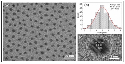
FIG. 1: TEM MICROGRAPH OF SYNTHESIZED CARBON DOTS SHOWING QUASI-SPHERICAL MORPHOLOGY AND UNIFORM SIZE DISTRIBUTION. SCALE BAR = 20 nm.
Dynamic Light Scattering and Zeta Potential: DLS analysis indicated a hydrodynamic diameter (Dh) of 7.2 ± 0.8 nm with aPDI of 0.18 ± 0.03, confirming the monodisperse nature of the carbon dot suspension. The hydrodynamic diameter was larger than the TEM-measured primary size due to the hydration shell and surface-bound solvent molecules. The PDI value below 0.25 indicates excellent colloidal stability and uniformity, which is critical for reproducible drug loading and analytical performance. Zeta potential measurements revealed a positive surface charge of +32.5 ± 2.1 mV at pH 7.4, attributed to protonated amine groups from PEI nitrogen doping. This high positive zeta potential ensures electrostatic stabilization of the colloidal suspension and promotes interaction with negatively charged cell membranes for enhanced cellular uptake.
TABLE 1: PHYSICOCHEMICAL CHARACTERIZATION OF SYNTHESIZED CARBON DOTS
| Parameter | Value | Method |
| Primary particle size (TEM) | 4.8 ± 1.2 nm | TEM |
| Hydrodynamic diameter (DLS) | 7.2 ± 0.8 nm | DLS |
| Polydispersity index (PDI) | 0.18 ± 0.03 | DLS |
| Zeta potential (pH 7.4) | +32.5 ± 2.1 mV | Laser Doppler electrophoresis |
| Quantum yield | 42.3 ± 3.1% | Comparative fluorescence method |
| λmax (UV-Vis) | 340 nm | UV-Vis spectroscopy |
| λex/λem (optimal) | 360/450 nm | Fluorescence EEM |
Fourier-Transform Infrared Spectroscopy: FTIR spectroscopy confirmed the presence of characteristic surface functional groups on the synthesized carbon dots. The broad absorption band at 3200–3600 cm⁻¹ was assigned to O–H stretching vibrations from hydroxyl and carboxyl groups. The sharp peak at 1720 cm⁻¹ corresponded to C=O stretching of carboxyl groups, while the peak at 1560 cm⁻¹ was attributed to N–H bending vibrations from PEI amine groups. The peak at 1400 cm⁻¹ was assigned to C–N stretching, confirming nitrogen incorporation into the carbon dot structure. The C–O stretching bands at 1000.The C–O stretching bands at 1000–1300 cm⁻¹ indicated the presence of ether and hydroxyl functionalities. The N–H stretching band at approximately 3300 cm⁻¹ further confirmed PEI nitrogen doping. These FTIR data collectively demonstrate successful synthesis of nitrogen-doped carbon dots with abundant oxygen-containing and nitrogen-containing surface functional groups, which are essential for curcumin loading through hydrogen bonding and π-π stacking interactions.
Fluorescence Spectroscopy: The carbon dots exhibited strong, excitation-dependent photoluminescence characteristic of nitrogen-doped CDs. Excitation-emission matrix mapping revealed optimal excitation at 360 nm with maximum emission at 450 nm. The fluorescence quantum yield was determined to be 42.3 ± 3.1% using quinine sulfate as a reference standard, which is consistent with literature values for citric acid/PEI-derived carbon dots. The excitation-dependent emission behavior, with emission wavelength shifting from 430 nm to 500 nm as excitation wavelength increased from 320 nm to 400 nm, is attributed to the heterogeneous surface states and different emissive sites on the carbon dot surface.
UV-Visible Absorption Spectroscopy: The UV-Vis absorption spectrum of carbon dots showed Fig. 2 broad-spectrum absorption spanning 200–700 nm, with a characteristic absorption peak at 340 nm attributed to the n→π* transition of surface functional groups and π→π* transition of the graphitic carbon core. Critically, significant absorbance was observed in the 350–500 nm region, with an absorbance value of 0.285 ± 0.032 at 425 nm for a 0.1 mg/mL CD suspension. This absorbance at the curcumin analytical wavelength confirms the potential for optical interference in spectrophotometric curcumin quantification, as the CD matrix contributes substantially to the apparent absorbance at 425 nm.
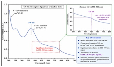
FIG. 2: UV-VISIBLE ABSORPTION SPECTROSCOPY
Thermogravimetric Analysis: TGA analysis of lyophilized carbon dots under nitrogen atmosphere showed initial weight loss of 8.5% below 150°C, attributed to adsorbed water and residual solvents. The major decomposition occurred between 200°C and 450°C, with a weight loss of 45.2% corresponding to pyrolysis of surface organic functional groups and decomposition of the carbon core. The thermal stability profile confirms that carbon dots are stable under the temperature conditions employed during synthesis, storage, and analytical processing.
Future Scope: Scale-up production of carbon dots for industrial and pharmaceutical applications. Improve batch-to-batch reproducibility and process optimization. Develop carbon dots for delivery of other phytochemicals and drugs. Conduct in-vivo pharmacokinetic and biodistribution studies of carbon dot formulations. Explore carbon dots as theranostic nanoplatforms combining drug delivery and fluorescence imaging. Investigate targeted carbon dots for cancer, inflammatory, and infectious diseases.Integrate artificial intelligence (AI) and Quality-by-Design (QbD) approaches for optimization of carbon dot synthesis and characterization. Study long-term stability, toxicity, and regulatory aspects of carbon dot-based nanomedicines. Explore multifunctional carbon dots for bioimaging, biosensing, diagnostics, and environmental monitoring. Develop green and sustainable synthesis methods with improved quantum yield and surface functionality.
CONCLUSION: The present study successfully demonstrated the synthesis of nitrogen-doped carbon dots using citric acid and polyethyleneimine through a simple and reproducible hydrothermal approach. The developed method produced highly dispersed carbon dots with nanoscale dimensions and desirable physicochemical characteristics. Comprehensive characterization confirmed the successful formation of quasi-spherical nanoparticles with a narrow size distribution, excellent colloidal stability, and abundant surface functional groups.TEM and DLS analyses verified the nanoscale particle size and monodisperse nature of the synthesized carbon dots, while zeta potential measurements indicated good suspension stability. FTIR studies confirmed the presence of oxygen- and nitrogen-containing functional groups that contribute to enhanced surface reactivity and potential drug-loading capability. Fluorescence analysis revealed strong excitation-dependent photoluminescence and a satisfactory quantum yield, highlighting the suitability of the synthesized carbon dots for imaging and sensing applications. UV–Visible spectroscopy demonstrated characteristic optical absorption behavior, and thermogravimetric analysis confirmed adequate thermal stability under experimental conditions. Overall, the synthesized nitrogen-doped carbon dots exhibited excellent structural, optical, and thermal properties, making them promising candidates for future pharmaceutical and biomedical applications. Their favorable characteristics suggest significant potential in drug delivery, bioimaging, biosensing, cancer diagnostics, and theranostic systems. The findings of this work provide a strong foundation for further research aimed at translating carbon dot technology into practical healthcare and nanomedicine applications.
ACKNOWLEDGEMENT: Nil
CONFLICT OF INTEREST: Nil
Top of Form
Bottom of Form
REFERENCES:
- Mishra V, Patil A, Thakur S and Kesharwani P: Carbon dots: emerging theranostic nanoarchitectures. Drug Discov Today 2018; 23(6): 1219-1232.
- Babar DG and Garje SS: Nitrogen and phosphorus co-doped carbon dots for selective detection of nitro explosives. ACS Omega 2020; 5(6): 2710-2717.
- Saravanan A, Maruthapandi M, Das P, Luong JHT and Gedanken A: Green synthesis of multifunctional carbon dots with antibacterial activities. Nanomaterials 2021; 11(2): 369.
- Li H, Kang Z, Liu Y and Lee ST: Carbon nanodots: synthesis, properties and applications. J Mater Chem 2012; 22(46): 24230-24253.
- Lim SY, Shen W and Gao Z: Carbon quantum dots and their applications. Chem Soc Rev 2015; 44(1): 362-381.
- Konar S, Samanta D, Mandal S, Shaw M, Mandal M and Pathak A: Selective and sensitive detection of cinnamaldehyde by nitrogen and sulphur co-doped carbon dots. RSC Adv 2018; 8: 42361-42373.
- Joshi PN, Mathias A and Mishra A: Synthesis of ecofriendly fluorescent carbon dots and their biomedical and environmental applications. Mater Technol 2018; 33(10): 672-680.
- Suryawanshi AR, Jagtap MR and Mujawar NK: Carbon Dots: New Promising Diagnostic Tool for Cancer Diagnosis. In: Recent Advances in Drug Discovery & Development. Vol 1. Weser Books 2021; 113-115.
- Elbordiny HS, Elonsy SM, Daabees HG and Belal TS: Assessment of greenness, blueness, and whiteness profiles of a validated HPLC-DAD method for quantitation of donepezil HCl and curcumin in their laboratory-prepared co-formulated nanoliposomes. BMC Chem 2025; 19(1): 15. doi:10.1186/s13065-024-01377-y.
- Holubowski T, Nowak T and Walczak M: Management of validation of HPLC method for determination of acetylsalicylic acid impurities using quality by design approach. Sci Rep 2022; 12(1): 17440. doi:10.1038/s41598-022-22350-y.
- Roshan F, Azarpoor-Masooleh M, Aghajanpour-Pasha M, Fateh E and Shahbazi-Azar A: Curcumin-encapsulated poly(lactic-co-glycolic acid) nanoparticles: a comparison of drug release kinetics from particles prepared via electrospray and nanoprecipitation. Macromol Mater Eng 2024; 309(10): 2400040. doi:10.1002/mame.202400040.
- Mujawar NK & Mulla SJA: Optimization and In Vitro Evaluation of Lapatinib-Encapsulated Lipomer Suspension for Effective Cancer Therapy. International Journal of Applied Pharmaceutics 2026; 18(2): 410–417. doi.org.10.22159/ijap.2026v18i2.57180
- Musmade DS, Gurav AS, Patil PB and Shinde SD: Box-Behnken design-assisted optimization of RP-HPLC method for the estimation of evogliptin tartrate by analytical quality by design. Future J Pharm Sci 2023; 9(1): 57. doi:10.1186/s43094-023-00509-w.
How to cite this article:
Kore P, Mahadik P, Patil M and Mujawar N: Hydrothermal synthesis and multifaceted characterization of fluorescent nitrogen-doped carbon dots for pharmaceutical and biomedical applications. Int J Pharmacognosy 2026; 13(8): 829-35. doi link: http://dx.doi.org/10.13040/IJPSR.0975-8232.IJP.13(8).829-35.
This Journal licensed under a Creative Commons Attribution-Non-commercial-Share Alike 3.0 Unported License.
Article Information
9
829-835
795 KB
162
English
IJP
Poonam Kore, Parag Mahadik, Mahesh Patil and Nigar Mujawar *
ASPM, College of Pharmacy, Sangulwadi, Sindhudurg, Maharashtra, India.
researchaspm@gmail.com
20 June 2026
15 July 2026
23 July 2026
10.13040/IJPSR.0975-8232.IJP.13(8).829-35
01 August 2026
 Download