


EVALUATION OF THE ANTIFUNGAL ACTIVITY OF HERBAL EXTRACT
HTML Full TextEVALUATION OF THE ANTIFUNGAL ACTIVITY OF HERBAL EXTRACT
Shubham Kumar *, Himanshu Kumar and Prashant Kumar
Himalaya College of Pharmacy, Chiksi, Paliganj, Patna, Bihar, India.
ABSTRACT: The present study evaluated the phytochemical composition and antifungal activity of ethanolic extracts of five medicinal plants: Azadirachta indica (Neem), Ocimum sanctum (Tulsi), Allium sativum (Garlic), Curcuma longa (Turmeric), and Syzygium aromaticum (Clove). Preliminary phytochemical screening revealed the presence of alkaloids, flavonoids, tannins, glycosides, terpenoids, steroids, saponins, and phenolic compounds, with flavonoids and phenolics being the predominant constituents. Antifungal activity was assessed against Candida albicans and Aspergillus niger using the agar well diffusion method. Garlic extract exhibited the highest antifungal activity among the plant extracts, followed by clove and neem, while tulsi showed the lowest activity. Fluconazole demonstrated the greatest inhibition against both fungal strains. The findings support the traditional use of these medicinal plants and suggest that garlic and clove possess promising potential as natural antifungal agents for future therapeutic applications.
Keywords: Medicinal plants, Antifungal activity, Candida albicans, Aspergillus niger, Phytochemical screening
INTRODUCTION: Fungal infections are among the most common microbial diseases affecting humans and have become a significant public health concern worldwide 1. Opportunistic fungi such as Candida albicans and Aspergillus niger are responsible for a wide range of infections, particularly in immunocompromised individuals 2. Although several synthetic antifungal drugs are available for treatment, their prolonged use is often associated with adverse effects, toxicity, high cost, and the emergence of drug-resistant fungal strains 3. These limitations have encouraged researchers to explore natural products as safer and more effective alternatives for the management of fungal infections.
Medicinal plants have been used in traditional systems of medicine for centuries because they are rich sources of bioactive phytochemicals with antimicrobial properties. Secondary metabolites such as alkaloids, flavonoids, tannins, phenolic compounds, terpenoids, glycosides, and essential oils have been reported to possess significant antifungal activity 4, 5. These phytochemicals inhibit fungal growth through various mechanisms, including disruption of the fungal cell membrane, inhibition of cell wall synthesis, interference with enzyme activity, and suppression of fungal metabolism 6. Consequently, herbal medicines have gained considerable attention as potential sources of novel antifungal agents.
The present study focuses on the evaluation of the antifungal activity of five widely used medicinal plants, namely Neem (Azadirachta indica), Tulsi (Ocimum sanctum), Garlic (Allium sativum), Turmeric (Curcuma longa), and Clove (Syzygium aromaticum). Different plant parts were selected based on their traditional medicinal uses, including neem leaves, tulsi leaves, garlic bulbs, turmeric rhizomes, and clove flower buds 7, 8, 9. These plants are known to contain pharmacologically important constituents such as azadirachtin, allicin, curcumin, eugenol, flavonoids, and phenolic compounds, which have been reported to exhibit broad-spectrum antimicrobial and antifungal activities.
Several scientific studies have demonstrated that these medicinal plants possess remarkable inhibitory effects against pathogenic fungi. Garlic is recognized for its potent antifungal activity due to the presence of allicin, while clove contains eugenol, a major phenolic compound with strong fungicidal properties 10. Neem exhibits antifungal effects mainly because of azadirachtin and nimbin, whereas turmeric contains curcumin, a natural polyphenol with antimicrobial and antioxidant properties. Tulsi is also rich in essential oils and phenolic compounds that contribute to its antimicrobial potential 11, 12.
The combined presence of these bioactive constituents makes these plants promising candidates for the development of herbal antifungal formulations. The findings of this research are expected to provide scientific evidence supporting the traditional use of these medicinal plants and may contribute to the development of safe, effective, and economical plant-based antifungal agents for the treatment of fungal infections.
MATERIALS AND METHODS:
Chemicals and Reagents: The chemicals and reagents used in the present study were of analytical grade and procured from reputed manufacturers. Ethanol (95%), hydrochloric acid (HCl), concentrated sulfuric acid (H₂SO₄), sodium hydroxide (NaOH), ferric chloride, Mayer's reagent, and Dragendorff's reagent were obtained from Merck Life Science Pvt. Ltd., Mumbai, India. Potato Dextrose Agar (PDA), Sabouraud Dextrose Agar (SDA), Sabouraud Dextrose Broth (SDB), and sterile cotton swabs were purchased from HiMedia Laboratories Pvt. Ltd., Mumbai, India. Fluconazole, used as the standard antifungal drug, was procured from HiMedia Laboratories Pvt. Ltd., Mumbai, India. Normal saline and distilled water were prepared in the laboratory using standard procedures and used throughout the experimental work.
Collection of Plant Materials: The plant materials used in the present study were collected from the Medicinal Garden, Himalaya College of Pharmacy, Chiksi, Paliganj, Patna, Bihar, India. Fresh and healthy plant parts, namely leaves of Neem (Azadirachta indica), leaves of Tulsi (Ocimum sanctum), bulbs of Garlic (Allium sativum), rhizomes of Turmeric (Curcuma longa), and flower buds of Clove (Syzygium aromaticum), were collected during the study period.
The collected plant materials were carefully examined to ensure they were free from disease, insect infestation, and mechanical damage. After collection, the samples were washed thoroughly with distilled water to remove adhering dust and other foreign matter, shade-dried at room temperature, and subsequently used for extraction and further phytochemical and antifungal studies.
Preparation of Plant Extracts: The collected plant materials were thoroughly washed with distilled water and shade-dried for 10–15 days. The dried samples were ground into a coarse powder using a mechanical grinder.
About 100 g of the powdered material was extracted with 500 mL of ethanol by maceration. The extract was filtered through Whatman No. 1 filter paper, and the filtrate was concentrated using a rotary evaporator or water bath to obtain the crude extract. The dried extract was stored in airtight containers at 4°C until further phytochemical and biological analyses 13.
Preliminary Phytochemical Screening: The prepared extracts were subjected to preliminary qualitative phytochemical screening using standard procedures to detect the presence of major classes of secondary metabolites 14. The analysis was carried out to identify the occurrence of alkaloids, flavonoids, tannins, saponins, glycosides, terpenoids, and phenolic compounds based on characteristic colour changes or precipitate formation in specific phytochemical tests.
Approximately 100 mg of the dried herbal extract was dissolved in 10 mL of the appropriate solvent (ethanol). The solution was filtered using Whatman No. 1 filter paper before carrying out the phytochemical tests.
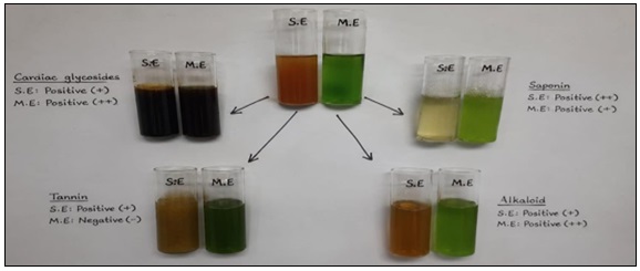
FIG. 1: PREPARATION OF SAMPLES FOR PHYTOCHEMICAL INVESTIGATION
Antifungal Activity: The antifungal activity of the different solvent extracts was evaluated against Candida albicans and Aspergillus niger using the agar well diffusion method. Pure fungal cultures were obtained from a recognized microbiology laboratory and maintained on Sabouraud Dextrose Agar (SDA) slants. Sabouraud Dextrose Agar was prepared according to the manufacturer's instructions, sterilized by autoclaving at 121°C for 15 minutes, poured into sterile Petri dishes, and allowed to solidify. Fresh fungal cultures were suspended in sterile normal saline, and the turbidity was adjusted to approximately 0.5 McFarland standard to obtain a uniform inoculum 15, 16.
The fungal suspension was evenly spread on sterile SDA plates using a sterile cotton swab. Wells of 6 mm diameter were prepared aseptically using a sterile cork borer. Different concentrations of the plant extracts (25, 50, and 100 mg/mL) were introduced into the respective wells. Fluconazole was used as the positive control, while sterile distilled water or the extraction solvent served as the negative control 17. The inoculated plates were incubated at 28–30°C for 48–72 hours, after which the zones of inhibition (mm) were measured to determine the antifungal activity of the extracts 18, 19.
Statistical Analysis: The results are expressed as Mean ± SD (n = 3). One-way ANOVA indicated statistically significant differences among the treatment groups (p < 0.05). Garlic and clove extracts showed significantly greater antifungal activity than tulsi and turmeric extracts, while fluconazole exhibited the highest inhibitory effect 20.
RESULTS AND DISCUSSION: The preliminary phytochemical screening of the selected medicinal plant extracts revealed the presence of various bioactive secondary metabolites, including alkaloids, flavonoids, tannins, saponins, glycosides, terpenoids, steroids, and phenolic compounds Table 1. The distribution and intensity of these phytoconstituents varied among the different plant extracts.
Neem (Azadirachta indica) showed a strong presence of flavonoids and phenolic compounds (+++), while alkaloids, tannins, glycosides, and terpenoids were present in moderate amounts (++). Saponins and steroids were detected in weak amounts (+). Tulsi (Ocimum sanctum) exhibited a strong presence of terpenoids (+++), moderate amounts of flavonoids, tannins, saponins, and phenolics (++), whereas alkaloids, glycosides, and steroids were weakly present (+). Garlic (Allium sativum) contained moderate levels of terpenoids and phenolic compounds (++), while alkaloids, flavonoids, saponins, and glycosides were weakly present (+). Tannins and steroids were absent (–). Turmeric (Curcuma longa) exhibited a strong presence of flavonoids and phenolic compounds (+++), moderate amounts of terpenoids (++), weak levels of tannins, glycosides, and steroids (+), whereas alkaloids and saponins were absent (–). Clove (Syzygium aromaticum) demonstrated a strong presence of tannins, terpenoids, and phenolic compounds (+++). Flavonoids and glycosides were moderately present (++), while alkaloids and steroids were detected in low amounts (+). Saponins were absent in clove extract. Overall, flavonoids, terpenoids, and phenolic compounds were the most abundant phytochemicals among the selected medicinal plants. Clove, neem, and turmeric exhibited comparatively richer phytochemical profiles, indicating their potential as important sources of bioactive compounds responsible for their reported medicinal and antifungal properties.
TABLE 1: PHYTOCHEMICAL SCREENING RESULTS OF SELECTED MEDICINAL PLANTS
| Phytochemical | Neem | Tulsi | Garlic | Turmeric | Clove |
| Alkaloids | ++ | + | + | – | + |
| Flavonoids | +++ | ++ | + | +++ | ++ |
| Tannins | ++ | ++ | – | + | +++ |
| Saponins | + | ++ | + | – | – |
| Glycosides | ++ | + | + | + | ++ |
| Terpenoids | ++ | +++ | ++ | ++ | +++ |
| Steroids | + | + | – | + | + |
| Phenolics | +++ | ++ | ++ | +++ | +++ |
+++ = Strong presence; ++ = Moderate presence; + = Weak presence; – = Absent.
Antifungal Activity by Agar Well Diffusion Method: The antifungal activity of the selected medicinal plant extracts was evaluated against Candida albicans and Aspergillus niger using the agar well diffusion method. The results demonstrated that all plant extracts exhibited varying degrees of antifungal activity, whereas the negative control showed no inhibition, confirming that the solvent had no antifungal effect. Fluconazole, used as the standard antifungal drug, exhibited the highest activity against both fungal strains, producing inhibition zones of 24.3 ± 0.6 mm against C. albicans and 22.8 ± 0.5 mm against A. niger. Among the plant extracts, garlic extract showed the strongest antifungal activity, with inhibition zones of 20.1 ± 0.7 mm against C. albicans and 18.7 ± 0.5 mm against A. niger. Clove extract also demonstrated marked antifungal activity, producing inhibition zones of 19.5 ± 0.6 mm and 18.3 ± 0.5 mm, respectively. Neem extract exhibited good inhibitory activity against both fungal organisms, with inhibition zones of 18.2 ± 0.5 mm against C. albicans and 17.0 ± 0.4 mm against A. niger. In contrast, turmeric and tulsi extracts displayed comparatively moderate antifungal effects. Tulsi extract showed the lowest inhibition among the tested plant extracts, recording 15.6 ± 0.4 mm against C. albicans and 14.8 ± 0.5 mm against A. niger.
The results indicate that Candida albicans was slightly more susceptible to the plant extracts than Aspergillus niger, as evidenced by the comparatively larger inhibition zones observed against C. albicans. The superior antifungal activity of garlic and clove extracts may be attributed to the presence of bioactive compounds such as allicin, eugenol, flavonoids, phenolics, and terpenoids, which are known to disrupt fungal cell membranes and inhibit fungal growth. Neem also exhibited appreciable activity due to the presence of azadirachtin, nimbin, and other phytochemicals with well-documented antimicrobial properties.
Overall, the antifungal activity of the plant extracts followed the order:
Fluconazole > Garlic > Clove > Neem > Turmeric > Tulsi.
These findings suggest that garlic and clove extracts possess significant antifungal potential and may serve as promising natural alternatives or complementary agents for the management of fungal infections. Further studies involving isolation of active constituents, determination of minimum inhibitory concentration (MIC), and in-vivo evaluation are recommended to validate their therapeutic efficacy.
TABLE 2: ANTIFUNGAL ACTIVITY OF SELECTED MEDICINAL PLANT EXTRACTS AGAINST CANDIDA ALBICANS
| Treatment | Concentration (mg/mL) | Zone of Inhibition (mm) (Mean ± SD) |
| Negative Control | — | 0.0 ± 0.0 |
| Fluconazole (Standard) | 25 | 24.3 ± 0.6 |
| Neem Extract | 100 | 18.2 ± 0.5 |
| Tulsi Extract | 100 | 15.6 ± 0.4 |
| Garlic Extract | 100 | 20.1 ± 0.7 |
| Turmeric Extract | 100 | 16.8 ± 0.5 |
| Clove Extract | 100 | 19.5 ± 0.6 |
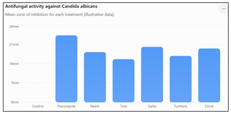
FIG. 2: ANTIFUNGAL ACTIVITY AGAINST CANDIDA ALBICANS
TABLE 3: ANTIFUNGAL ACTIVITY OF SELECTED MEDICINAL PLANT EXTRACTS AGAINST ASPERGILLUS NIGER
| Treatment | Concentration (mg/mL) | Zone of Inhibition (mm) (Mean ± SD) |
| Negative Control | — | 0.0 ± 0.0 |
| Fluconazole (Standard) | 25 | 22.8 ± 0.5 |
| Neem Extract | 100 | 17.0 ± 0.4 |
| Tulsi Extract | 100 | 14.8 ± 0.5 |
| Garlic Extract | 100 | 18.7 ± 0.5 |
| Turmeric Extract | 100 | 15.9 ± 0.5 |
| Clove Extract | 100 | 18.3 ± 0.5 |
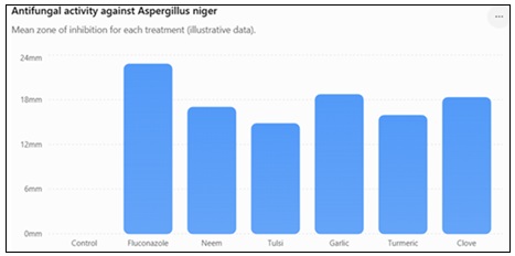
FIG. 3: ANTIFUNGAL ACTIVITY AGAINST ASPERGILLUS NIGER
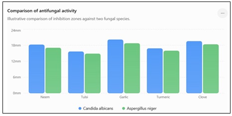
FIG. 4: ANTIFUNGAL ACTIVITY COMPARISON
CONCLUSION: The present study demonstrated that the selected medicinal plants possess diverse phytochemical constituents responsible for their antifungal activity. Flavonoids, phenolics, terpenoids, tannins, and glycosides were widely distributed among the extracts and are likely to contribute to their biological effects. Among the tested plants, garlic exhibited the strongest antifungal activity, followed by clove and neem, whereas tulsi and turmeric showed comparatively moderate inhibition. Although fluconazole remained the most effective antifungal agent, the promising activity of garlic and clove highlights their potential as natural alternatives or adjuncts in the management of fungal infections. These findings provide scientific support for the traditional use of these medicinal plants and encourage further studies on the isolation of active compounds, MIC determination, toxicity evaluation, and in-vivo efficacy.
ACKNOWLEDGEMENT: Nil
CONFLICT OF INTEREST: Nil
REFERENCES:
- Akinpelu DA: Antimicrobial activity of Anacardium occidentale bark. Fitoterapia 2001; 72: 286-287.
- Al Ashaal HA, Farghaly AA, Abd El Aziz MM and Ali MA: Phytochemical investigation and medicinal evaluation of fixed oil of Balanites aegyptiaca fruits (Balantiaceae). J Ethnopharmacol 2010; 127: 495–501.
- Barman S, Sahu N, Deka S, Dutta S and Das S: Anti-inflammatory and analgesic activity of leaves of Spilanthes acmella (ELSA) in experimental animal models. Pharmacologyonline 2009; 1: 1027-1034.
- National Committee for Clinical Laboratory Standards (NCCLS). 2002. Reference method for broth dilution antifungal susceptibility testing for conidium-forming filamentous fungi; approved standard. NCCLS Document M38-A. Wayne, PA, USA.
- Ojo OO, Nadro MS and Tella IO: Protection of rats by extracts of some common Nigerian trees against acetaminopheninduced hepatotoxicity. Afr J Biotechnol 2006; 5: 755-760.
- Panghal M, Kaushal V and Yadav JP: In-vitro antimicrobial activity of ten medicinal plants against clinical isolates of oral cancer cases. Ann Clin Microbiol Antimicrob 2011; 10: 21-32.
- Parekh J and Chanda S: Antibacterial Activity of Aqueous and Alcoholic Extracts of 34 Indian Medicinal Plants against Some Staphylococcus Species. Turk J Biol 2008; 32: 63-71.
- Phongpaichit S, Subhadhirasakul S and Wattanapiromsakul C: Antifungal activities of extracts from Thai medicinal plants against opportunistic fungal pathogens associated with AIDS patients. Mycoses 2005; 48: 333-338.
- Prachayasittikul V, Prachayasittikul S, Ruchirawat S and Prachayasittikul V: High Therapeutic potential of Spilanthes acmella: A review. EXCLI Journal 2013; 12: 291-312.
- Rani SA and Murty SU: Antifungal potential of flower head extract of Spilanthes acmella Linn. Afri J Biomed Res 2006; 9: 67-68.
- Ratnasooriya WD, Pieris KPP, Samaratunga U and Jayakody JRAC: Diuretic activity of Spilanthes acmella flowers in rats. J Ethnopharmacol 2004; 91: 317-320.
- Reddy BU: Antimicrobial activity of Datura Stramonium L. and Tylophora indica (Burm.f.) Merr. Pharmacologyonline 2009; 1: 1293- 1300.
- Samy RP and Ignacimuthu S: Antibacterial activity of some folklore medicinal plants used by tribals in western ghats of India. J. Ethnopharmacol 2000; 69: 63-71.
- Harborne JB: Phytochemical Methods: A Guide to Modern Techniques of Plant Analysis. 3rd ed. London: Chapman & Hall 1998.
- Trease GE and Evans WC: Trease and Evans Pharmacognosy. 16th ed. Edinburgh Elsevier 2009.
- Kokate CK, Purohit AP and Gokhale SB: Pharmacognosy. 56th ed. Pune: Nirali Prakashan 2021.
- Singh I and Singh VP: Antifungal properties of aqueous and organic extracts of seed plants against Aspergillus flavus and A. niger. Phytomorphology 2000; 50: 151-157.
- Edeoga HO, Okwu DE and Mbaebie BO: Phytochemical constituents of some Nigerian medicinal plants. Afr J Biotechnol 2005; 4(7): 685–688.
- Parekh J and Chanda S: In-vitro antimicrobial activity of medicinal plants against bacterial and fungal pathogens. Afr J Biomed Res 2007; 10: 175–181.
- Mohamed AM, Wolf D and Spiess WE: Recovery and characterization of Balanites aegyptiaca Del. Kernel proteins: Effect of defatting, air classification, wet sieving and aqueous ethanol treatment on solubility, digestibility, amino acid composition and sapogenin content. Nahrung 2000; 44: 7-12.
How to cite this article:
Kumar S, Kumar H and Kumar P: Evaluation of the antifungal activity of herbal extract. Int J Pharmacognosy 2026; 13(9): 980-85. doi link: http://dx.doi.org/10.13040/IJPSR.0975-8232.IJP.13(9).980-85.
This Journal licensed under a Creative Commons Attribution-Non-commercial-Share Alike 3.0 Unported License.
Article Information
10
980-985
741 KB
235
English
IJP
Shubham Kumar *, Himanshu Kumar and Prashant Kumar
Himalaya College of Pharmacy, Chiksi, Paliganj, Patna, Bihar, India.
mspharma8081@gmail.com
15 July 2026
25 August 2026
27 August 2026
10.13040/IJPSR.0975-8232.IJP.13(9).980-85
01 September 2026
 Download