


ESTROGENIC EFFECTS OF RICINODENDRON HEUDOLOTII (EUPHORBIACEAE) SEEDS IN FEMALE RATS
HTML Full TextESTROGENIC EFFECTS OF RICINODENDRON HEUDOLOTII (EUPHORBIACEAE) SEEDS IN FEMALE RATS
Kouassi Emile Begbin *, Prisca Joëlle Djoman Doubran, Mataphouet Emmanuel Affy, Koffi Roger Kouakou and Koffi Kouakou
Laboratory of Animal Biological Sciences, Faculty of Sciences and Technologies, Alassane OUATTARA University, BP V18 Bouake 01, Côte d’Ivoire.
ABSTRACT: Disorders related to female hormonal imbalance can profoundly and permanently affect quality of life. This study aimed to evaluate the estrogenic effects of an aqueous extract of Ricinodendron heudelotii almonds in ovariectomized rats. The seeds, harvested in Andé (Ivory Coast), were dried, ground, and then extracted with distilled water. A phytochemical screening was conducted to identify the main groups of secondary metabolites present in the extract. Acute toxicity was evaluated in six Swiss mice in accordance with OECD Guideline 423. Estrogenic activity was studied in ovariectomized Wistar rats using the OECD 440 uterine trophism bioassay. The animals were divided into four groups: control, ethinyl estradiol (0.02 mg/kg), 100 mg/kg of extract, and 300 mg/kg of extract. The treatments were administered orally daily for seven days. Body weight, as well as the relative weights of the uterus, cervix, and adrenal glands, were assessed. Vaginal smears were also collected to analyze cytological changes in the vaginal epithelium. Phytochemical analysis revealed the presence of sterols, polyterpenes, and alkaloids. No deaths or obvious signs of toxicity were observed at a dose of 2,000 mg/kg. The 100 mg/kg extract significantly reduced body weight gain and increased the weight of the uterine horns and cervix, with a high presence of eosinophils. These results suggest that R. heudelotii possesses estrogenic activity, particularly at a dose of 100 mg/kg, and may serve as a source of compounds with phytoestrogenic potential.
Keywords: Ricinodendron heudelotii, Estrogen, Phytoestrogen, Ovariectomy
INTRODUCTION: Disorders related to hormonal imbalance in women, particularly those associated with declining estrogen levels, can profoundly and permanently affect quality of life 1.
These disorders manifest as infertility 2, 3, osteoporosis, an increased risk of cardiovascular disease 4, memory, mood, and sleep disturbances, weight gain, accelerated skin aging, as well as hot flashes and night sweats 5, 6, 7.
Although hormone replacement therapy (HRT) is widely used, it is often associated with significant adverse effects, such as an increased risk of thrombosis, endometrial cancer, and biliary disorders 8, 9, 10.
This situation has heightened scientific interest in natural alternatives, particularly phytoestrogens, which can help regulate the menstrual cycle, protect against certain hormone-dependent cancers 11, reduce postmenopausal bone loss 12 and the risk of cardiovascular disease 13, improve insulin sensitivity 14, and protect tissues such as the liver, the heart, and the brain through their antioxidant and anti-inflammatory activities 15.
In this context, African herbal medicine is an important source of bioactive compounds traditionally used in the management of gynecological disorders. Among these plants, Ricinodendron heudelotii (Euphorbiaceae) is renowned for its gynecological and obstetric properties. In Benin, the leaves of R. heudelotii are used to support pregnancies; the stem bark is used to relieve abdominal pain during menstruation and to prevent miscarriages; and the seeds, when used for therapeutic purposes, induce ovulation16. In southern Côte d’Ivoire, in the Adzopé department, both the stem bark and the seeds of this plant are used individually or in combination to treat certain causes of infertility. However, despite its empirical use in treating certain female fertility disorders, experimental evidence demonstrating the estrogenic effects of R. heudelotii stem bark remains limited. Thus, the present study aims to evaluate the estrogenic effects of R. heudelotii seeds in female rats.
MATERIALS AND METHODS:
Biological Material: The seeds of R. heudelotii constituted the plant material used in this study. They were purchased in Andé, a village in the Agou Subprefecture, Adzopé Department, Mé Region (Ivory Coast).
The experiments were conducted on rodents. The species used were Rattus norvegicus (Muridae) of the Wistar strain and Mus musculus (Muridae) of the Swiss strain. They were obtained from the vivarium at the École Normale Supérieure d’Abidjan (Ivory Coast). These animals were housed in a room maintained at a temperature of 28 ± 2°C with a photoperiod of 12 hours of natural light and 12 hours of darkness. Humidity ranged from 50 to 55% and the animals had free access to water and food (56.9% carbohydrates, 16.6% protein, 3.9% fat, 13.2% fiber, and 9.4% minerals) provided by the Ivorian Compound Feed Manufacturing Company. Mice were used for the acute toxicity test, while rats were used for the pharmacology study.
These animals were nulliparous and non-pregnant. The mice were approximately 9 weeks old, and their body weight ranged from 25 to 29 grams. The rats were ovariectomized at 7 weeks of age and weighed between 90 and 110 g.
Preparation of the Aqueous Extract from R. heudelotii Almonds: The R. heudelotii kernels, dried out of direct sunlight, were pulverized using an electric grinder, and the resulting powder was used to prepare the aqueous extract according to the method described by Yapo et al. 17. Fifty grams (50 g) of powder were mixed with 1 liter of distilled water in a blender. After a series of three blending cycles, each lasting three minutes at room temperature, the resulting homogenate was filtered twice through a square of white cloth (poplin), then five times through cotton wool. The resulting filtrate is evaporated in an oven at 50°C for 3 days until a dry extract is obtained.
Phytochemical Screening of the Aqueous Extract of R. heudelotii Seeds: The various chemical groups in the aqueous extract of R. heudelotii seeds were characterized using the techniques described in the studies by Wagner & Bladt 18 and Békro et al.19. The phytochemical analyses were conducted at the Pharmacognosy Laboratory of the Faculty of Pharmaceutical and Biological Sciences at Félix Houphouët-Boigny University.
Sterols and polyterpenes were detected using the Liebermann reaction. The ferric chloride (FeCl₃) reaction was used to characterize the polyphenols. Flavonoids were detected using the “cyanidin” reaction. Catechin tannins were identified using Stiasny’s reagent, while gallic tannins were detected using FeCl₃. Borntraegen’s reagent was used to detect free and bound quinone compounds. Alkaloids are characterized using Dragendorff’s reagent (potassium iodobismuthate) and Bouchardat’s reagent (iodo-iodide). To test for saponosides, 15 mL of the aqueous extract of R. heudelotii almonds was poured into a test tube 15 cm long and 15 mm in diameter, then the tube was shaken vigorously for 10 seconds and allowed to stand for 10 minutes. If the foam remains at a height of more than 3 cm, saponins are present.
Acute Toxicity Study: The acute toxicity study was conducted in accordance with the Organization for Economic Cooperation and Development (OECD) Guideline 423 for the Testing of Chemicals 20. The mice were 9 weeks old, and their body weights ranged from 26 to 31 grams
The limit test with a dose of 2000 mg/kg body weight was conducted. Six young, nulliparous, non-pregnant mice with body weights ranging from 25 to 29 grams and approximately 9 weeks of age were used. They were divided into two groups of three mice each: one group treated with the extract and one control group. They were individually marked, fasted for 4 hours, and weighed prior to the experiment. Subsequently, a single dose of 2,000 mg/kg was administered orally to each mouse in the treated group using an esophageal tube. After treatment, the mice were again deprived of food for 4 hours. They were observed individually during the first 30 minutes and once daily for 14 days. The observation of the animals consisted of noting symptoms of pathology or behavioral changes.
Bilateral Oophorectomy Technique: The female rats underwent ovariectomy at 7 weeks of age, when their body weights ranged from 90 to 110 g. For this procedure, the animals were anesthetized with ether. Once anesthesia took effect, the dorsal lumbar region was shaved bilaterally, and the skin was cleaned with 96° ethyl alcohol. A dorsal incision of approximately 3/4 cm was made just below the last rib, penetrating the abdominal cavity. The periovarian adipose tissue was identified and isolated. The exteriorized ovary was transected at its junction with the uterine horns 21. The peritoneum and skin were then sutured after the organs were returned to their original positions. An antibiotic injection (penicillin) was administered immediately after the operation and repeated every three days for one week. Penicillin ointment and liquid Betadine were used to disinfect the wounds and prevent infection until complete healing. The ovariectomized rats were used for experiments after a 15-day postoperative recovery period 22.
Vaginal Smear Technique: Vaginal smears were prepared using the method described in Kouakou 23. The procedure was carried out in three phases: collection and smearing of vaginal cells onto slides, staining of the slides, and examination of the slides.
Collection of Vaginal Cells: A cotton swab moistened with physiological saline (9‰ NaCl) is gently inserted into the female rat’s vagina without causing her stress, then gently rotated in the same direction until slight resistance is felt. The swab is then removed, and the sample is smeared onto a clean microscope slide.
Staining of the Slides: The samples were stained with methylene blue. To do this, a drop of 1% methylene blue solution diluted to one-tenth strength was placed on the slide, which was then covered with a coverslip. The sample was allowed to stand for 10 to 15 minutes to allow the vaginal epithelial cells to stain.
Slide Examination: Slide examination was performed under an optical microscope based on the different proportions of stained cells on the slide. Three cell types were observed: basophils, eosinophils, and leukocytes. The percentage of each cell type was determined to identify the different phases of the estrous cycle.
Experimental Design: For this study, the rodent uterotrophicity bioassay was used in accordance with OECD Guideline 440 22. The method using young adult ovariectomized females, known as the adult-Ovx method, was employed. A total of 30 young adult female rats, ovariectomized at 7 weeks of age and weighing between 90 and 110 g, were divided into four groups of five subjects each. Treatments were administered as follows:
- Group 1 (control): distilled water;
- Group 2: 0.02 mg/kg ethinyl estradiol;
- Group 3: 100 mg/kg of aqueous extract of heudelotii almonds;
- Group 4: 300 mg/kg of aqueous extract of heudelotii almonds.
A volume of 1 mL of the test substance was administered orally to each animal daily for 7 days. The body weight of each animal was recorded daily. The day after treatment ended-on the 8th day-vaginal smears were performed to determine the nature of the cells in the vaginal epithelium of the female rats, which were then euthanized after ether anesthesia. An incision was made in the abdominal cavity to remove and weigh the uterus, cervix, and adrenal glands.
Data Analysis: Statistical analyses of the experimental results were performed using GraphPad Prism 5.01 software (Microsoft, USA). Values are presented as mean ± standard error. The data were analyzed using one-way ANOVA followed by Tukey’s multiple comparison test at the 5% significance level to assess the significance of the observed differences.
RESULTS:
Phytochemical Screening: Phytochemical analysis of the aqueous extract of R. heudelotii almonds revealed the presence of sterols, polyterpenes, and alkaloids Table 1.
TABLE 1: CHEMICAL COMPOSITION OF THE AQUEOUS EXTRACT OF R. HEUDELOTII ALMONDS
| Chemical Groups | Observations |
| Sterols and Polyterpenes | + |
| Polyphenols | - |
| Flavonoids | - |
| Tannins | - |
| Quinones | - |
| Alkaloids | + |
| Saponins | - |
(+): present; (–): absent
Acute Toxicity Study: Oral administration of 2,000 mg/kg of the aqueous extract of R. heudelotii almonds to mice did not result in any deaths. No signs of toxicity were observed. Weight gains in the treated and control groups were statistically identical (p > 0.05) Table 2.
TABLE 2: WEIGHT GAINS IN CONTROL AND TREATED MICE AT THE END OF THE ACUTE TOXICITY TEST
| Experimental groups | ||
| Control | AERh2000 | |
| Weight on Day 0 (g) | 25,75 ± 1,59 | 28,27 ± 1,047 |
| Weight on Day 14(g) | 30,19 ± 1,63 | 30,52 ± 0,99 |
| Weight gain (g) | 2,44 ± 0,46 | 2,25 ±0,06 |
Values are expressed as mean ± standard error of the mean (n=3), AERh2000: Aqueous extract of R. heudelotii almonds at a dose of 2000 mg/kg
Effects of the Aqueous Extract of R. heudelotii Almonds on the Body Weight of Female Rats: During the treatment period, the body weights of both the control female rats and those treated with the aqueous extract of R. heudelotii almonds increased gradually over time. However, body weight gains in rats treated with a 100 mg/kg dose of the plant extract were significantly lower (p<0.05) than those of the control group starting on the 6th day of treatment. Furthermore, the average weight of the animals treated with ethinyl estradiol remained relatively constant and was significantly lower (p < 0.001) than that of the control group until the end of the study Fig. 1.
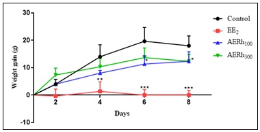
FIG. 1: EFFECTS OF THE AQUEOUS EXTRACT OF R. HEUDELOTII ALMONDS AND ETHINYL ESTRADIOL ON THE BODY WEIGHT OF RATS OVER TIME. Values are expressed as mean ± standard error of the mean (n=5), (*) P<0.05: marginally significant difference; (**): P<0.01: significant difference; (***): P<0.001: highly significant difference. AERh100: 100 mg/kg of aqueous extract of R. heudelotii almonds. AERh300: 300 mg/kg of aqueous extract of R. heudelotii almonds. EE2: 0.02 mg/kg of ethinyl estradiol.
Effects of the Aqueous Extract of R. heudelotii Almonds on Organ Weight: Treatment of ovariectomized female rats with EE2 resulted in a highly significant (p < 0.001) increase in the relative weight of the uterine horns and cervix compared to the controls. As for the animals treated with the aqueous extract of R. heudelotii almonds, a highly significant increase (p < 0.001) in the weight of the uterine horns and cervix was observed at the 100 mg/kg dose, as well as a significant increase (p < 0.05) at the 300 mg/kg dose. The relative weight of the adrenal glands was increased (p < 0.05) only at the 100 mg/kg dose Table 3.
TABLE 3: EFFECTS OF ETHINYL ESTRADIOL AND R. HEUDELOTII ALMONDS ON ORGAN WEIGHT
| Organ Weight (mg/100 g of body weight) | Experimental groups | |||
| Control | EE2 | AERh100 | AERh300 | |
| Adrenal glands | 20,92 ± 1,30 | 22,94 ± 0,50 | 27,12 ± 2,08* | 20,56 ± 1,49 |
| Cervix | 6,99 ± 0,34 | 97,44 ± 9,93**** | 75,19 ± 5,36*** | 27,89 ± 1,01* |
| Uterine horns | 10,49 ± 0,51 | 146,20 ± 14,89**** | 112,80 ± 8,05*** | 41,83 ± 1,52* |
Values are presented as the mean ± standard error of the mean (n=5). (*) p<0.05: marginally significant difference; (**): p<0.01: very significant difference; (****): p<0.001: highly significant difference. AERh100: 100 mg/kg of aqueous extract of R. heudelotii almonds. AERh300: 300 mg/kg of aqueous extract of R. heudelotii almonds. EE2: 0.02 mg/kg of ethinyl estradiol.
Effects of the Aqueous Extract of R. heudelotii Almonds on Eosinophils in the Vaginal Epithelium: A high number of eosinophils was observed in vaginal smears from rats treated with ethinyl estradiol as well as those treated with 100 mg/kg (dry weight) of the aqueous extract of R. heudelotii almonds Table 4.
TABLE 4: EFFECTS OF THE AQUEOUS EXTRACT OF R. HEUDELOTII ALMONDS ON EOSINOPHILS IN THE VAGINAL EPITHELIUM
| Experimental groups | ||||
| Control | EE2 | AERh100 | AERh300 | |
| Observations | - | ++ | ++ | + |
(-): absent; (+): low levels; (++): high levels. AERh100: 100 mg/kg of aqueous extract of R. heudelotii almonds. AERh300: 300 mg/kg of aqueous extract of R. heudelotii almonds. EE2: 0.02 mg/kg of ethinyl estradiol.
DISCUSSION: The objective of this study was to evaluate the estrogenic effects of the aqueous extract of R. heudelotii almonds in ovariectomized rats, a standard experimental model for studying substances with estrogenic activity. The results show that the extract possesses estrogenic properties while exhibiting low acute toxicity.
A phytochemical analysis revealed the presence of sterols, polyterpenes, and alkaloids in the aqueous extract of R. heudelotii seeds. These secondary metabolites are widely recognized for their biological activities. In particular, certain plant-derived sterols (phytosterols) have a chemical structure similar to that of cholesterol, a precursor of steroid hormones capable of interacting with estrogen receptors or modulating their signaling. Terpenes and their derivatives are also believed to possess endocrine activities, while certain alkaloids can influence hormonal pathways through various mechanisms. Thus, the phytochemical composition observed in this study provides an initial indication that may explain the estrogenic effects demonstrated in-vivo. These observations are consistent with the work of Cowan 24, who emphasizes that plant secondary metabolites are responsible for numerous pharmacological activities, as well as with that of Kuiper et al.25, who demonstrated that several plant compounds are capable of binding to estrogen receptors.
The acute toxicity study showed that no deaths or clinical signs of toxicity were observed following oral administration of 2,000 mg/kg of aqueous extract to mice. Furthermore, weight gain in the treated animals remained comparable to that of the control group. In accordance with OECD Guideline 42320, the absence of mortality at this dose suggests that the LD₅₀ is greater than 2,000 mg/kg, indicating low acute toxicity of the extract. These results are consistent with several studies conducted on medicinal plant extracts rich in secondary metabolites, which report good tolerability following oral administration at high doses. This safety profile represents a significant advantage for the potential therapeutic use of R. heudelotii. With regard to body weight, rats treated with the extract at a dose of 100 mg/kg showed significantly less weight gain than the control group, while treatment with ethinylestradiol virtually prevented any weight gain. This observation is consistent with the known physiological effects of estrogens. Indeed, following ovariectomy, estrogen deficiency generally leads to an increase in body weight associated with increased food intake and decreased energy expenditure. The administration of exogenous estrogens generally reverses this phenomenon by reducing the accumulation of fat reserves and improving energy metabolism. The results obtained with the R. heudelotii extract therefore suggest that it partially replicates the metabolic effects of estrogens. These observations are consistent with the work of Rogers et al.26, who showed that estrogens play a major role in regulating body weight, as well as with that of Villa et al.27, which indicate that phytoestrogens also help limit weight gain in animals with estrogen deficiency.
The uterotrophic test is the gold standard recommended by OECD guidelines 440 22 for detecting estrogenic activity. In this study, ethinyl estradiol caused a highly significant increase in the weight of the uterine horns and the cervix, thereby confirming the validity of the experimental model. The aqueous extract of R. heudelotii seeds also induced a significant increase in the weight of these organs, particularly at a dose of 100 mg/kg, indicating stimulation of the growth of estrogen-sensitive genital tissues. The increase in uterine weight resulted primarily from epithelial cell hypertrophy, glandular hyperplasia, increased vascularization, and water retention induced by the activation of estrogen receptors. These results suggest that the extract contains compounds capable of exerting agonist activity on these receptors. Similar observations have been reported for several medicinal plants rich in phytoestrogens, notably Pueraria mirifica 28, Glycine max, and Trifolium pratense, whose isoflavones also increase uterine weight in ovariectomized rats 29, 30.
The increase in the relative weight of the adrenal glands, observed only at the 100 mg/kg dose, may reflect a moderate stimulation of the endocrine activity of these organs. The adrenal glands are involved in the synthesis of several steroid hormones, including precursors that can be converted into estrogens in peripheral tissues. However, this change was not observed at the 300 mg/kg dose, which could indicate a nonlinear response or a biphasic effect of certain components of the extract. Such a dose-response relationship is frequently described for phytoestrogens, whose activity depends on their concentration, their affinity for the ERα and ERβ receptors, and the presence of cellular cofactors. Further studies on hormone levels and estrogen receptor expression would be necessary to clarify the mechanisms involved.
The results obtained from vaginal smears support the hypothesis that the extract has estrogenic activity. Indeed, the abundant presence of eosinophils in smears from female rats treated with ethinyl estradiol and the extract at a dose of 100 mg/kg indicates stimulation of vaginal epithelial maturation. In rodents, exposure to estrogens induces proliferation of the vaginal epithelium followed by keratinization and an increase in the number of superficial eosinophilic cells, which are characteristic of the estrous phase. Thus, the cytological changes observed serve as a reliable marker of estrogenic activity. These results are consistent with those of Diel et al.31 and Owens & Ashby 32, who demonstrated that phytoestrogens induce vaginal cytological changes similar to those caused by synthetic estrogens.
CONCLUSION: In conclusion, the results of this study show that the aqueous extract of R. heudelotii seeds exhibits potential estrogenic activity under the experimental conditions used, with a more pronounced response at a dose of 100 mg/kg than at 300 mg/kg. This difference between doses suggests that the response does not necessarily follow a linear dose-response relationship and warrants further research. These results provide an initial experimental basis for better understanding the pharmacological properties of this plant. However, they are not sufficient on their own to identify the responsible compounds or to elucidate the molecular mechanism of this activity. Further studies, particularly those focusing on the characterization of the active constituents, their interaction with estrogen receptors, and the assessment of subchronic toxicity, would be necessary to confirm and expand upon these observations.
ACKNOWLEDGEMENT: Nil
CONFLICT OF INTEREST: The authors declare that there is no conflict of interest regarding the publication of this paper.
REFERENCES:
- Desreux J, Gaspard U, Bleret V, Van Cauwenberge JR, Thille A, Herman P and Lifrange E: Le cancer du sein en Belgique : pourquoi sommes-nous les premiers en Europe? [Breast cancer in Belgium: why are we the first in Europe?]. Rev Med Liege 2011; 66(5-6): 385-392.
- Nelson LM: Clinical practice. Primary ovarian insufficiency. N Engl J Med 2009; 360(6): 606-14. doi: 10.1056/NEJMcp0808697
- Klein DA, Paradise SL and Reeder RM: Amenorrhea: A Systematic Approach to Diagnosis and Management. Am Fam Physician 2019; 100(1): 39-48.
- Rivera CM, Grossardt BR, Rhodes DJ, Brown RD, Roger VL, Melton LJ and Rocca WA:. Increased cardiovascular mortality after early bilateral oophorectomy. Menopause 2009; 16(1): 15-23. doi: 10.1097/gme.0b013e31818888f7.
- Rocca WA, Bower JH, Maraganore DM, Ahlskog JE, Grossardt BR, De Andrade M and Melton LJ: Increased risk of parkinsonism in women who underwent oophorectomy before menopause. Neurology 2008; 70(3): 200-209. doi: 10.1212/01.wnl.0000280573.30975.6a
- Brincat M, Muscat Baron Y and Galea R: Estrogens and the skin. Climacteric 2005; 8(2): 110–123. doi: 10.1080/13697130500118100
- Kaufmann M, Jonat W, Blamey R, Cuzick J, Namer M, Fogelman I, de Haes JC, Schumacher M and Sauerbrei W: Survival analyses from the ZEBRA study: goserelin (Zoladex™) versus CMF in premenopausal women with node-positive breast cancer. Eur J Cancer 2003; 39(12): 1711-1717. doi : 10.1016/S0959-8049(03)00392-7
- Cirillo DJ, Wallace RB, Rodabough RJ, Greenland P, LaCroix AZ, Limacher MC and Larson JC: Effect of estrogen therapy on gallbladder disease. JAMA 2005; 293(3): 330-339. doi: 10.1001/jama.293.3.330.
- Canonico M, Oger E, Plu-Bureau G, Conard J, Meyer G, Lévesque H, Trillot N, Barrellier MT, Wahl D, Emmerich J and Scarabin PY: Hormone Therapy and Venous Thromboembolism Among Postmenopausal Women: Impact of the Route of Estrogen Administration and Progestogens: The ESTHER Study. Circulation. 2007; 115(7): 840-845. doi: 10.1161/CIRCULATIONAHA.106.642280
- Duralde ER, Sobel TH and Manson JE: Management of perimenopausal and menopausal Symptoms. BMJ 2023; 382: e072612. doi: 10.1136/bmj-2022-072612
- Wei Y, Lv J, Guo Y, Bian Z, Gao M, Du H, Yang L, Chen Y, Zhang X, Wang T, Chen J, Chen Z, Yu C, Huo D and Li L: China Kadoorie Biobank Collaborative Group. Soy intake and breast cancer risk: a prospective study of 300,000 Chinese women and a dose-response meta-analysis. Eur J Epidemiol 2020; 35(6): 567-578. doi: 10.1007/s10654-019-00585-4.
- Inpan R, Na Takuathung M, Sakuludomkan W, Dukaew N, Teekachunhatean S and Koonrungsesomboon N: Isoflavone intervention and its impact on bone mineral density in postmenopausal women: a systematic review and meta-analysis of randomized controlled trials. Osteoporos Int 2024; 35: 413–430. doi: 10.1007/s00198-023-06944-y
- Yeung J and Yu TF: Effects of isoflavones (soy phyto-estrogens) on serum lipids: a meta-analysis of randomized controlled trials. Nutr J 2003; 2: 15. doi: 10.1186/1475-2891-2-15.
- Barańska A, Błaszczuk A, Kanadys W, Baczewska B, Jędrych M, Wawryk-Gawda E and Polz-Dacewicz M: Effects of Soy Protein Containing of Isoflavones and Isoflavones Extract on Plasma Lipid Profile in Postmenopausal Women as a Potential Prevention Factor in Cardiovascular Diseases: Systematic Review and Meta-Analysis of Randomized Controlled Trials. Nutrients 2021; 13(8): 2531. doi: 10.3390/nu13082531.
- Nadia ME, Nazrun AS, Norazlina M, Isa NM, Norliza M and Ima Nirwana S: The Anti-Inflammatory, Phytoestrogenic, and Antioxidative Role of Labisia pumila in Prevention of Postmenopausal Osteoporosis. Adv Pharmacol Sci 2012; 1-7. doi: 10.1155/2012/706905.
- Akpovo AH and Fandohan AB: Usages, distribution des connaissances traditionnelles et valeur économique de Ricinodendron heudelotii au Bénin. Rev Mar Sci Agron Vét 2021; 9(2): 274-285.
- Yapo CVY, Konkon DG, Coulibaly K, Camara D and Zirihi GN: Étude botanique, évaluation de l’activité antifongique sur la croissance in-vitro de Candida albicans etdela toxicitésur des cellules HFF de feuilles de Mallotus oppositifolius (Geiseler) Müller. Arg (Euphorbiaceae). Journal of Animal & Plant Sciences 2016; 28(1): 4330-4339.
- Wagner H and Bladt S: Plant Drug Analysis. In: A thin Layer Chromatography Atlas (2nd ed.). Springer, Berlin (Allemagne) 2001; 349-364.
- Békro YA, Békro JAM, Boua BB, Tra BFH and Ehilé EE: Etude ethnobotanique et screening phytochimique de Caesalpinia benthamiana (Baill.) Herend. et Zarucchi (Caesalpiniaceae). [Ethnobotanical Study and Phytochemical Screening of Caesalpinia benthamiana (Baill.) Herend and Zarucchi (Caesalpiniaceae).] Sciences & Nature 2007; 4: 217-225.
- Organisation de Coopération et de Développement Économiques (OCDE). Test No. 423: Acute Oral Toxicity – Acute Toxic Class Method. OECD Publishing 2001.
- Keshri G, Singh M M, Lakshmi V and Kamboj VP: Post coital contractive efficacy of the seed of Nigella sativa in rats. Indian J. Physiol. Pharmacol 1995; 39(1): 59-62.
- Organisation de Coopération et de Développement Économiques (OCDE). Test No. 440: Uterotrophic Bioassay in Rodents: A Short-term Screening Test for Oestrogenic Properties. OECD Publishing 2007. doi: 10.1787/9789264067417-en
- Kouakou K: Etude des effets antifertilisants de l’extrait de deux champignons (Daldinia concentrica, Bolt.1863 et Psathyrella efflorescens, Berk, 1977) de la pharmacopée ivoirienne chez la ratte. Thèse de Doctorat 3ème cycle, Université de Cocody-Abidjan 2000; 122.
- Cowan MM: Plant products as antimicrobial agents. Clin Microbiol Rev 1999; 12(4): 564-82. doi: 10.1128/CMR.12.4.564.
- Kuiper GG, Lemmen JG, Carlsson B, Corton JC, Safe SH, van der Saag PT, van der Burg B and Gustafsson JA: Interaction of estrogenic chemicals and phytoestrogens with estrogen receptor beta. Endocrinology 1998; 139(10): 4252-63. doi: 10.1210/endo.139.10.6216.
- Rogers NH, Li JWP, Strissel KJ, Obin MS and Greenberg AS: Reduced energy expenditure and increased inflammation are early events in the development of ovariectomy-induced obesity. Endocrinology 2009; 150(5): 2161–2168. doi: 10.1210/en.2008-1405
- Villa P, Costantin, B, Suriano R, Perri C, Macrì F, Ricciardi L, Panunzi S and Lanzone A: The Differential Effect of the Phytoestrogen Genistein on Cardiovascular Risk Factors in Postmenopausal Women: Relationship with the Metabolic Status. J of Clinical Endocrinology and Metabolism 2009; 94: 552-558. doi: 10.1210/jc.2008-0735
- Malaivijitnond S: Medical applications of phytoestrogens from the Thai herb Pueraria mirifica. Front Med 2012; 6: 8–21. Doi: 10.1007/s11684-012-0184-8
- Ososki AL and Kennelly EJ: Phytoestrogens: a review of the present state of research. Phytother Res 2003; 17: 845-869. doi: 10.1002/ptr.1364
- Patisaul HB and Jefferson W: The pros and cons of phytoestrogens. Frontiers in Neuroendocrinology 2010; 31(4): 400–419. doi: 10.1016/j.yfrne.2010.03.003
- Diel P, Smolnikar K, Schulz T, Laudenbach-Leschowski U, Michna H and Vollmer G: Phytoestrogens and carcinogenesis-differential effects of genistein in experimental models of normal and malignant rat endometrium. Human Reproduction (Oxford, England). 2001; 16(5): 997-1006. doi: 10.1093/humrep/16.5.997.
- Owens JW and Ashby J: Critical review and evaluation of the uterotrophic bioassay for the identification of possible estrogen agonists and antagonists: in support of the validation of the OECD Uterotrophic Protocols for the Laboratory Rodent. Critical Reviews in Toxicology 2002; 32(6): 445–520. doi: 10.1080/20024091064291
How to cite this article:
Begbin KE, Doubran PJD, Affy ME, Kouakou KR and Kouakou K: Estrogenic effects of Ricinodendron heudolotii (Euphorbiaceae) seeds in female rats. Int J Pharmacognosy 2026; 13(9): 964-71. doi link: http://dx.doi.org/10.13040/IJPSR.0975-8232.IJP.13(9).964-71.
This Journal licensed under a Creative Commons Attribution-Non-commercial-Share Alike 3.0 Unported License.
Article Information
8
964-971
504 KB
160
English
IJP
Kouassi Emile Begbin *, Prisca Joëlle Djoman Doubran, Mataphouet Emmanuel Affy, Koffi Roger Kouakou and Koffi Kouakou
Laboratory of Animal Biological Sciences, Faculty of Sciences and Technologies, Alassane OUATTARA University, BP V18 Bouake 01, Côte d’Ivoire.
kbegbin@gmail.com
11 August 2026
24 August 2026
25 August 2026
10.13040/IJPSR.0975-8232.IJP.13(9).964-71
01 September 2026
 Download