


EFFECTS OF THE AQUEOUS STEM BARK EXTRACT OF VOACANGA AFRICANA STAPF (APOCYNACEAE) ON NIPPLE LENGTH AND MAMMARY GLAND HISTOLOGY IN FEMALE RATS AFTER TOPICAL APPLICATION
HTML Full TextEFFECTS OF THE AQUEOUS STEM BARK EXTRACT OF VOACANGA AFRICANA STAPF (APOCYNACEAE) ON NIPPLE LENGTH AND MAMMARY GLAND HISTOLOGY IN FEMALE RATS AFTER TOPICAL APPLICATION
Kouassi Emile Begbin *, Koné Oumar, Doh Arioste Delchinor Gneho and Koffi Kouakou
Faculty of Sciences and Technologies, Alassane Ouattara University, BP V18 Bouake 01, Côte d’Ivoire.
ABSTRACT: Insufficient milk production can limit exclusive breastfeeding. In Côte d’Ivoire, the stem bark of Voacanga africana Stapf (Apocynaceae) is traditionally applied topically to the breasts to promote lactation. This study aimed to evaluate the effects of an aqueous extract of V. africana stem bark on nipple length and mammary gland histology in rats. The stem bark was harvested in Bin-Houyé (Tonkpi region, Côte d’Ivoire). The aqueous extract was prepared from 50 g of powder in 1 L of distilled water, filtered, and then dried in an oven. A qualitative phytochemical screening was performed. Acute dermal toxicity was assessed according to the principles of OECD Guideline 402. In the pharmacological study, fifteen Wistar rats were divided into three groups of five (untreated control; 100 mg/kg; 300 mg/kg). The extract was applied daily to the mammary glands for 14 days. Nipple length was measured, and the mammary glands were examined histologically. Sterols, polyterpenes, alkaloids, and saponins were detected. A single topical application at a dose of 2,000 mg/kg resulted in no mortality, no clinical signs of toxicity, no skin reactions, and no macroscopic abnormalities over a 14-day period. Nipple length was significantly greater than that of the controls, by 57.64% at the 100 mg/kg dose and by 59.77% at the 300 mg/kg dose (P < 0.01). Histological examination suggested increased development of the ductal and glandular compartments in the treated animals.
Keywords: Voacanga Africana, Mammary gland, Nipple length, Histology, Topical application
INTRODUCTION: Breastfeeding is the natural and recommended method of feeding for infants during the first months of life 1, 2. Breast milk provides the nutrients necessary for an infant’s growth and development and helps protect the infant against infections 3, 4, 5, 6. Milk is produced by the mammary glands, whose development and function are regulated by several hormones, including prolactin, estrogen, and progesterone 8, 9.
The integrity and proper development of breast tissue are therefore essential for effective lactation. However, some women experience insufficient milk production, which can compromise exclusive breastfeeding. In Côte d’Ivoire, medicinal plants play an important role in traditional healthcare and are widely used by local communities and traditional healers 9.
Plants traditionally used to promote milk production are commonly referred to as “galactagogues,” and several plant extracts have been reported to influence lactation or the function of mammary cells 10, 11, 12. Voacanga africana Stapf (Apocynaceae) is a medicinal tree widely distributed throughout tropical Africa.
Its roots, leaves, fruits, and especially its bark are used in traditional medicine, particularly to treat diarrhea, leprosy, generalized edema, convulsions in children, and certain genitourinary disorders 13. More recent ethnomedical data also report its use against malaria, parasitic infections, ulcers, pain, certain cardiovascular diseases, gynecological disorders, and complications of childbirth 14. Experimental studies have demonstrated antidiarrheal, antiulcer, antimicrobial, anti-inflammatory, antiparasitic, and neuro-pharmacological properties, which have been attributed to its bioactive compounds 13, 14, 15. The plant’s uses in the areas of reproduction and breast disorders deserve special attention. Ethnomedical data mention its use for gynecological and reproductive conditions 14. In the Tonkpi region of Côte d’Ivoire, women with insufficient milk production are said to apply a paste made from the bark of the V. africana stem directly to their breasts to stimulate milk production. To our knowledge, the effect of this practice on breast tissue has not been the subject of experimental studies, although milk production depends on the development and functional activity of the mammary gland.
We therefore hypothesized that the topical application of an aqueous extract of V. africana stem bark would alter the morphology of the mammary gland in rats. The primary objective of this study was to evaluate the effects of this extract on nipple length and mammary gland histology in female rats. The secondary objectives were to characterize the extract through qualitative phytochemical screening and to evaluate its acute dermal toxicity.
MATERIALS AND METHODS:
Plant Material: The stem bark of V. africana Stapf (Apocynaceae) Fig. 1 was collected in Bin-Houyé, in the Tonkpi region of Côte d’Ivoire. The species was identified at the National Center for Floristics at Félix Houphouët-Boigny University in Abidjan. A reference specimen was deposited under the number UCJ002266. In accordance with traditional local practices, the bark was harvested using a stone tool. It was then pounded in a mortar and dried in the shade for two weeks. The dried material was ground into a powder using an electric grinder.
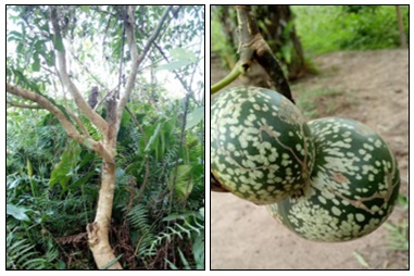
FIG. 1: TREE AND FRUITS OF V. AFRICANA STAPF (APOCYNACEAE)
Preparation of the Aqueous Extract: The extract was prepared according to the method described by Yapo et al. 16. Fifty grams of powder were suspended in 1 L of distilled water (5% w/v) and homogenized in a blender by three successive blending cycles of 3 minutes each. The homogenate was filtered twice through a white cloth, then five times through absorbent cotton. The filtrate was dried in a Memmert oven at 55 °C for three days. The dry extract was stored until use.
Phytochemical Screening: The classes of secondary metabolites present in the aqueous extract were detected using standard colorimetric and precipitation reactions, according to the methods of Tona et al. 17, Longanga et al. 18, and Békro et al. 19. Sterols and polyterpenes were detected using the Liebermann reaction; polyphenols using ferric chloride (FeCl₃); flavonoids using the cyanidin reaction; catechin tannins using Stiasny’s reagent; gallic tannins using FeCl₃; free and bound quinones using Bornträger’s reagent; alkaloids using the Dragendorff and Bouchardat reagents; and saponins using the foam test. For the foam test, 15 mL of aqueous extract was placed in a test tube (15 cm × 15 mm), shaken vigorously for 10 seconds, and then allowed to stand for 10 minutes. A persistent foam height greater than 3 cm was considered a positive result.
Acute Dermal Toxicity: Acute dermal toxicity was evaluated in accordance with the principles of OECD Guideline No. 402 20, using a dose limit of 2,000 mg/kg. Six adult, nulliparous, non-pregnant Wistar rats, approximately eight weeks old (155–163 g), were divided into a control group (n = 3) and a treatment group (n = 3). After acclimatization and clinical examination, the animals were weighed, and the dorsal region was shaved 24 h before application. A single dose of 2,000 mg/kg body weight was applied uniformly to the shaved skin of the treated rats and then covered with a compress for 24 hours. The control animals underwent the same procedure without the extract. After removing the compress and cleaning the area, the animals were observed individually during the first few hours, then daily for 14 days to assess mortality, general condition, behavior, clinical signs, and skin reactions. Body weight was recorded on days 0, 7, and 14. Surviving animals underwent a macroscopic examination at the end of the observation period.
Experimental Protocol: Fifteen nulliparous, non-pregnant Wistar rats (approximately 10 weeks old, weighing between 170 and 202 g) were randomly assigned to three groups of five: an untreated control group (Group 1) and two groups treated with the aqueous extract at doses of 100 mg/kg (Group 2) and 300 mg/kg (Group 3). The extract was applied daily to the mammary glands for 14 consecutive days. At the end of the treatment, nipple length was measured.
The animals were then euthanized by decapitation. The mammary glands were removed and fixed in 10% formalin.
Histology: The fixed mammary glands (10% formalin, 48 h) were placed in labeled cassettes, dehydrated using ethanol at increasing concentrations (80%, 90%, 100%), clarified in three toluene baths, and embedded in paraffin (two baths at 58–60 °C; Memmert oven, Germany). 5-µm sections were cut using a Leica RM2125 RTS microtome (Germany), floated in a water bath (at approximately 40 °C), mounted on slides, and dried at a temperature between 58 and 60 °C for 30 minutes. After deparaffinization with toluene and rehydration using ethanol at increasing concentrations (100%, 95%, 75%), the sections were stained with modified Harris’s hematoxylin and 3% yellow eosin, dehydrated, clarified with toluene, and mounted with Eukitt. The sections were examined and photographed using an Olympus CK41SF trinocular microscope (Philippines) connected to a computer running Videomet v8.0.9 software (France). The method followed that of Zougrou et al. 21.
Statistical Analysis: Data are expressed as mean ± standard error of the mean (SEM). Differences between groups were analyzed using one-way analysis of variance (ANOVA), followed by Tukey-Kramer’s multiple comparison test using GraphPad Prism 8 software (San Diego, California, United States). Histological observations were descriptive.
RESULTS:
Phytochemical Analysis: The aqueous extract contained sterols and polyterpenes, alkaloids (positive results with Dragendorff’s and Bouchardat’s reagents), and saponins. No polyphenols, flavonoids, tannins, or quinones were detected Table 1.
TABLE 1: PHYTOCHEMICAL SCREENING OF THE AQUEOUS EXTRACT OF V. AFRICANA STEM BARK
| Sterols and Polyterpenes | Polyphenols | Flavonoids | Tannins | Quinones | Alkaloids | Saponins | ||
| Gal | Cat | D | B | |||||
| + | - | - | - | - | - | + | + | + |
(+) : present, (-): absent, Gal: Gallic, Cat: catechic, D: Dragendorff, B: Bouchardat
Acute Skin Toxicity: A single topical application at a dose of 2,000 mg/kg resulted in no deaths or clinical signs of toxicity during the 14-day observation period. Body weight increased in both groups, rising from 158 ± 4.36 g (day 0) to 188 ± 8.08 g (day 14) in the control group and from 158 ± 1.00 g to 187 ± 1.00 g in the treated group Table 2; no significant differences were observed between the groups (P > 0.05). No abnormal changes were observed in behavior, general condition, skin, coat, eyes, or mucous membranes, and necropsy revealed no macroscopic abnormalities. Under these conditions, no signs of acute dermal toxicity were observed.
TABLE 2: BODY WEIGHT (G) OF RATS DURING THE ACUTE DERMAL TOXICITY TEST
| Day 0 | Day 7 | Day 14 | |
| Control | 158 ± 4,36 | 176 ± 8,96 | 188 ± 8,08 |
| AEVa2000 | 158 ± 1,00 | 172 ± 1,53 | 187 ± 1,00 |
Values are expressed as mean ± standard error of the mean, (n=3).
Effect of the Extract on Nipple Length: Topical application of the extract at doses of 100 and 300 mg/kg resulted in a significant increase in nipple length compared to the control group (P < 0.01), by 57.64% and 59.77%, respectively.
The mean nipple length was 437.7 ± 2.40 µm in the control group, 690.0 ± 6.66 µm at the 100 mg/kg dose, and 699.3 ± 23.39 µm at the 300 mg/kg dose Fig. 2 after 14 days of treatment.
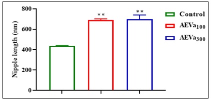
FIG. 2: AVERAGE SIZES OF THE NIPPLES OF CONTROL RATS AND THOSE TREATED WITH THE AQUEOUS EXTRACT OF V. AFRICANA STEM BARK AT THE END OF THE STUDY. Values are expressed as mean ± standard error of the mean, (n=5), (*) P<0.05; (**) P<0.01; (***) P<0.001. AEVa100: 100 mg/kg of aqueous extract of V. africana stem bark. AEVa300: 300 mg/kg of aqueous extract of V. africana stem bark.
Histology of Breast Tissue: The qualitative histological results are presented in Fig. 3. The sections from the control group consisted mainly of dense and relatively abundant connective tissue, with underdeveloped glandular structures scattered throughout the tissue, which is consistent with breast tissue exhibiting a low degree of glandular development. At the 100 mg/kg dose, the sections showed more rounded or tubular structures, interpreted as ductal and glandular structures, distributed within the connective and adipose tissue; the adipose tissue was well represented. Compared to the controls, this organization suggested a larger glandular compartment. At a dose of 300 mg/kg, ductal and glandular structures were also visible within the connective tissue, exhibiting a more pronounced organization in the observed areas, as well as clusters of adipose tissue and a relatively well-developed connective tissue network.
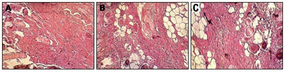
FIG. 3: MICROGRAPH OF THE MAMMARY GLANDS OF CONTROL RATS AND RATS TREATED WITH THE AQUEOUS EXTRACT OF V. AFRICANA STEM BARK. A: Control, B: 100 mg/kg of aqueous extract of V. africana stem bark, C: 300 mg/kg of aqueous extract of V. africana stem bark, Magnification: ×100; Staining: Hematoxylin-eosin
DISCUSSION: Topical application of an aqueous extract of V. africana stem bark at doses of 100 and 300 mg/kg for 14 days was associated with significantly greater nipple length and histological features suggesting increased development of the ductal and glandular compartments, compared with untreated controls.
Both doses resulted in similar increases (57.64% and 59.77%). This similarity may reflect a plateau within the tested dose range. Nipple length is an indirect morphological indicator and, on its own, does not allow conclusions to be drawn about lactation. It may be related to histological changes, as mammary morphology is highly dependent on the hormonal environment 7, 8, 22.
In rodents, estrogens contribute to the growth and elongation of the ducts, while progesterone and prolactin contribute to branching and alveolar development 23, 24. Mammary development involves a phase of duct elongation and branching, followed by alveolar development associated with pregnancy and lactation 25, 26. Histological observations are consistent with stimulation of the epithelial compartment, though they do not prove it.
These changes could reflect hormone-like activity or an indirect modulation of mammary development, but this hypothesis has not been verified: concentrations of estradiol, progesterone, and prolactin, as well as the expression of mammary ER/PR receptors, were not measured.
Studies conducted on other plants indicate that certain secondary metabolites can alter mammary morphology or hormone-related pathways; this is the case, for example, with saponins derived from Phytolacca in a rat model of mammary hyperplasia, where they reduced proliferation and influenced the expression of sex hormone receptors 27. This study reported an anti-hyperplastic effect, which differs from a stimulatory effect; its relevance in the present context is therefore limited.
Preliminary analysis detected sterols/polyterpenes, alkaloids, and saponins, but no polyphenols, flavonoids, tannins, or quinones. The presence of alkaloids is consistent with the literature on V. africana 28, 29, 30. Babiaka et al. isolated several alkaloids from the bark of the trunk, including voacangine, voacristine, coronaridine, tabernanthine, ibogaine, voacamine, and voacorine, while Chen et al. reported the presence of voacamine, vobasine, voacangine, and voacristine 28, 31.
Since, the screening was qualitative and the extract was not standardized, the compounds responsible for the effects, as well as their relative contribution compared to sterols or saponins, remain unknown.
A single dermal dose of 2,000 mg/kg resulted in no deaths or clinical changes over a 14-day period, and body weight gain was comparable between groups. This result is consistent with the absence of overt acute toxicity following a single exposure. However, it does not rule out the possibility of local irritation or systemic effects following repeated applications.
CONCLUSION: In female rats, topical application of an aqueous extract of V. africana bark for 14 days was associated with an increase in nipple length and histological features suggesting increased development of the ducts and glands, with no signs of acute skin toxicity at a dose of 2,000 mg/kg following a single application. These results are preliminary. In the absence of quantitative histological and endocrine data, it is not possible to conclude that there is a hormonal, estrogenic, or galactagogue effect, and the safety of the extract for breastfeeding women and infants remains to be evaluated. These results warrant further research.
ACKNOWLEDGEMENT: Nil
CONFLICT OF INTEREST: The authors declare that they have no conflicts of interest.
REFERENCES:
- Khan J, Vesel L, Bahl R and Martines JC: Timing of breastfeeding initiation and exclusivity of breastfeeding during the first month of life: effects on neonatal mortality and morbidity a systematic review and meta-analysis. Matern Child Health J 2015; 19: 468–479. doi:10.1007/s10995-014-1526-8.
- Patnode CD, Henrikson NB, Webber EM, Blasi PR, Senger CA and Guirguis-Blake JM: Breastfeeding and health outcomes for infants and children: a systematic review. Pediatrics 2025; 156(1): e2025071516. doi:10.1542/peds.2025-071516.
- Jakaitis BM and Denning PW: Human breast milk and the gastrointestinal innate immune system. Clin Perinatol 2014; 41(2): 423–435. doi:10.1016/j.clp.2014.02.011.
- Le Doare K, Holder B, Bassett A and Pannaraj PS: Mother’s milk: a purposeful contribution to the development of the infant microbiota and immunity. Front Immunol 2018; 9: 361. doi:10.3389/fimmu.2018.00361.
- Lyons KE, Ryan CA, Dempsey EM, Ross RP and Stanton C: Breast milk, a source of beneficial microbes and associated benefits for infant health. Nutrients 2020; 12(4): 1039. doi:10.3390/nu12041039.
- Richard C, Lewis ED and Field CJ: Evidence for the essentiality of arachidonic and docosahexaenoic acid in the postnatal maternal and infant diet for the development of the infant’s immune system early in life. Appl Physiol Nutr Metab 2016; 41(5): 461–475. doi:10.1139/apnm-2015-0660.
- Brisken C and O’Malley B: Hormone action in the mammary gland. Cold Spring Harb Perspect Biol 2010; 2(12): a003178. doi:10.1101/cshperspect.a003178.
- Hannan FM, Elajnaf T, Vandenberg LN, Kennedy SH and Thakker RV: Hormonal regulation of mammary gland development and lactation. Nat Rev Endocrinol 2023; 19(1): 46–61. doi:10.1038/s41574-022-00742-y.
- Malan DF, Neuba DF and Kouakou KL: Medicinal plants and traditional healing practices in Ehotile people, around the a by Lagoon (eastern littoral of Côte d’Ivoire). J Ethnobiol Ethnomed 2015; 11: 21. doi:10.1186/s13002-015-0004-8.
- Kelleher SL, Burkinshaw S and Kuyooro SE: Polyphenols and lactation: molecular evidence to support the use of botanical galactagogues. Mol Nutr Food Res 2024; 68: 2300703. doi:10.1002/mnfr.202300703.
- Intan PR, Alegantina S, Isnawati A, Yunarto N, Ekawasti F and Rinendyaputri R: Effects of a blend extract of Sauropus androgynus, Moringa oleifera, and Coleus amboinicus on milk production in lactating rats. Open Vet J 2024; 14(12): 3630–3639. doi:10.5455/OVJ.2024.v14.i12.44.
- Chen J, Pan Z, Li Q, Wu Y, Li X and Wang X: The aqueous extract of Hemerocallis citrina Baroni improves the lactation-promoting effect in bovine mammary epithelial cells through the PI3K-AKT signaling pathway. Foods 2024; 13(17): 2813. doi:10.3390/foods13172813.
- Tan PV, Penlap VB, Nyasse B and Nguemo JDB: Anti-ulcer actions of the bark methanol extract of Voacangaafricana in different experimental ulcer models in rats. J Ethnopharmacol 2000; 73(3): 423–428. doi:10.1016/S0378-8741(00)00292-0.
- Bekoe EO, Opare JAA, Lartey M and Amoateng P: Ethnomedicinal uses, biological activities, and toxicity of Voacanga africana Stapf ex Scott-Elliot. Adv Tradit Med 2024; 24: 431–448. doi:10.1007/s13596-023-00709-y.
- Koroch AR, Juliani HR, Kulakowski D, Arthur H, Asante-Dartey J and Simon JE: Voacanga africana: chemistry, quality and pharmacological activity. In: Juliani HR, Simon JE, Ho CT, editors. African Natural Plant Products: New Discoveries and Challenges in Chemistry and Quality. Washington, DC: American Chemical Society 2010; 363–380. doi:10.1021/bk-2009-1021.ch020.
- Yapo CVY, Konkon DG, Coulibaly K, Camara D and Zirihi GN: Étude botanique, évaluation de l’activité antifongique sur la croissance in-vitro de Candida albicans et de la toxicité sur des cellules HFF de feuilles de Mallotus oppositifolius (Geiseler) Müll.Arg. (Euphorbiaceae). J Anim Plant S 2016; 28(1): 4330–4339.
- Tona L, Kambu K, Ngimbi N, Cimanga K and Vlietinck AJ: Antiamoebic and phytochemical screening of some Congolese medicinal plants. J Ethnopharmacol 1998; 61(1): 57–65. doi:10.1016/S0378-8741(98)00015-4.
- Longanga Otshudi A, Vercruysse A and Foriers A: Contribution to the ethnobotanical, phytochemical and pharmacological studies of traditionally used medicinal plants in the treatment of dysentery and diarrhoea in Lomela area, Democratic Republic of Congo (DRC). J Ethnopharmacol 2000; 71(3): 411–423. doi:10.1016/S0378-8741(00)00167-7.
- Békro YA, Mamyrbekova JA, Boua BB, Tra BFH and Ehilé EE: Étude ethnobotanique et screening phytochimique de Caesalpinia benthamiana (Baill.) Herend. et Zarucchi (Caesalpiniaceae). Sci Nat 2007; 4(2): 217–225. doi:10.4314/scinat.v4i2.42146.
- OECD. Test No. 402: Acute Dermal Toxicity. OECD Guidelines for the Testing of Chemicals, Section 4. Paris: OECD Publishing 2017. doi:10.1787/9789264070585-en.
- Zougrou NE, Blahi AN, Kouassi KD and Kouakou K: Effects of the aqueous extract of Cnestis ferruginea on the histological structure of female rat ovary and uterine horns. Biomed J Sci Tech Res 2018; 2(1): 2073–2078. doi:10.26717/BJSTR.2018.02.000625.
- Zhou X, Ullah A, Shi L, Dou M, Wang C and Khan MZ: Molecular regulatory mechanisms of mammary gland development: a review. Animals 2025; 15: 3480. doi:10.3390/ani15233480.
- Atwood CS, Hovey RC, Glover JP, Chepko G, Ginsburg E and Robison WG: Progesterone induces side-branching of the ductal epithelium in the mammary glands of peripubertal mice. J Endocrinol 2000; 167(1): 39–52. doi:10.1677/joe.0.1670039.
- Halperin J, Dorfman VB, Fraunhoffer N and Vitullo AD: Estradiol, progesterone and prolactin modulate mammary gland morphogenesis in adult female plains vizcacha (Lagostomus maximus). J Mol Histol 2013; 44(3): 299–310. doi:10.1007/s10735-012-9477-0.
- Hovey RC, Trott JF and Vonderhaar BK: Establishing a framework for the functional mammary gland: from endocrinology to morphology. J Mammary Gland Biol Neoplasia 2002; 7(1): 17–38. doi:10.1023/A:1015766322258.
- Macias H and Hinck L: Mammary gland development. Wiley Interdiscip Rev Dev Biol 2012; 1(4): 533–557. doi:10.1002/wdev.35.
- Li X, Wang Z, Wang Y, Zhang Y, Lei X and Xin P: Anti-hyperplasia effects of total saponins from Phytolaccae Radix in rats with mammary gland hyperplasia via inhibition of proliferation and induction of apoptosis. Front Pharmacol 2018; 9: 467. doi:10.3389/fphar.2018.00467.
- Chen HM, Yang YT, Li HX, Cao ZX, Dan XM and Mei L: Cytotoxic monoterpenoid indole alkaloids isolated from the barks of Voacanga africana Stapf. Nat Prod Res 2016; 30(10): 1144–1149. doi:10.1080/14786419.2015.1046132.
- Fouotsa H, Le Pogam P and Mkounga P: Voatria fricanines A and B, trimeric vobasine-aspidosperma-aspidosperma alkaloids from Voacanga africana. J Nat Prod 2021; 84(10): 2755–2761. doi:10.1021/acs.jnatprod.1c00812.
- Qin M, Gao W, Wang H, Yin S, Hu J, Gao W and Ding C: Monoterpenoid indole alkaloids from the stem barks of Voacanga africana and their chemotaxonomic significance. Biochem Syst Ecol 2024; 116: 104863. doi:10.1016/j.bse.2024.104863.
- Babiaka SB, Simoben CV, Abuga KO, Mbah JA, Karpoormath R and Ongarora D: Alkaloids with anti-onchocercal activity from Voacanga africana Stapf (Apocynaceae): identification and molecular modeling. Molecules 2020; 26(1): 70. doi:10.3390/molecules26010070.
How to cite this article:
Begbin KE, Oumar K, Gneho DAD and Kouakou K: effects of the aqueous stem bark extract of Voacanga africana Stapf (Apocynaceae) on nipple length and mammary gland histology in female rats after topical application. Int J Pharmacognosy 2026; 13(11): 100-07. doi link: http://dx.doi.org/10.13040/IJPSR.0975-8232.IJP.13(11).1165-71.
This Journal licensed under a Creative Commons Attribution-Non-commercial-Share Alike 3.0 Unported License.
Article Information
6
1141-1147
812 KB
4
English
IJP
Kouassi Emile Begbin *, Koné Oumar, Doh Arioste Delchinor Gneho and Koffi Kouakou
Faculty of Sciences and Technologies, Alassane Ouattara University, BP V18 Bouake 01, Côte d’Ivoire.
kbegbin@gmail.com
06 October 2026
29 October 2026
31 October 2026
10.13040/IJPSR.0975-8232.IJP.13(11).1165-71
01 November 2026
 Download