


SUBSTANTIATION OF USING CHLORELLA GENUS MICROALGAE AS A RAW MATERIAL FOR PREPARATION OF CHEMOPREVENTIVE SUBSTANCES
HTML Full TextSUBSTANTIATION OF USING CHLORELLA GENUS MICROALGAE AS A RAW MATERIAL FOR PREPARATION OF CHEMOPREVENTIVE SUBSTANCES
E. V. Тretiakova * 1, P. G. Beliaeva 2 and A. I. Saralov 2
Department of Pharmaceutical Technology 1, Perm State Pharmaceutical Academy - 2, Polevaya Str., Perm, 614990, Russia.
Institute of Ecology and Genetics of Microorganisms 2 Ural Branch Russian Academy of Sciences -13, Goleva Str., Perm, 614081, Russia.
ABSTRACT: The purpose of this study was to substantiate the use of suspension of Chlorella genus microalgae as the most valuable raw material for the preparation of carotene and chlorophyll а. The method is proposed for ultrasonication of Chlorella vulgaris microalgae suspension to increase the availability of biologically active substances and improve the microbiological performance of the prepared product. Before and after ultrasonication of samples, we determined the content of chlorophylls (a, b, c) and carotenoids using the spectrophotometric method, the level of total bacterial contamination using the method of limiting dilutions on MacConkey agar and microscopic study of microalgae and bacteria cell viability. It has been established that the ultrasonication method at wave amplitude 16 µm allows to transfer pigments into the water phase, inhibit the viability of bacteria and obtain homogenization of the Chlorella vulgaris microalgae suspension with preservation of carotenoids and green chlorophyll а. Thus, the combined use of ultrasonication and parallel plating on MacConkey agar seems to be promising given preparation and quality control of axenic concentrate of Chlorella vulgaris microalgae, which can be used for preparation of chemopreventive pharmaceuticals.
| Keywords: |
Chlorella vulgaris microalgae, Carotenoids, Chlorophyll, Ultrasonication
INTRODUCTION: Dietary phytochemical substances - chlorophyll and carotene - are widespread plant pigments that have a physiological effecton the treatment of chronic diseases. Nowadays, they are especially interesting due to their ability to improve the resistance of the human body to mutagenesis and carcinogenesis, which is of great importance in the prevention of malignant diseases 1, 2.
Multiple epidemiological and experimental studies around the world confirm that carotene and chlorophyll manifest anti-carcinogenic, anti-mutagenic, anti-oxidant, immunomodulating, and anti-inflammatory properties. On molecular and cellular levels, they are capable of preventing transformations induced by oxidants, genotoxic substances, X-ray and UV irradiation, and increase immunocompetence 1, 2, 3, 4, 5, 6.
The role of such chemopreventive agents is important in primary prevention of cancer, whereas the availability of additional anti-toxic and anti-inflammation properties enables their use for reduction of toxicity of basic therapy 7. Natural complex substances are at least as efficient as the synthetic compounds; however, their advantage is potentially lower toxicity, whereas the use of combinations of such agents improves the degree of manifestation and the range of chemopreventive effect 1, 5. Search for new raw material sources of carotene and derivatives of chlorophyll is caused by their low content in natural sources, which is related to the dependence of their total content on natural climatic conditions. The disadvantages of chemical synthesis are the possibility of residual content of semi-products and products of by-reactions in the target product 1, 5.
In this field, the promising area is the use of the raw material base of lower plants-green unicellular microalgae, specifically the Chlorella genus, which are unique producers of biologically active substances. High speed of reproduction of microalgae, their indiscriminateness to life environment and the possibility of correction of biochemical content of cells through conditions of cultivation allow using controlled cultivation of microalgae in artificially recreated conditions 8, 9. This enables production, over a short period, of large quantities of environmentally clean biomass characterized by a high content of chlorophyll and carotenoids, which can serve as a basis for the preparation of valuable vitamin and food additives, as well as pharmaceuticals.
Presently, Chlorella microalgae are mainly used as a food additive for correction of diet. Effectiveness of their preventive use for strengthening overall immunity, elimination of products of intoxication and heavy metals, as well as, in the long-term, reduction of the occurrence rate of radiation-induced forms of cancer, is confirmed by research 9, 10, 11, 12, 13. The natural state of Chlorella microalgae is suspension, i.e., microalgae cells dispersed in their culture medium. Mostly they are used in dry form (microalgae powder).
However, the use of powder as a raw material is less promising, due to the loss of a part of biologically active substances in the process of drying and its high cost due to the low output of dry biomass. To reduce the cost, cultivation is performed in large volume reservoirs using sunlight energy, often in the open air, which results in contamination of the culture with fungi, bacteria and other species of algae 14. The microalgae suspension is a more promising source of biologically active substances since the cells are in a live state and can maintain their biochemical content. Growing a unialgal culture of microalgae is possible through the use of purified water and sealed photo-bio-reactors for cultivation, while correction of conditions of cultivation and feeding helps control the accumulation of target products in the microalgae cells. In the process of cultivation, the microalgae cells are located in a nutrient medium with the required temperature (27-32 ºС) and lighting 14, 15. Such conditions are favorable for the growth of the microalgae, but also the growth of extraneous bacterial microflora, whereas due to the live state of cells and instability of chlorophyll, standard methods of conservation and sterilization are not applicable 9, 14.
The second challenge in the use of microalgae suspension is its digestion by the human body. The cell wall of a mature microalga contains microfibers of cellulose (poly-β-1.4-D-N- acetyl-glucosamine) and structurally polymerized carotenoid substrate (sporopolenine). It ensures the strength of the cell wall, but the human body is not capable to effectively digest it 8, 16, 17, 18. Therefore, the use of microalgae suspension is only rational after disruption of its cell wall, to enable maximum output of target products. The purpose of this study was to substantiate the use of a suspension of Chlorella genus microalgae as the most valuable raw material for the preparation of carotene and chlorophyll а. The method is proposed for ultrasonication of the microalgae suspension to increase the availability of biologically active substances and improve the microbiological performance of the prepared product.
MATERIALS AND METHODS: The research examines the unicellular Chlorella vulgaris Beyerinck [Beijerinck] microalgae, strain IMBR-19, provided by the Kovalevsky Institute of Marine Biological Research of the Russian Academy of Sciences, Sevastopol. The provided culture has been grown on a rich nutrient media for intensive cultivation of green algae in a sealed photo-bio-reactor. Two specimens of culture were used for the research, corresponding in terms of quantitative content of microalgae cells to 40-50 million cells/ ml: 1st specimen-1-month-old suspension, 2nd specimen- as grown suspension.
For storage of the culture, nutrient-poor liquid and solid agarized Knop medium was used (g/l, for green algae diluted 1:2): Ca(NO3)2 - 0.25; MgSO4 × 7H2O - 0.06; KH2PO4 - 0.06; KCl - 0.08; FeCl3 × 6H2O - 0.001. All manipulations with the culture met the conditions of sterility common for microbiological practice. Re-inoculation of cultures was performed in a laminar safety hood pre-sterilized for at least 30 min using bactericide UV lamps BUV- 40. Inoculation was performed over the spirit lamp flame.
Preparation of the Algae Suspension Homogentisate: Disruption of the microalgae cell wall was performed using an ultrasonic disintegrator UD-20 (Techpan, Poland). The operating frequency of the disintegrator constitutes 22 ± 1.65 KHz. Introduction of ultrasonic energy in the liquid cause’s formation of cavitation bubbles which cause shock waves when collapsing. The intensity of cavitation processes is controlled through modification of oscillation amplitude in 5 ranges (1-8 µm, 2-10 µm, 3-12 µm, 4-14 µm, 5-16 µm). Average energy in the medium volume of approximately 50 ml (kg × m2/sec2 or J) at wave amplitude 8 × 10⁻⁶ m constitutes 6.1 × 10⁻⁴, at amplitude 16 × 10-6 m - 24.4 × 10⁻⁴. Further research was conducted on specimens before and after ultrasonication.
Determination of Plant Pigments: Content of chlorophylls a, b, c, and carotenoids were determined using spectrophotometry, modified based on the specifics of their determination in the water phase of the suspension after deposition of сells with centrifuging, on the spectrophotometer Cary100 (Agilent Technologies, Malaysia) 19. Comparative evaluation of the content of chlorophylls and carotenoids before and after ultrasonication was performed based on the formulas of standard spectrophotometric method for determination of pigments 20.
Determination of Total Bacterial Content: Bacterial count in the specimens under study has been determined using the method of limiting dilutions on MacConkey agar. For isolation and identification of enteral gram-negative bacteria, the following substances were used (g/l): gelatin peptone- 17.0; bile salts no. 3-1.5; neutral red-0.03; polypeptide- 3.0; sodium chloride- 5.0; crystal violet- 0.001; lactose- 10.0; agar- 13.5. The end value constituted рН 7.1 ± 0.2. Inoculations were incubated at 37 ºС until the emergence of individual colonies in 2-3 days.
Gram-positive microorganisms are inhibited by bile salts and crystal violet. Other gram-negative microorganisms which are not enteral bacteria (Pseudomonas, Aeromonas) grow in the form of small colonies, from colorless to greenish-brown. Pathogenic Proteus genus bacteria form colorless and transparent colonies. Lactose-fermenting enteral bacteria form red or pink colonies and reduce рН of the medium, which can be determined using neutral red indicator: Escherichia сoli form even pink non-mucoid colonies, Klebsiella spp. -big red mucoid colonies 21.
Study of Viability of Microalgae and Bacteria Cells: Study of the viability of microalgae and bacteria cells was performed using a confocal laser scanning microscope Olympus Corporation (Japan). A drop (20 µl) of cell suspension was placed on the cover glass, mixed with an equivalent volume of fluorescent dye LIVE/DEAD® BacLightTM, Bacterial Viability Kit (Invitrogen, USA), and dried in the air in the dark during 10-15 min. The product was scanned using the microscope with an immersion lens.
To induce fluorescence of SYTO 9 and propidium iodide, argon laser (wavelength 488 nm) with 505/ 525 nm barrier filter and helium-neon laser (wavelength 543 nm) with 560/660 nm barrier filter were used. Image analysis was performed using FV10-ASW3.1 program (Olympus Corporation, Japan). Alive, actively reproducing cells had green color, dead cells-red color, wilting- yellow-green or yellow-orange color.
RESULTS AND DISCUSSION: Chlorella vulgaris microalgae contain carotenoids and chlorophylls typical for higher plants. Their concentration was determined before and after ultrasonication in the water phase after the deposition of cells with centrifuging. Spectra of plant pigments before and after ultrasonication of specimen 1 are shown in Fig. 1А, specimen 2- in Fig. 1B. Content and concentration of chlorophylls and carotenoids in specimens before and after ultrasonication are shown in Table 1.
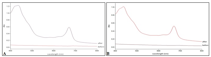
FIG. 1: SPECTRA OF PLANT PIGMENTS IN WATER PHASE OF SPECIMEN 1 (А) AND 2 (B) BEFORE AND AFTER ULTRASONICATION
TABLE 1: CONTENT AND CONCENTRATION OF PLANT PIGMENTS IN WATER PHASE OF SPECIMENS BEFORE AND AFTER ULTRASONICATION (Chl - Chlorophyll, Karot – Carotenoids)
| Specimen | Chl
a |
Chl
b |
Chl
c |
Karot | E480/
E665 |
ΣChl | % Chl
a |
%Chl
b |
% Chl
c |
Karot/
Chl a |
| 1 before | 0.22±0.08 | 0.34±0.11 | 0.98±0.26 | 0.16±0.04 | 2.21 | 1.53±0.47 | 13.31±1.19 | 21.25±1.05 | 65.44±2.24 | 0.87 |
| 1 after | 5.18±0.10 | 3.94±0.16 | 8.52±0.17 | 2.79±0.04 | 1.45 | 17.63±0.42 | 29.39±0.15 | 22.30±0.37 | 48.31±0.22 | 0.54 |
| 2 before | 0.25±0.09 | 0.38±0.11 | 1.11±0.28 | 0.20±0.04 | 2.11 | 1.74±0.48 | 13.57±1.51 | 21.16±0.82 | 65.28±2.33 | 0.95 |
| 2 after | 4.55±0.10 | 3.56±0.14 | 9.02±0.36 | 2.53±0.04 | 1.49 | 17.13±0.60 | 26.59±0.35 | 20.78±0.10 | 52.64±0.26 | 0.56 |
The data is provided as a mean value with standard deviation, M ±SD.
Based on the available data, it was established that after ultrasonication, chlorophylls and carotenoids exit the disrupted cells into the water phase. Content of carotenoids in the water phase after ultrasonication increased in specimen 1 by 17.4 times, in specimen 2 by 12.6 times, the content of chlorophylls increased by 11.5 times and by 9.8 times, respectively, whereas the proportion of chlorophyll an increased by 2 times, and proportion of chlorophyll с decreased by 1.3 times. Cells of one-month-old culture 1 have been easier to disrupt with ultrasound than those of the as-grown culture. Going by the pigment indexes Karot/Chla and E480 / E665, chlorophylls are much easier disengaged from ultrasound-disrupted pigment-albumen complexes than the more hydrophobic carotenoids. Preliminary microscopic study of cell suspension based on the standard method on cover glass using the microscope ZEISS Axiostar Plus (Germany) at 700x magnification allowed to trace the effectiveness of disruption of algae and bacteria cells depending on the power and duration of ultrasonication. The minimum power of ultrasound (in range 1) during 1 min resulted in the observed disruption of microcolonies of bacteria aggregated in the organo-mineral dispersion. Ultrasonication of the suspension during 4 min at maximum amplitude (in range 5) resulted in full disappearance of bacteria and free-floating microalgae cells from the field of view, and in the exit of pigments into the water phase.
TABLE 2: IMPACT OF ULTRASOUND ON ENTERAL BACTERIA COUNTS IN SPECIMENS BEFORE AND AFTER ULTRASONICATION
| Ultrasonic
disintegration mode |
Micro-
organisms |
Count, cells/ml | |
| Specimen 1 | Specimen 2 | ||
| Initial, before ultrasonication
|
Enterobacteriaceae Escherichia coli Klebsiella spp. |
32000-34000
30000-32000 2200-2500 |
35000-36000
30000-31000 5000-5500 |
| 1 min in range 3 | 14000-16000
13500-14000 800-1000 |
110-130
110-120 40-70 |
|
| 3 min in range 5
|
150-160
145-155 30-60 |
2-5
2-5 0 |
|
| 4 min in range 5 | 0 | 0 | |
Results obtained in the course of quantitative analysis of enteral bacteria on MacConkey agar in the samples of initial suspensions and after their ultrasonication are shown in Table 2. In the initial specimens, enteral bacteria count appeared to be approximately equally high in both samples (32 - 36 thousand cells/ml). Their composition was predominated by lactose-fermenting Escherichia coli, forming even non-mucoid pink colonies of average size. Total enteral bacteria count in specimen 2 appeared to be somewhat higher, mainly due to the representatives of Klebsiella genus, forming large mucoid red colonies. There were no relatively hazardous pathogenic enteral bacteria of Proteus, Salmonella genus or non-enteral gram-negative bacteria of Aeromonas, Pseudomonas genus. After ultra-sonication in specimens 1 and 2 all bacteria present in the microalgae suspension were fully dead after 4 min of ultrasonication with the laboratory ultrasonic disintegrator due to the effect of cavitation processes at maximum ultrasound power (range 5). Whereas after ultrasonication of specimen 2 cell suspension, at maximum ultra-sound power during 3 min, several bacterial cells preserved their viability (2-5 cells/ml). After reduction of ultrasonication by 1 more min (2 min, range 5) and in case of ultrasonication during 1 min at average power (range 3), the count of viable cells increased by 30-50 times (or up to 0.3-0.4% fromthe initial count in the suspension). Effect of ultra-sonication of specimen 1 during 3 min at average ultrasound power (range 3) was the same as the effect of ultrasound in range 5 during 3 min (viable cell count 0.4-0.5% from the initial count in the suspension). Ultrasonication of the suspension in range 3 during only 1 min appeared to have low efficiency, as the viability of almost 50% of bacterial cells was preserved.
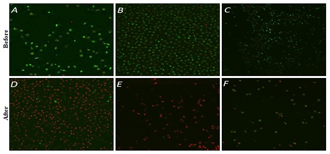
FIG. 2: MICROPHOTOGRAPHS OF SPECIMEN 1 (А, D) AND SPECIMEN 2 (В, C, E, F) OF THE MICROALGAE SUSPENSION BEFORE (А, В, С) AND AFTER (D, E, F) ULTRASONICATION
Study of microorganisms colored with fluorescent dye LIVE/DEAD using the laser scanning microscope allowed to assess the viable state of cells under the effect of ultrasonication. Microphotographs of specimens before and after ultrasonication are shown in Fig. 2. In the initial suspension specimens, micro-photographs show the apparent prevalence of live cells, green colored algae, and bacteria, Fig. 2А, B. In some fields of vision, in accumulations of smaller bacterial cells, their count was comparable to the count of free-floating microalgae cells Fig. 2C. In these samples, the count of dead microalgae and bacteria cells painted red did not exceed 0.1%. The bacterial cells were mostly rod forms capable of forming loose aggregates.
Ultrasonication of specimen 1 at maximum amplitude during 4 min resulted in the death of 97.5% microalgae cells: in the field of vision, the count of dead red cells amounted to 400, while that of viable green cells-only 10 Fig. 2D. In case of the maximum power of ultrasound effecting specimen 2 (the as-grown suspension), after ultrasonication during 2 min, the cell nuclei of microalgae were almost completely dead (red) Fig. 2E.
3 min of ultra-sonication resulted in a rapid decrease of the count of microalgae cells, nuclei of all cells turned dead (red), but in whole and semi-disrupted cells, most of the volume of green chlorophyll was retained Fig. 2F. Taking into account the strong negative impact of ultrasound on the microalgae cell nuclei, inoculation on liquid Knop medium was performed using suspensions of microalgae after ultra-sonication during 2-4 min. Samples have been incubated in the light at 20-23 ºС during 10 days. However, in 8 inoculated replications (platings) the growth of microalgae did not reoccur.
CONCLUSION: The method of ultrasonication of Chlorella vulgaris suspension at wave amplitude 16 µm allows obtaining an axenic microalgae concentrate containing easily available chemo-preventive biologically active substances. Compared to the initial microalgae suspension, the quantity of carotenoids increases in average by 15 times, the quantity of free chlorophyll- by 10 times, mostly due to the increase in the proportion of chlorophyll а, which represents a substantiation of prospective use of this method of ultrasonication for preparation of chlorophyll а and carotenoids.
The one-month old microalgae culture was more prone to disruption by ultrasound than the cells of the as-grown culture. At the same time, in the as-grown microalgae suspension it was easier to eliminate bacteria content. Thus, the combined use of ultrasonication and parallel plating on MacConkey agar seems to be promising in view of preparation and quality control of axenic concentrate of Chlorella vulgaris microalgae, which can be used for preparation of pharmaceuticals and biologically active additives.
The obtained concentrate cannot be used for further microalgae cultivation due to the death of cell nuclei. More detailed identification of pigments, the stability of microalgae concentrate in the process of storage, toxicity, and pharmacological activity can be examined in further studies.
ACKNOWLEDGEMENT: The authors express their gratitude to the General Director of OOO “Les” (Limited Liability Company) Alexander Fedyuk for the provided culture of Chlorella vulgaris microalgae and funding of the study.
CONFLICT OF INTEREST: The authors declare that the conflict of interest is absent.
REFERENCES:
- Shashkina MY, Shashkin PN and Sergeev AV: Carotenoids in human health and prevention of diseases. Russian Biotherapeutic Journal 2010; 9 (1): 77-86.
- Chupahina GN, Maslennikov PV, Skrypnik LN, Chupahina NJ and Feduraev PV: Antioxidant properties of cultural plants in the Kaliningrad region: monograph. The Baltic Federal University named after Immanuel Kant Kaliningrad: 2016.
- Shvidko EA, Malyavina VV and Sampiev AM: Pharmaceutical grade rang carotene of the containing medicines. Kuban Scientific Medical Bulletin 2010; 3-4(117-118): 215-220.
- Sergeev AV, Anan'ev VS, Kapitanov AB, Korostуlev SA, Bukreev YM and Vlasenkova NK: Pharmacokinetics of carotenoids and carotene containing compounds. Russian Biotherapeutic Journal 2017; 16(3): 92-106.
- Mishra VK, Bacheti RK and Husen A: Chlorophyll: structure, function and medicinal uses. Medicinal uses of Chlorophyll: a critical overview. Nova Science Publishers, Inc, Hauppauge 2011: 177-196.
- Chernomorsky S, Segelman A and Poretz RD: Effect of dietary chlorophyll derivatives on mutagenesis and tumor cell growth. Teratog Carcinog Muta 1999; 19(5): 313-22.
- Dyshlova SA, Shubin LK, Fedorov SN, Kuzmich AS, Radchenko OS and Krasokhin VB: Patent of the Russian Federation no. 2429840; 2011.
- Auzhanova NB: Morphological and systematic characterization of Chlorella. Its production and use. Scientific Bulletin 2014; 1(1): 113-126.
- Hosikian A, Lim S, Halim R and Danquah MK: Chlorophyll extraction from microalgae: A Review on the process engineering aspects. International Journal of Chemical Engineering 2010; 1-11.
- Knizhnikov VA, Shandala NK, Komleva VA, Yarmonenko SP and Tutelyan VA: Decrease of radiogenic cancer and leukemia risk by adding selenium in trace amounts and green alga Momotaru E-25 to diets. Medical Radiology and Radiation Safety 1998; 43(5): 6-13.
- Tumanova AL: Innovative methods of prevention and rehabilitation of end ecological health using food concentrate “Living Chlorella”. Modern aspects of sanatorium-and-spa treatment and rehabilitation at the stages of rendering medical care to children and adults 2017; 1: 27-30.
- Chaplygin OS and Sukhikh SA: Functional drink based on Chlorella vulgaris cell suspension. Actual Questions of the Beverage Industry 2017; 1: 134-136.
- Bengwayan PT, Laygo JC, Pacio AE, Poyaoan JLZ, Rebugio JF and Yuson ALL: A comparative study on the antioxidant property of Chlorella (Chlorella sp.) Tablet and Glutathione Tablet. E- International Scientific Research Journal 2010; 2(1): 25-35.
- Görs M, Schumann R, Hepperle D and Karsten U: Quality analysis of commercial Chlorella products used as dietary supplement in human nutrition. Journal of Applied Phycology 2010; 22(3): 265-276.
- Pulz O: Photobioreactors: Production systems for phototrophic microorganisms. Applied Microbiology and Biotechnology 2001; 57(3): 287-293.
- Cheng YS, Zheng Y and Labavitch JM: The impact of cell wall carbohydrate composition on the chitosan flocculation of Chlorella. Process Biochemistry 2011; 46: 1927-1933.
- Gerken HG, Donohoe BS and Knoshaug EP: Enzymatic cell wall degradation of vulgaris and other microalgae for biofuels production. Planta 2013; 237(1): 239-253.
- Takeda H: Sugar composition of the cell wall and the taxonomy of Chlorella (Chlorophyceae). Journal of Phycology 1991; 27(2): 224-232.
- Monographs on Oceanographic Methodology. Determination of photosynthetic pigments in seawater. SCOR-UNESCO Working Group No. 17, UNESCO, Pari, 1966.
- Jeffrey SW and Humphrey GF: New spectrophotometric equations for determining chlorophylls a, b, c1 and c2 in higher plants algae and natural phytoplankton. Biochemie und Physiologie der Pflanzen 1975; 167(2): 191-194.
- Kuznetsov SI and Dubinina GA: Methods of studying aquatic microorganisms. Nauka, Moscow 1989.
How to cite this article:
Тretiakova EV, Beliaeva PG and Saralov AI: Substantiation of using Chlorella genus microalgae as a raw material for preparation of chemopreventive substances. Int J Pharmacognosy 2018; 5(12): 774-80. doi link: http://dx.doi.org/10.13040/IJPSR.0975-8232.IJP.5(12).774-80.
This Journal licensed under a Creative Commons Attribution-Non-commercial-Share Alike 3.0 Unported License.
Article Information
4
774-780
858
1488
English
IJP
E. V. Тretiakova *, P. G. Beliaeva and A. I. Saralov
Department of Pharmaceutical Technology, Perm State Pharmaceutical Academy - 2, Polevaya Str., Perm, 614990, Russia.
tiloket@mail.ru
31 October 2018
22 November 2018
27 November 2018
10.13040/IJPSR.0975-8232.IJP.5(12).774-80
01 December 2018