POLY HERBAL SEMISOLID DOSAGE FORM DEVELOPMENT FOR WOUND HEALING
HTML Full TextPOLY HERBAL SEMISOLID DOSAGE FORM DEVELOPMENT FOR WOUND HEALING
Sankaradoss Nirmala * 1, M. Sudhakar 1, P. Anusri 1 and P. Nadanasabapathi 2
Department of Pharmacognosy 1, Malla Reddy College of Pharmacy (Affiliated OU), Maisammaguda, Secunderabad - 500014, Telangana, India.
Aizant Drug Research Solutions 2, Hyderabad - 500100, Telangana, India.
ABSTRACT: Purpose: The aim of the present study was to investigate the wound healing activity of the selected Indian medicinal plants Terminalia chebula, Terminalia bellarica, Ficus racemosa, Piper nigrum, Zingiber officinalis. Method: Excision and incision models for diabetes-induced rats at specified varying doses. Result: The plants showed a definite, positive effect on wound healing. Conclusion: The efficacy of these plants in wound healing may be due to its action on antioxidant enzymes, thereby justifying the traditional claim.
| Keywords: |
Terminalia chebula, Cucimum cyminum, Trachyspermum ammi, Piper nigrum, Zingiber officinalis
INTRODUCTION: Wound healing or wound repair, is an intricate process in which the skin (or another organ) repair itself after injury. In normal skin, the epidermis (out outer most layers) and dermis (the innermost layer) exists in steady state equilibrium, forming a protective barrier against the external environment. Once the protective barrier is broken, the normal (physiologic) process of wound healing is immediately set in motion. Piper longum and Terminalia chebula plants were found to offer protection against these stressors, and black piper (Piper nigrum) were investigated for their antioxidant and radical scavenging activities, Terminalia bellerica and finally identified as the compound was having hepato-protective activity and the management of wounds.
Triphala (dried fruits of Terminalia chebula, Terminalia bellarica, and Phyllanthus emblica) and the treated group has shown significantly improved wound closure. Atsushi Kato et al., (2006) investigated on Ginger (Zingiber officinale Roscoe) continues to be used as an important cooking spice and herbal medicine around the world.
Scientific research has gradually verified the antidiabetic effects of ginger 1, 2, 3, 4. Based on the earlier claims the following plants were selected for the proposed study diabetic wound healing activity plants- Terminalia chebula (Combretaceae), Terminalia bellerica (Combretaceae), Ficus racemosa (Moraceae), Piper nigrum (Piperaceae), Zingiber officinalis (Zingiberaceae).
MATERIALS AND METHODS:
Plant Collection: Terminalia bellerica (fruit), Terminalia chebula (fruit), Cuminum cyminum (seed), Piper nigrum (fruit), Zingiber officinalis (rhizome) and are collected from authorized suppliers.
Poly Herbal Formulation: The collected materials are dried in sunlight for few days and ground all together to make into powder form. The polyherbal powder was mixed with simple ointment in a porcelain tile and transferred to a tightly-closed amber colored container. For topical administration, PHF 5% w/w was prepared by adding 5 gm of polyherbal powder to the 100 gm of simple ointment base I.P. and PHF 10% w/w ointment was prepared by adding 10 gm of polyherbal powder in 100 gm of simple ointment base i.p, packed in an airtight container and stored at room temperature.
TABLE 1: COMPOSITION OF POLYHERBAL POWDER
| S. no. | Ingredient | Parts of the plant used | Quantity for 1000 gm |
| 1 | Terminalia bellerica | Fruit | 100gm |
| 2 | Terminalia chebula | Fruit | 100gm |
| 3 | Cuminum cyminum | Seed | 100gm |
| Piper nigrum | Fruit | 100gm | |
| 5 | Zingiber officinalis | Rhizome | 100gm |
TABLE 2: COMPOSITION FOR SIMPLE OINTMENT
| S. no. | Ingredients | for 100gm of simple ointment base | PHF 5%w/w | PHF 10%w/w |
| 1 | Wool fat | 5gm | 5gm | 5gm |
| 2 | Hard paraffin | 5gm | 5gm | 5gm |
| 3 | Yellow soft paraffin | 85gm | 85gm | 85gm |
| 4 | Cetostearyl alcohol | 5gm | 5gm | 5gm |
| 5 | Polyherbal powder | - | 5gm | 10gm |
Selection and Procurement of Animals: After taking permission for animal studies from the Institutional Animals Ethics Committee (IAEC) healthy Albino Rats (XII/VELS/PCOL/25/2000/ CPCSEA/IAEC/08.08.12) were procured. The rats of both sex weighing 150 - 200 gm and 1.5 - 2 kg of Rabbits were selected for the present study. They kept in plastic cages and maintained at 24 - 28 ºC. All the animals are housed individually with free access to food and water ad libitum. They are fed with a standard diet and kept in well-ventilated animal house. They also maintained with alternate dark-light cycle of 12 h throughout the studies. The animals were closely observed for any infection, and if they showed signs of infection, they are separated or excluded from the study and replaced. The animal experiment is performed by the legislation on welfare.
Evaluation of Wound Healing Activity: 5, 6 For assessment of wound healing activity, excision and incision diabetic wound models were used.
Excision Wound Model with Diabetes: Overnight fasted animals received freshly prepared alloxan (Sigma Co. St Louis, USA) in normal saline intraperitoneally as a single dose of 120 mg/kg body weight. Forty-eight hours after alloxan administration, blood samples were obtained via tail bleeding, and blood glucose levels were determined to confirm diabetes. The blood glucose level of all the rats checked for the diabetic condition was determined. After confirming diabetes, the rats were depilated on the back, and circular cutaneous wound of 8 mm diameter was inflicted on the pre-shaved sterile dorsal surface of the animal by cutting in each group each animal received four wounds. Animals were housed individually in metallic cages. The wound was left undressed to the open environment. Then the treatment was started in the following pattern:
Group I: control (Simple ointment only).
Group II: Standard (Povidone-iodine 5% w/w ointment).
Group III: PHF 5% w/w.
Group IV: PHF 10% w/w.
The drug was applied once a day after cleaning with surgical cotton.
Parameters Used to Assess Wound Healing Activity: 7, 8 To assess the area of the healing wound the surface area was measured by tracing the boundary on semitransparent paper and calculation was done using a graph paper. The percentage of wound closure was recorded on day 0, 5, 10 and 14. The wound area is traced and measured planimetrically with the help of sq. mm graph paper. The number of days required for falling of the scar without any residual raw wound gave the period of epithelization. Histopathological studies of wound tissues stained with erosion and hematoxylin were studied for angiogenesis, fibrogenesis, and epithelization.
Incision Wound Model with Diabetes: Similar as above, animals in each group diabetes was induced, and then the rats were anesthetized, and one paravertebral long incisions were made through the skin and cutaneous muscle at a distance of about 1.5 cm from the midline on each side of the depilated back of the rat. Full aseptic measures were not taken, and no local or systemic antimicrobials were used throughout the experiment. All the groups were treated in the same manner as mentioned in the case of excision wound model. No ligature was used for stitching.
RESULT AND DISCUSSION:
Antimicrobial Property: Antimicrobial properties are useful tools in the control of microorganisms especially in the treatment of infections and food spoilage. PHF were screened for their in-vitro inhibitory effects against certain strains of fungi and bacteria. Many plants contain microbial inhibitors.
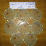
MIC values obtained for the tested microorganisms are reported in Table 3 and Fig. 1. The PHF produced significant activity against Candida albicans, Pseudomonas aeroginosa, Salmonella paratyphi, and Aspergillus niger.
FIG. 1: ANTIMICROBIAL ACTIVITY OF POLY-HERBAL FORMULATION ON VARIOUS MICROORGANISMS
TABLE 3: ANTIMICROBIAL ACTIVITY OF PHF
| Antimicrobial Agent | Inhibition zones in diameter (mm) | ||||
| Concentration | Escherichia coli | Pseudomonas aeroginosa | Salmonella paratyphi | Candida albicans | Aspergillus niger |
| Control | 07 | 10 | 09 | 07 | 09 |
| Test 1-25 µg/ml | 09 | 08 | 05 | 04 | 04 |
| Test 2-50 µg/ml | 12 | 12 | 10 | 08 | 09 |
| Test 3-75 µg/ml | 03 | 07 | 05 | 06 | 07 |
Poly herbal formulation Showed maximum inhibition for the species Escherichia coli (12 mm), Pseudomonas aeroginosa (12 mm), Candida albicans (8 mm), Salmonella paratyphi (10 mm) and Aspergillus niger (9 mm) at the concentration of 50 µg/ml.
Wound Healing Property: A wound which is disrupted the state of tissue caused by physical, chemical, microbial or immunological insult ultimately heals either by regeneration or fibroplasia. Wound healing is the process of repair that follows injury to the skin and other soft tissues. Wounds are still a major problem in developing countries, often having severe complications and involving high costs for therapy.
An important aspect of the use of traditional medicinal remedies and plants in the treatment of burns and wounds is the potential to improve healing and the same time to reduce the financial burden. Several plants and herbs have been used experimentally to treat skin disorders, including wound injuries, in traditional medicine. Collagen is a major protein of the extracellular matrix and is the component that ultimately contributes to wound strength 9, 10.
In excision wound model, on days 5 and 10 the wound area of standard and test ointment treated groups was found to be significant (p < 0.05) in comparison to control diabetic group animals treated with simple ointment base.
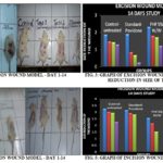
The activity is maximum on the day 14 and there was a statistically significant difference in the wound area relative in diabetic animals treated with PHF 5% w/w and PHF 10% w/w showed 52.59% and 52.33% healing respectively compared to standard drug group Table 4 Fig. 2, 3, 4, 6. It was also observed that epithelialization periods of PHF 5% w/w and PHF 10% w/w group were shorter in comparison to the control group.
On the day 14, the study of the histological structure showed excellent tissue regeneration in the skin wound treated with PHF 5% w/w and PHF 10% w/w and comparable with povidone ointment treated standard group.
TABLE 4: EFFECT OF PHF ON EXCISED WOUND IN DIABETIC RATS
| Treatment | Wound contraction (%) | ||||
| Day 1 | Day 5 | Day 10 | Day 14 | Epithelialization (day) | |
| Control | 1.87 ± 0.5 | 1.47 ± 0.7 (21.39%) | 1.49 ± 0.2 (24.06%) | 1.3 ± 0.5 (30.04%) | 28.12 ± 0.84 |
| Standard | 1.9 ± 0.8 | 0.85 ± 0.2 (55.26%) | 0.67 ± 0.2 (64.73%) | 0.25 ± 0.3 (86.84%) | 14.11 ± 0.67** |
| PHF 5% w/w | 2.32 ± 0.5 | 1.8 ± 0.4 (22.41%) | 1.72 ± 0.6 (25.86%) | 1.22 ± 0.5 (47.41%) | 18.20 ± 0.74a** |
| PHF 10% w/w | 1.72 ± 0.7 | 1.27 ± 1.1 (26.16%) | 1.12 ± 0.6 (34.18%) | 0.9 ± 0.8 (47.67%) | 21.25 ± 0.66a** |
Values are mean ± SEM (n=6); **p < 0.05; aP<0.01 (Comparison made between wounded control and standard).
In excision wound model the wound contraction of PHF 5% w/w and PHF 10% w/w is effective when compared to standard (aP<0.01) and control (**P<0.05).
On the day 14, excision and incision type of wounds in groups of PHF 5% w/w shown 100% healing and PHF 10% w/w shown 80.39% wound healing in diabetic rats as in collagenation, fibroblasts cells, whereas the skin wound treated with simple ointment base presented edema with cellular necrosis that was not observed in the test drug and standard drug-treated group of diabetic animals. Wound healing is a process by which damaged tissue is restored as closely as possible to its normal state and wound contraction is the process of shrinkage of the area of the wound Table 5, 6, 7.
TABLE 5: EFFECT OF PHF ON INCISION WOUND MODEL IN DIABETIC RATS
| Treatment | Breaking Strength (g) | Hydroxyproline mg/g |
| Control | 212 ± 10.82 | 12.18 ± 2.89NS |
| Standard | 376 ± 12.37** | 23.66 ± 4.10NS |
| PHF 5% w/w | 285 ± 10.96a** | 17.10 ± 3.00NS |
| PHF 10% w/w | 321 ± 12.20a** | 19.64 ± 3.12NS |
Values are mean ± SEM for groups of six animals each.
(**P<0.05; aP<0.01) Vs control and standard, (NSP>0.01) not significant to standard. In incision wound model the wound contraction of PHF 5% w/w and PHF 10% w/w is effective when compared to control (**P<0.05) and standard (aP<0.01) but the hydroxyproline mg/g is Nonsignificant (NS) when compared to standard and control.
TABLE 6: EFFECT OF PHF ON COLLAGEN CONTENT OF THE GRANULOMA TISSUE IN DIABETIC RATS (INCISION WOUND MODEL)
| Day | Collagen in mg/g | ||||
| Negative control | Positive control | Standard | PHF 5% w/w | PHF 10% w/w | |
| 5 | 1.23±0.17 | 0.14±0.05** | 0.34 ±0.07** | 0.27±0.04* | 0.29±0.05** |
| 10 | 1.25±0.12 | 0.17±0.04** | 0.67±0.06** | 0.48±0.05* | 0.55±0.06** |
| 14 | 1.23±0.02 | 0.19±0.06** | 0.99±0.09** | 0.75±0.08** | 0.80±0.05** |
Values are represented as Mean ± SEM for groups of six animals each. (**P<0.01; *P<0.05) vs. control. In incision wound model collagen content of PHF 10% w/w is significant to standard (**P<0.01) from the day 5 onwards but the collagen content of PHF 5% w/w is significant to standard only (**P<0.01) from DAY 14.
TABLE 7: PERCENTAGE WOUND CLOSURE OF INCISION WOUNDED RATS
| Group | Treatment | Wound closure (%) | |||
| Day 1 | Day 5 | Day 10 | Day 14 | ||
| I | Control-untreated | 3.25±0.50 | 3.02±0.5 (7.07%) | 2.95±0.05 (9.23%) | 2.25±0.50 (30.76%) |
| II | Standard-povidone | 3.15±0.30 | 2.92±0.65 (7.30%) | 2.82 ±0.05 (10.47%) | (100%) |
| III | PHF 5% w/w | 2.55±0.10 | 2.15±0.30 (15.68%) | 1.52±0.05a** (40.39%) | (100%) |
| IV | PHF 10% w/w | 2.55±0.10 | 2.07±0.45 (18.82%) | 1.62±0.05a** (36.47%) | 0.5(80.39%) |
Values are as mean ± S.E.M. for groups of six animals each. (**P<0.05) vs. Control; (aP<0.01) vs. standard. In the incision wound model the percentage of wound closure in PHF 5% w/w is 40.39% and comparable with standard (aP<0.01) and control (**P<0.05) and the percentage of wound closure in PHF 10% w/w is 36.47% and comparable with standard (aP<0.01) and control (**P<0.05)
It is mainly dependent upon the type and extent of damage, the general state of health and the ability of the tissue to repair. The main objectives in these processes are to regenerate and reconstruct the disrupted anatomical continuity and functional status of the skin. In the maturational phase, the final phase of wound healing, the wound undergoes contraction, resulting in a smaller amount of apparent scar tissue. Granulation tissue formed in the final part of the proliferative phase is primarily composed of fibroblasts, collagen, edema, and new small blood vessels. In the present study, the wound healing potential in diabetic animals for and PHF 5% w/w and was evident on the day 5 onwards; this potential was further confirmed in the histological evaluation on day 14. No remarkable healing effect was observed within control group diabetic rats.
On days 0, 5, 10 and 14 animals treated with the PHF, 5% w/w showed results similar to animals treated with povidone HCl, with improving the wound healing process. The results in this study are in support that the wound healing and repair is accelerated by applying PHF 5% w/w and PHF 10% w/w, which was highlighted by the full thickness coverage of the wound area by an organized epidermis in the presence of mature scar tissue in the dermis.
CONCLUSION: The present study indicates the wound healing activity of PHF in an experimental animal using incision, excision wound models. Wound contracture is a process that occurs throughout the healing process, commencing in the fibroblastic stage whereby the area of the wound undergoes shrinkage. Collagen, the major component which strengthens and supports extracellular tissue, is composed of the amino acid, hydroxyproline, which has been used as a biochemical marker for tissue collagen. The histologic study also substantiates the results and indicates that test drugs stimulate and enhances the faster lay down of collagen fibers with the PHF received diabetic animals than the untreated diabetic control wound.
Hence, based on the results it can be concluded that the test drug used in this study in different concentrations were effective and showing highly beneficial healing responses in the diabetic condition.
ACKNOWLEDGEMENT: We are greatly thankful to the School of pharmaceutical sciences, Vels University and Malla Reddy college pharmacy for providing a platform to complete the research.
CONFLICT OF INTEREST: We declare that we have no conflict of interest.
REFERENCES:
- Kato A, Higuchi Y, Goto H, Kizu H, Okamoto T, Asano N, Hollinshead J, Nash RJ and Adachi I: Inhibitory effects of Zingiber officinale Roscoe derived components on aldose reductase activity in-vitro and in-vivo. Journal of Agricultural and Food Chemistry 2006; 54(18): 6640-44.
- Rege NN, Thatte UM, and SA: Adaptogenic properties of six Rasayana herbs used in Ayurvedic Medicine, Phyto Therapy Research 1999; 13(4): 275-291.
- Gulcin I: The antioxidant and radical scavenging activities of black pepper (Piper nigrum) seeds. International Journal of Food Sciences and Nutrition 2005; 56(7): 491-99.
- Chatterjee S, Niaz Z, Gautam S, Adhikari S, Variyar PS and Sharma A: Antioxidant activity of some phenolic constituents from green pepper (Piper nigrum) and fresh nutmeg mace (Myristica fragrans). Food Chemistry 2007; 101(2): 515-23.
- Anand KK, Singh B, Saxena AK, Chandan BK, Gupta VN and Bhardwaj V: 3, 4, 5-Trihydroxy benzoic acid (Gallic acid), The hepatoprotective principle in the fruits of Terminalia bellerica-bioassay guided activity. Pharmacological Research 1997; 36(4): 315-21.
- Kumar MS, Kirubanandan S, Sripriya R and Sehgal PK: Triphala Promotes Healing of Infected Full-Thickness Dermal Wound. J of Surg Research 2008; 144(1): 94-01.
- Ahmad I, Mehmood Z and Mohammad F: Screening of some Indian medicinal plants for their antimicrobial properties. Journal of Ethnopharmacology 1998; 62(2): 183-93.
- Ehrlich HP and Hunt TK: The effect of cortisone and anabolic steroids on the tensile strength of healing wounds. Ann Surg 1968; 167: 117.
- Neumann RE and Logan MA: The determination of collagen and elastin in tissues. J Biochem 1972; 186: 549-56.
- Chithra, Sajithlal PG and Chandrakasan G: Influence of Aloe vera on collagen characteristics in healing dermal wounds in rats. Mol Cell Biochem 1998; 181: 71-76.
How to cite this article:
Nirmala S, Sudhakar M, Anusri P and Nadanasabapathi P: Poly herbal semisolid dosage form development for wound healing. Int J Pharmacognosy 2018; 5(8): 461-66. doi link: http://dx.doi.org/10.13040/IJPSR.0975-8232.IJP.5(8).461-66.
This Journal licensed under a Creative Commons Attribution-Non-commercial-Share Alike 3.0 Unported License.
Article Information
4
461-466
499
1366
English
IJP
S. Nirmala *, M. Sudhakar, P. Anusri and P. Nadanasabapathi
Department of Pharmacognosy, Malla Reddy College of Pharmacy (Affiliated OU), Maisammaguda, Secunderabad, Telangana, India.
nirmala.cognosy@gmail.com
28 April 2018
08 June 2018
13 June 2018
10.13040/IJPSR.0975-8232.IJP.5(8).461-66
01 August 2018


