


PHYTOCHEMICAL SCREENING, ACUTE TOXICITY, ANTIBACTERIAL AND ANTITUSSIVE EFFECTS OF THE AQUEOUS EXTRACT OF THE LEAVES OF HYMENOCARDIA ACIDA TUL.
HTML Full TextPHYTOCHEMICAL SCREENING, ACUTE TOXICITY, ANTIBACTERIAL AND ANTITUSSIVE EFFECTS OF THE AQUEOUS EXTRACT OF THE LEAVES OF HYMENOCARDIA ACIDA TUL.
G. F. Nsonde Ntandou *, D. P.-D. Louzolo, B. A. E. Loufoua, S. Makemba, G. Tsiba, S. S. Landou Pandzou, G. T. D. Nombo Lassy, D. R. Mban, A. P. Nzoumba Pandi and N. Isso
Laboratory of Animal Physiology and Pathophysiology, Faculty of Science and Technology, Marien NGOUABI University, Brazzaville, P.O. Box 69, Republic of the Congo.
ABSTRACT: This study aims at valorizing the leaves of Hymenocardia acida Tul., a medicinal plant traditionally used in the Republic of Congo and other African countries, for healing cough and respiratory infections. The aqueous extract, prepared by decoction, showed a low acute toxicity in mice (LD50 > 5000 mg/kg). The phytochemical screening revealed the presence of flavonoids, condensed tannins, saponins, mucilages, reducing sugars, and anthraquinones. The oral administration of the extract at 200 mg/kg and 500 mg/kg significantly inhibited ammonia-induced cough in guinea pigs, with a prolonged reaction latency and a reduction of post-exposure cough bouts, comparable to the effect of dextromethorphan (0.5 mg/kg). Conversely, no significant effects were observed on cigarette smoke-induced cough. The extract also exhibited an in vitro antibacterial activity against a clindamycin-resistant strain of staphylococcus aureus, at concentrations of 1% and 10%, but no activity was observed against Escherichia coli. These findings validate the traditional use of Hymenocardia acida Tul. and highlight its natural antitussive and antibacterial potential.
Keywords: Hymenocardia acida Tul., Antitussive, Antibacterial, Phytochemistry, Acute toxicity
INTRODUCTION: Respiratory infections constitute a major global public health issue, accounting for more than 4 million deaths worldwide each year and ranking as the third most deadly disease in the world.
Moreover, they are also the leading cause of death in developing countries, including the Republic of the Congo 1, 2.
Furthermore, although resulting from miscellaneous causes (infection, irritation, allergy, etc.), cough is the most frequent symptom of respiratory diseases. Indeed, it is a major cause of outpatient pediatric visits, affecting 50 to 70% of children under one year old and 30 to 60% of school-aged children 3. Indeed, for the treatment of respiratory infections and coughs, modern medicine provides us with antibiotics and cough suppressants, which are very often available over the counter.
However, the frequent and sometimes inappropriate use of antibiotics has led to the emergence of bacteria resistant to certain conventional antibiotics, thus complicating the treatment of common infections and increasing morbidity and mortality worldwide 4, 5. In fact, approximately 80% of the African population relies on traditional medicine 6. In the Republic of Congo, numerous medicinal plants, including Hymenocardia acida Tul., are traditionally used in the management of various respiratory disorders 7.
In this context, Hymenocardia acida Tul., whose antitussive and antimicrobial properties are widely recognized in traditional medicine, represents a promising opportunity for the development of local therapeutic alternatives that are more accessible, safer, and better adapted to the needs of the population 8.
Accordingly, the present study was conducted to address the following research question: does the aqueous leaf extract of Hymenocardia acida Tul. exhibit antitussive activity and inhibitory effects on the growth of certain bacteria responsible for respiratory infections, while remaining non-toxic at high doses?
MATERIALS AND METHODS:
Plant Material: The leaves of Hymenocardia acida Tul. were collected in May 2024 in the Republic of Congo, in the village of Makana 2 (Latitude: −4.3375570; Longitude: 15.0910900), Pool Department, on the outskirts of Brazzaville. Botanical identification was carried out by a qualified specialist, the Professor Jean Marie Moutsambote, thereby ensuring the authenticity and reliability of the plant material used. This identification was further confirmed by comparison with a reference specimen deposited at the National Herbarium of Belgium. The specimen was originally collected in the Republic of Congo, in the Mouyondzi district, specifically in the village of Kolo, by Markström on November 17, 1975 (specimen no. 036; catalogue BR0000021701283).
Extraction: The leaves of H. a. T. were air-dried and subsequently ground into a fine powder.
Fifty grams of the powder were mixed with 500 mL of distilled water and heated at 70 °C for 25 minutes. The mixture was then filtered through absorbent cotton, and the resulting filtrate was evaporated at 70 °C in a drying oven. The dry residue obtained was collected and stored in a sterile flask. For experimental use, this residue was reconstituted in distilled water, taking into account the desired final concentrations required for the assays 7. After each preparation of the aqueous extract, the extraction yield was calculated using the formula presented below:
Yield = (Mass of the extract) / (Mass of the powder from the leaves) x 100
Experimental Animals: To conduct this study, female albino mice weighing between 20 g and 23 g and aged at least three months were used. In addition, guinea pigs of both sexes, with body weights ranging from 440 g to 500 g and aged at least four months, were included. All animals were fed with a paste composed of wheat flour and soy flour mixed with commercial livestock feed, supplemented with a small amount of palm oil and salt. Water was provided ad libitum throughout the experimental period.
Bacterial Strains: Bacteria responsible for respiratory infections, namely Staphylococcus aureus and Escherichia coli, were used to evaluate the in vitro antibacterial activity of the aqueous extract of H. a. T.
Acute Toxicity Assessment: The acute toxicity of the aqueous extract of H. a. T. was evaluated in accordance with the method described in OECD Guideline No. 425 (2022), 9 with slight modifications. In compliance with OECD requirements, the limit test procedure was applied at doses of 2000 mg/kg and 5000 mg/kg.
For the limit test at the 2000 mg/kg dose, a female albino mouse was fasted for 3 hours and subsequently weighed to determine the exact volume of extract to be administered. The aqueous extract was then administered orally at a dose of 2000 mg/kg. Following administration, signs of toxicity, including mobility, posture, and fecal output, were carefully monitored for 4 consecutive hours. After confirming the survival of this animal, the same procedure was successively repeated in two additional mice. These three animals constituted group 1. Each mouse in group 1 was subsequently weighed every two days, at the same time as the initial weighing, over a 14-day period in order to detect any delayed toxic effects. The same experimental procedure was applied for the limit test at the 5000 mg/kg dose, thereby constituting group 2. In addition, three mice receiving distilled water at a dose of 10 mL/kg, administered orally by gavage, were used as the control group.
To check for changes in behavior, we looked for signs of increased drowsiness or unusual agitation. The reactivity was assessed by observing the reaction to the sound of a finger click. The evacuation was assessed by observing changes in the nature, quantity, and frequency of defecation. The alertness was assessed by observing the presence or absence of a corneal reflex by gently touching the cornea with a moistened finger. The observation of the animal's reactions to slight, unexpected noises or movements, or to light pressure on the tail (stimuli), allowed us to assess alterations in alertness or pain sensitivity.
Evaluation of Antitussive Activity: The antitussive activity of the aqueous extract was evaluated according to the method described by Loufoua et al. (2015) 7. The experimental animals were exposed to two different cough-inducing irritants: cigarette smoke for one group and ammonia for the other. To this end, four groups of five guinea pigs of both sexes, weighing between 440 g and 500 g and aged at least four months, were constituted. Prior to experimentation, all animals were fasted for 18 hours in order to standardize their physiological conditions.
The treatments were administered orally as follows: 200 mg/kg of aqueous H. a. T. extract for the first group, 500 mg/kg for the second group, 0.5 mL/100 g of distilled water for the negative control group, and 0.5 mg/kg of dextromethorphan for the positive control group. One hour after treatment, each animal was individually exposed to the cough-inducing irritant for 10 minutes. During this phase, two parameters were assessed: the latency time to the first cough, measured using a stopwatch, and the total number of coughing episodes recorded during the 10 minutes of exposure and the subsequent 5 minutes following exposure.
Evaluation of Antibacterial Activity:
Enhanced Version: The antibacterial activity of the aqueous extract was evaluated using the method described by Sofidiyia et al. (2009). Clindamycin and levofloxacin were used as positive controls against Staphylococcus aureus and Escherichia coli, respectively.
The aqueous extract was prepared at different concentrations (0.01%, 0.1%, 0.5%, 1%, and 10%). The bacterial strains were reactivated, and a bacterial suspension was prepared by emulsifying a single isolated colony in 2 mL of sterile distilled water contained in a hemolysis tube. The optical density of each suspension was measured at 625 nm using a spectrophotometer and adjusted between 0.08 and 0.10 absorbance units (AU), corresponding to an approximate concentration of 10^8 CFU/mL. The recorded values were 0.126 AU for E. coli and 0.092 AU for S. aureus.
The antibacterial activity was then determined using the agar well diffusion method on Plate Count Agar (PCA). The standardized bacterial suspensions were inoculated onto the surface of PCA plates using sterile swabbing to obtain a uniform bacterial lawn.
Wells were then created in the agar and filled with 100 µL of the different concentrations of the aqueous extract (0.01%, 0.1%, 0.5%, 1%, and 10%). Sterile distilled water was added to a dedicated well (100 µL) to serve as a negative control. Simultaneously, antibiotic discs corresponding to the positive controls were carefully placed on the agar surface.
The plates were kept at cold temperature for 15 minutes to allow diffusion of the extracts into the medium, then incubated at 37 °C for 24 hours.
After 24 hours of incubation, the antibacterial activity against E. coli and S. aureus was evaluated. Activity was considered significant when zones of inhibition (halos) with a diameter greater than or equal to 6 mm were observed around the wells or discs.
Phytochemical Screening of the Aqueous Extract of Hymenocardia acida Tul. Leaves: To determine the phytochemical composition of the aqueous extract of H. a. T., a qualitative phytochemical screening was performed targeting seven major classes of bioactive compounds of pharmacological interest, namely alkaloids, catechins, flavonoids, saponins, mucilages, reducing compounds, and anthraquinones. Classic colorimetric and precipitation tests were employed as described by Sofowora (1996), 11 Kouchadé et al. (2017),12 and Nsonde et al. (2018) 13.
Alkaloid Test: Five milliliters of the aqueous extract were dispensed into three test tubes. Subsequently, 1 mL of 1 N hydrochloric acid was added to each tube, followed by a few drops of Dragendorff’s, Bouchardat’s, and Mayer’s reagents.
The presence of alkaloids was indicated by the formation of a red precipitate with Dragendorff’s and Bouchardat’s reagents, or a yellowish to brown precipitate with Mayer’s reagent.
Condensed Tannins Test: Five milliliters of a 10% decoction were mixed with 1 mL of concentrated hydrochloric acid. The mixture was boiled for 15 minutes and then filtered. The formation of a red precipitate soluble in isoamyl alcohol indicated the presence of condensed tannins.
Flavonoid Test: In a test tube, 5 mL of a 10% decoction were mixed with 5 mL of hydrochloric acid solution, 1 mL of isoamyl alcohol, and a few magnesium shavings. The presence of flavonoids was confirmed by a color change in the upper layer: orange-pink for flavones, purplish-pink for flavanones, and red for flavonols.
Mucilage Test: One milliliter of the 10% decoction was mixed with 5 mL of alcohol in a test tube. The formation of a flaky precipitate after 10 minutes indicated the presence of mucilages.
Reducing Compounds Test: Five milliliters of the 10% aqueous decoction were evaporated to dryness in a water bath. One milliliter of Fehling’s solution was added to the residue. The appearance of a brick-red precipitate indicated the presence of reducing compounds.
Saponin Test: Two milliliters of the aqueous extract were placed in a test tube and distilled water was added to obtain a final volume of 5 mL. The tube was shaken lengthwise for 15 seconds and then allowed to stand for 20 minutes. A positive result was recorded when the height of the persistent foam exceeded 1 cm.
RESULTS:
Extraction Yield: Table 1 presents the relationship between the mass of powdered dry leaves of Hymenocardia acida Tul. (H. a. T.), the mass of dry aqueous extract obtained, and the extraction yield achieved by decoction. The yields ranged from 9.65% to 15.76%, with a mean yield of 11.36 ± 2.55%. The mean mass of dry extract obtained was 6.35 ± 4.74 g, corresponding to an average mass of powdered dry leaves of 55.41 ± 43.58 g.
TABLE 1: RELATIONSHIP BETWEEN THE MASS OF POWDERED DRY LEAVES OF HYMENOCARDIA ACIDA TUL. (H. A. T.), THE MASS OF DRY AQUEOUS EXTRACT OBTAINED, AND THE EXTRACTION YIELD ACHIEVED BY DECOCTION
| Test | Mass of pulverizeddry leaves (g) | Mass of dry extract obtained (g) | Yield (%) |
| 1 | 100 | 9.77 | 9.77 |
| 2 | 100 | 11.36 | 11.36 |
| 3 | 50 | 7.88 | 15.76 |
| 4 | 20 | 2.05 | 10.25 |
| 5 | 7.07 | 0.682 | 9.65 |
| Mean | 55.41 | 6.35 | 11.36 |
| Standard deviation | 43.58 | 4.74 | 2.55 |
Acute Toxicity:
Observation of the General Condition of Mice and Effects on Mortality: Table 2 presents the behavioral and physiological observations recorded in mice during the first four hours following the administration of the different treatments. The results indicate that oral administration of the aqueous extract of H. a. T. leaves at both tested doses did not alter the general condition of the animals and did not induce mortality. However, a reduction in fecal output and an absence of urine were observed in mice treated with the aqueous extract compared with those in the control group.
TABLE 2: BEHAVIORAL AND PHYSIOLOGICAL OBSERVATIONS OF MICE DURING THE FIRST FOUR HOURS AFTER ADMINISTRATION OF THE DIFFERENT TREATMENTS
| Parameters | Control group | Group 1 (H.a.T. at 2000 mg/kg) | Group 2 (H.a.T. at 5000 mg/kg) |
| Mobility | Normal | Normal | Normal |
| Ptosis | Absent | Absent | Absent |
| Convulsions | Absent | Absent | Absent |
| Vomiting | Absent | Absent | Absent |
| Feces | 1–5 stools (formed) | 0–1 stool (formed) | No stools |
| Urination | Present at 2 h | Absent | Absent |
| Vocalization | Absent | Absent | Absent |
| Piloerection | Absent | Absent | Absent |
| Alertness | Present | Present | Present |
| Sleep | Absent | Absent | Absent |
| Aggressiveness | Absent | Absent | Absent |
| Number of deaths | 0 | 0 | 0 |
| Posture | Normal | Normal | Normal |
| Pain sensitivity | Present | Present | Present |
Effect of the Aqueous Extract of H.a.T. Leaves on the Body Weight of Mice: Fig. 1 illustrates changes in the relative body weights of mice over a 14-day observation period. Compared with Day 0, both the control and treated groups exhibited variations in body weight. However, according to the Mann–Whitney U test, no significant differences were observed between the control group (distilled water, 10 mL/kg) and the treated groups from Day 0 to Day 14. Nevertheless, a suggestive, although not statistically significant, trend was observed in the group receiving the H.a.T. extract at a dose of 2000 mg/kg, with mice appearing to gain slightly more weight than those in the control group from Day 6 to Day 14. Furthermore, mice treated with the H.a.T. extract at 5000 mg/kg also showed a sudden, but non-significant, increase in body weight on the final day of the experiment.
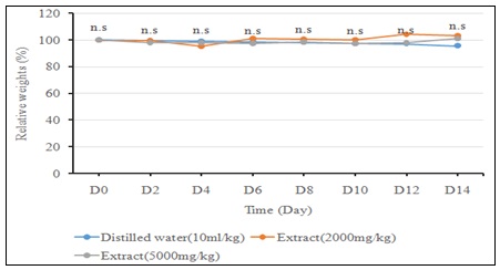
FIG. 1: CHANGES IN THE RELATIVE WEIGHTS OF MICE FROM DIFFERENT GROUPS DURING THE 14 DAYS OF OBSERVATION. NS: NOT SIGNIFICANT
Evaluation of Antitussive Activity:
Against Ammonia-induced Cough: Fig. 2 illustrates the effects of the aqueous extract of H.a.T. on cough latency and the number of cough bouts in guinea pigs exposed to ammonia. Regarding reaction latency, the results show that the group treated with the aqueous extract of H.a.T. at 500 mg/kg (26.71 ± 10.14 s) and the group treated with dextromethorphan (DM) at 0.5 mg/Kg (25.33 ± 3.18 s) exhibited highly significant prolongations of latency compared with the group receiving distilled water (D.W) at 0.5 mL/100 g (0.67 ± 0.37 s), with p = 0.008 (Mann–Whitney U test). In contrast, the group treated with the aqueous extract of H.a.T. at 200 mg/kg (1.67 ± 0.73 s) showed a significant but more limited increase in latency (p = 0.032). For the number of cough bouts during ammonia exposure, no significant differences were observed between any of the treated groups and the group receiving distilled water (p > 0.05 for all comparisons), although the latter exhibited a slightly higher mean value (0.53 ± 0.45) compared with the other groups (0.00 ± 0.00). Finally, with regard to the number of post-exposure cough bouts, a highly significant difference was observed between all treated groups (0.00 ± 0.00) and the negative control group (2.00 ± 0.63), with p = 0.008.
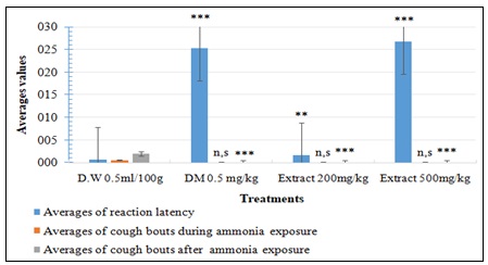
FIG. 2: EFFECTS OF AQUEOUS EXTRACT OF H.A.T. ON REACTION LATENCY AND NUMBER OF COUGH BOUTS IN GUINEA PIGS EXPOSED TO AMMONIA. THE RESULTS ARE EXPRESSED AS MEAN ± ES WITH N = 5. NS: NOT SIGNIFICANT, **: SIGNIFICANT, ***: VERY SIGNIFICANT
Against Cigarette Smoke-Induced Cough: Fig. 3 illustrates the effects of H.a.T. aqueous extract on reaction latency and the number of cough bouts in guinea pigs exposed to cigarette smoke. No significant differences were observed between the treated groups and the control group for any of the parameters measured. However, a trend toward statistical significance (p = 0.056) was noted for the group receiving 500 mg/kg of H.a.T. aqueous extract, characterized by a reduction in the number of cough bouts during and after exposure, compared to the negative control (distilled water, 0.5 mL/kg).
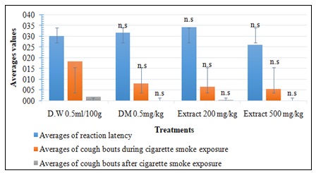
FIG. 3: EFFECTS OF H.A.T. AQUEOUS EXTRACT ON REACTION LATENCY AND THE NUMBER OF COUGH BOUTS IN GUINEA PIGS EXPOSED TO CIGARETTE SMOKE. THE RESULTS ARE EXPRESSED AS MEAN ± ES WITH N = 5, NS: NOT SIGNIFICANT
Evaluation of Antibacterial Activity: The results of the evaluation of the antibacterial activity of the aqueous extract of Hymenocardia acida Tul. leaves against Staphylococcus aureus and Escherichia coli are presented in Table 3 and illustrated in Fig. 4. Inhibition of bacterial growth was assessed based on the presence and diameter of inhibition zones surrounding the wells containing the extract or the disc impregnated with the reference antibiotic (positive control).
Regarding S. aureus, inhibition zones were observed at extract concentrations of 1% and 10%, as well as with clindamycin. At 10%, the inhibition zones measured 14 mm vertically, 13 mm horizontally, and 14 mm obliquely. At 1%, the recorded diameters were 7 mm in both the vertical and horizontal directions and 8 mm obliquely. For clindamycin, all three measurements were 16.8 mm.
In contrast, no inhibition zone was observed for Staphylococcus aureus at concentrations below 1%.
Regarding E. coli, no inhibition zone was observed at any extract concentration, with the exception of the positive control (levofloxacin), which exhibited an inhibition zone of 19.4 mm (vertical, horizontal, oblique).
TABLE 3: COMPARATIVE TABLE OF INHIBITION ZONES PRODUCED BY THE AQUEOUS EXTRACT OF H.A.T. AGAINST STAPHYLOCOCCUS AUREUS AND ESCHERICHIA COLI
| Treatments | S. aureus (Inhibition diameter, mm) | E. coli (Inhibition diameter, mm) |
| Distilled water (100 µL) | No inhibition zone | No inhibition zone |
| H.a.T. extract 0.01% (100 µL) | No inhibition zone | No inhibition zone |
| H.a.T. extract 0.1% (100 µL) | No inhibition zone | No inhibition zone |
| H.a.T. extract 0.5% (100 µL) | No inhibition zone | No inhibition zone |
| H.a.T. extract 1% (100 µL) | 7 mm (vertical), 7 mm (horizontal), 8 mm (oblique) | No inhibition zone |
| H.a.T. extract 10% (100 µL) | 14 mm (vertical), 13 mm (horizontal), 14 mm (oblique) | No inhibition zone |
| Positive control (clindamycin / levofloxacin) | 16.8 mm (vertical, horizontal, oblique) | 19.4 mm (vertical, horizontal, oblique) |
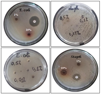
FIG. 4: RESULTS OF THE EVALUATION OF THE ANTIBACTERIAL ACTIVITY OF THE AQUEOUS EXTRACT OF H.A.T. LEAVES, TESTED AT DIFFERENT CONCENTRATIONS ON S. AUREUS AND E. COLI
Phytochemical Screening of the Aqueous Leaf Extract of Hymenocardia acida Tul.: Table 4 presents the results of the phytochemical screening of the aqueous extract of Hymenocardia acida Tul. leaves. The analysis of the extract revealed the presence of saponins, condensed tannins, flavonol-type flavonoids, reducing compounds (particularly reducing sugars), mucilages, and anthraquinones.
However, tests for alkaloids were negative, confirming their absence (–) in the extract.
TABLE 4: RESULTS OF THE PHYTOCHEMICAL SCREENING OF THE AQUEOUS EXTRACT OF H.A.T. LEAVES
| Secondary metabolites | Results |
| Saponins | + |
| Condensed tannins | + |
| Flavonoids (flavonols) | + |
| Mucilages | + |
| Alkaloids | – |
| Reducing compounds (reducing sugars) | + |
| Anthraquinones | + |
+ : Presence; - : Absence
DISCUSSION:
Extraction Yield: In our study, the mean yield obtained was 11.36 ± 2.55 %, which is slightly lower than the 11.4 % w/w reported by Sofidiya et al. (2010), 14 who carried an aqueous extraction of the same plant’s leaves. That marginal difference could be explained by variations in extraction methods used or environmental conditions.
Acute Toxicity: Up to the tested oral dose of 5000 mg/kg, the aqueous extract induced neither mortality nor any clinical signs of severe toxicity. These results align with those obtained by Sofidiya et al. (2010), 15 who, utilizing an aqueous extract of H.a.T. leaves, observed no mortality up to 3200 mg/kg, as well as those reported by Koffi et al. (2022), who noted an oral LD50 exceeding 12,000 mg/kg for the aqueous extract of H.a.T. roots 15. Furthermore, despite the absence of mortality, some slight physiological changes were observed in the animals administered with the extract; notably, a decrease in stool output and an absence of urine during the first four hours post-administration, as well as a non-significant weight gain at the conclusion of the study. The reduced fecal excretion could be related to the plant's antidiarrheal properties. Indeed, Koné et al. (2005) confirmed through an ethnobotanical survey that H.a.T. is traditionally used to treat diarrhea in Ivory Coast, 16 an observation supported by the ethnobotanical survey conducted by Loufoua et al. (2015) in Congo 7. From a pharmacological perspective, Usman et al. (2021) demonstrated that the methanolic extract of H.a.T. bark possesses significant antidiarrheal properties 17. Furthermore, this seems corroborated by the presence of catechin tannins, which are well-known for their astringent and consequently antidiarrheal properties, 18 identified within our extract. Regarding the anuria, no chemical class revealed by the phytochemical screening of our extract exhibits antidiuretic properties; this may ultimately be attributed to random fluctuations or to secondary metabolites not targeted during the phytochemical screening. Concerning the weight gain in the extract-treated groups, this is inconsistent with the ethnobotanical use of H.a.T., reported for the management of obesity in Cameroon by Epoh et al. (2020), 19 particularly as Wang et al. (2024) and Wink (2015) reported that certain flavonols reduce adipose tissue accumulation and improve insulin sensitivity 20, 21.
Phytochemical Screening: The phytochemical screening of the aqueous extract of H.a.T. leaves revealed the occurrence of saponins, catechuic tannins, and anthraquinones, thus corroborating the observations of Iyadi et al. (2003); 22 the presence of flavonol-type flavonoids, consistent with the results of Sofidiya et al. (2009); 10 the presence of reducing compounds (reducing sugars), in alignment with the findings of Agbidye et al. (2020) who detected them in the methanolic extract of H.a.T. leaves; 23and the presence of mucilages, which is neither confirmed nor refuted by any existing literature, suggesting that few, if any, studies have yet specifically investigated their occurrence in Hymenocardia acida Tul.; conversely, the absence of alkaloids was noted, contradicting Danladi et al. (2021) who had reported the contrary for the same plant 24. This absence represents a particularly noteworthy result. Indeed, by employing phytochemical screening methodologies based on chromogenic and precipitation reactions, similar to those conducted in the present study, Nsonde Ntandou et al. (2005) and Nkundineza et al. (2021) demonstrated the presence of alkaloids in the aqueous extract of Cassia alata leaves as well as in the aqueous extract of Brenaniabrieyi bark 25, 26. Furthermore, several secondary metabolites identified in H.a.T. could be the underlying drivers of the antibacterial and/or antitussive activities, as confirmed by various experimental studies. Flavonoids exhibit proven antibacterial activity along with a demonstrated antitussive effect 27, 28, 29. Alkaloids also exhibit these two pharmacological profiles, reported respectively by Wélé et al. (2010) for the antibacterial effect, and by Liu et al. (2015) and Loufoua et al. (2015) for the antitussive effect 7, 30, 31. Mucilages are described as active against specific bacterial strains and furthermore exert an antitussive effect 32, 33. Saponins possess a dual activity as well: antibacterial and antitussive 34, 35, 36. Condensed tannins have demonstrated antimicrobial efficacy and antitussive activity 37, 38, 39. In contrast, regarding anthraquinones, only antibacterial activity has been reported; 40 no data pertaining to an antitussive effect was identified in the available literature. Finally, reducing sugars appear to exert a hormetic-type antibacterial effect, 41 whereas no antitussive activity has been reported in the consulted literature.
Evaluation of Antitussive Activity:
Against Ammonia-induced cough: The results obtained highlight significant effects of the aqueous extract of Hymenocardia acida Tul. on the latency of the reaction to ammonia and the frequency of cough bouts following exposure to ammonia, as well as a non-significant difference between all experimental groups and the negative control group regarding the number of cough bouts in the presence of ammonia.
The administration of the aqueous extract at 500 mg/kg (Lot 2) prolonged the latency period prior to the ammonia-induced reaction very significantly (26.71 ± 10.14 seconds; p = 0.008), with performances comparable to the positive control group treated with dextromethorphan (25.33 ± 3.18 seconds; p = 0.008). This similarity suggests that the high-dose extract may exert an inhibitory effect on cough receptors (TRPs, RARs, or even C-fibers), possibly via bioactive compounds such as flavonoids, mucilages, saponins, condensed tannins, and mucilages known for their antitussive properties. In contrast, the effect observed for Lot 1 (200 mg/kg) remained significant but more modest (1.67 ± 0.73 seconds; p = 0.032), indicating a potential dose-dependent relationship. These data substantiate the hypothesis that the concentration of the extract directly influences its antitussive efficacy.
After exposure to ammonia, the highly significant difference between the negative control (2.00 ± 0.63 cough bouts) and all other groups (0.00 ± 0.00; p = 0.008) underscores the marked protective effect of the aqueous extract of H.a.T. This result reinforces the idea of a long-lasting action of the extract, comparable to that of dextromethorphan. The non-significant difference in cough bouts between all groups during ammonia exposure suggests that the extract does not have an immediate inhibitory effect on the triggering of the ammonia-induced cough reflex. This stands in contrast to the results of Ge et al. (2015),42 who, having evaluated the antitussive activity of aqueous extracts of the leaves of three Elaeagnus species (E. pungens, E. lanceolata and E. henryi) in mice for ammonia-induced cough, stated that all the aqueous extracts induced a period of cough latency and reduced the frequency of cough in a dose-dependent manner during exposure to ammonia; all the more so as the chemical analysis by HPLC-DAD of the three Elaeagnus species suggested the presence of flavonoid compounds, also found in our extract.
Against Cigarette Smoke-Induced Cough: The results obtained with this test highlight non-significant effects of the aqueous extract of H.a.T. on reaction times and the number of cough bouts in guinea pigs exposed to cigarette smoke, with a trend toward significance for group 2 (500 mg/kg) regarding the number of cough bouts during and after exposure to cigarette smoke (p-values = 0.056).
The results obtained do not allow us to conclude that the aqueous extract of H.a.T. is significantly effective against cigarette smoke-induced cough, although a trend is observed. These results are similar to those obtained by Zhong et al. (2015),43 who, although not using the same method as ours, had demonstrated that the aqueous extract of Schisandra chinensis did not have a significant effect on cough induced by cigarette smoke and that only the ethanolic and hydroethanolic extracts had significantly reduced the frequency of coughing; which could indicate that the aqueous extract, administered orally, would be less effective against cough induced by cigarette smoke than other extracts. It is noteworthy that, given the trend toward significance for Lot 2 (500 mg/kg) regarding the number of cough bouts during and after exposure to cigarette smoke, our extract could potentially have a dose-dependent effect and could consequently be more effective at higher doses.
In summary, the difference in efficacy of our aqueous extract between the two cough models could be due to the fact that cigarette smoke activates a greater number of receptors than ammonia, thus making the cough more difficult to inhibit. These observations suggest that the aqueous extract of H.a.T. could constitute an interesting alternative to synthetic antitussives. However, further analyses on the mechanism of action of the molecules involved, as well as studies on more diverse animal models and over prolonged exposure durations, would be necessary to confirm these preliminary results.
In this study, we induced cough in two ways. The difference in response to treatment with the extract between the two types of cough lies in the pathophysiological mechanisms of cough induction and thus reveals a mechanism of action specific to ammonia-induced cough.
Ammonia cough is an acute and violent irritation of the upper and lower respiratory tract, which can lead to bronchospasm or pulmonary edema, especially at high concentrations 7, 42. Smoker's cough, on the other hand, even though ammonia is present, is generally chronic, linked to the paralysis of cilia and an overproduction of mucus, resulting in wheezing, crackling sounds, and morning expectoration, and can progress to COPD 43.
It is possible that the extract does not act through a local antiinflammatory mechanism, but rather acts against irritations and inhibits the cough reflex through a central mechanism, similar to opiates such as codeine. Indeed, opiates also have a central analgesic effect via µ-opioid receptors, 44 and the extract also has a central analgesic effect 45.
Evaluation of Antibacterial Activity: The objective of this section was to evaluate the antibacterial activity of the extract against respiratory pathogens (E. coli and S. aureus). The results obtained in this study reveal, on the one hand, an antibacterial activity of the aqueous extract of Hymenocardia acida Tul. against Staphylococcus aureus, proportional to the concentration of the extract. Furthermore, although clindamycin exhibits a slightly greater halo of inhibition than the extract up to 10%, this strain of S. aureus is still not susceptible to it, in accordance with the recommendations of EUCAST 2024 V.1.0 June, which sets the acceptable range of the diameter of inhibition at 23 to 29 mm for clindamycin against S. aureus. On the other hand, no antibacterial activity was observed against Escherichia coli, either for the extract or for levofloxacin, whose halo of inhibition had a diameter smaller than the acceptable range (29–37 mm) established by EUCAST 2024 V.1.0 June. These observations were compared to the work of Sofidiya et al. (2009), 10 who studied the activity of aqueous and methanolic extracts of H.a.T.
In our study, the aqueous extract showed inhibition of S. aureus only at concentrations of 1% (10 mg/mL) and 10% (100 mg/mL). In contrast, Sofidiya et al. (2009) reported a minimum inhibitory concentration (MIC) of 2.5 mg/mL (0.25%) for the aqueous extract 10. This difference suggests that, to achieve a similar antibacterial effect, our extract requires much higher concentrations, which could be related to a lower content of bioactive compounds, different extraction conditions, or a variation in the bacterial strain used.
Regarding E. coli, our results indicate a complete absence of inhibitory effect, even at the maximum tested concentration of 10% (100 mg/mL). For comparison, Sofidiya et al. (2009) observed a MIC of 1.0 mg/mL (0.1%) for the aqueous extract and 0.5 mg/mL (0.05%) for the methanolic extract against E. coli 10. These results demonstrate significantly greater antibacterial activity in their study, suggesting that the extract they used was more concentrated in bioactive compounds. The phytochemical analysis performed by Sofidiya et al. (2009) detected the presence of flavonoids, phenols, steroids, and triterpenoids in H.a.T. extracts 10.
They also found that the methanolic extract had higher levels of bioactive compounds, with total phenols reaching 35.6 mg/g compared to 20.0 mg/g for the aqueous extract. This difference in composition could partly explain the superior efficacy of the methanolic extract, particularly against E. coli. As Sofidiya et al. (2009) point out, the low activity of the extracts against Gram-negative bacteria is partly explained by the presence of an outer membrane and a periplasmic space 10. These structures limit the penetration of antibacterial agents, thus reducing the susceptibility of E. coli compared to S. aureus, which, as a Gram-positive bacterium, does not possess these barriers.
CONCLUSION: This study has shown that the aqueous extract of H.a.T. leaves is tolerable at high doses and possesses both antitussive and antibacterial activity, which can vary depending on the dose or concentration of the extract and the preparation method used. H.a.T. is therefore a plant to consider for the development of antitussives and drugs to treat bacterial infections, particularly those caused by S. aureus.
Funding Declaration: This work, which was made possible by individual contributions from the various authors, did not receive any funding from any state or any organization responsible for funding research.
ACKNOWLEDGEMENTS: The authors wish to thank Professor Jean Marie MOUTSAMBOTE of the National Institute for Research in Exact and Natural Sciences (IRSEN) for the botanical identification and authentication, as well as the traditional practitioners who contributed to the collection of plant material.
CONFLICTS OF INTEREST: No conflicts of interest to declare.
REFERENCES:
- Forum of International Respiratory Societies. The global impact of respiratory disease. Third Edition. European Respiratory Society 2021; 1-52. firsnet.org/images/publications/FIRS_Master_09202021.pdf
- Schluger NW: Acute respiratory infections atlas: First edition. Independent Publisher 2010; 123.
- Krutikhina SB, Meleshkina AV & Yablokova EA: Cough in children: the most common problem in pediatrics. Meditsinskiysovet = Medical Council 2020; (18): 53–57.
- Carle S: La résistance aux antibiotiques: un enjeu de santé publique important. Pharmactuel 2009; 42(2): 6-21.
- Brüssow H: The antibiotic resistance crisis and the development of new antibiotics. Microbial Biotechnology 2024; 14510: 1-17.
- Oyebode O, Kandala NB, Chilton PJ & Lilford RJ: Use of traditional medicine in middle-income countries: a WHO-SAGE study. Health Policy and Planning 2016; 31(8): 984–991.
- Loufoua BAE, Bassoueka DJ, Nsonde Ntandou GF, Nzonzi J, Etou-Ossibi AW, Ouamba JM & Abena AA: Étude ethnobotanique, pharmacologique et phytochimique de quelques plantes médicinales congolaises à potentialité antitussive [Ethnobotanical, pharmacological, and phytochemical study of some Congolese plants withpotential antitussive properties]. Phytothérapie 2015; 13: 377–383.
- Amom TT, Yahwe SR & Vershima AJ: Phytochemical and medicinal activities of Hymenocardia acida (Euphorbiaceae): A review. Journal of Natural Products and Plant Resources.2013; 3: 11–16.
- OECD: Test Guideline 425: Acute Oral Toxicity: Up-and-Down Procedure, OECD Guide. OECD Publishing, Paris 2022; 1-8.
- Sofidiya MO, Odukoya OA, Afolayan AJ & Familoni OB: Phenolic contents, antioxidant and antibacterial activities of Hymenocardia acida. Natural Product Research 2009; 23(2): 168–177.
- Sofowora A: Plantes médicinales et médecines traditionnelles d’Afrique. Karthala, Paris 1996; 378.
- Kouchadé SA, Adjatin AR, Adomou AC, Dassou HG & Akoègninou A: Phytochimiques des plantes médicinales utilisées dans la prise en charge des maladies infantiles au Sud-Bénin. European Scientific Journal 2017; 13(3): 471–488.
- Nsonde Ntandou GF, Boumba SL & Abena AA: Chemical screening, acute toxicity and analgesic effect of the aqueous extracts of Vitex madiensis (Lamiaceae–Viticoïdeae) and Phytolacca dodecandra L’ Hérit. (Phytolaccaceae) leaves. International Journal of Sciences 2018; 7(01): 1–9.
- Sofidiya MO, Adedapo AA, Jimoh FO, Masika PJ, Afolayan AJ, Odukoya OA & Familoni OB: Safety evaluation of Hymenocardia acida leaf extracts in rats and mice. Journal of Chemical and Pharmaceutical Sciences 2010; 3(2): 91–95.
- Koffi S, Soro TY, Bégbin KE, Abizi G, Ahebié ME & Zougrou NE: Acute and subchronic toxicities of the aqueous extract of the Hymenocardia acida roots in rodents. European J of Med Plants 2022; 33(1): 39–48.
- Koné WM, Atindehou KK, Dossahoua T & Betschart B: Anthelmintic activity of medicinal plants used in northern Côte d'Ivoire against intestinal helminthiasis. Pharmaceutical Biology 2005; 43(1): 72–78.
- Usman AM, Danjuma NM, Ya’u J, Ahmad MM, Alhassan Z, Abubakar YM & Ahmad MH: Antidiarrhoeal potentials of methanol bark extract of Hymenocardia acida Tul (Euphorbiaceae) in laboratory animals. Bulletin of the National Research Centre 2021; 45(1): 118.
- Ibingou Dibala C: Composés phénoliques et propriétés biologiques de deux plantes de la pharmacopée traditionnelle utilisées contre les toxi-infections alimentaires au Burkina Faso. Thèse de doctorat, Université Ouaga I Pr Joseph KI-ZERBO, Burkina Faso, Soutenue le 13 Janvier 2017: 192. DICAMES.https://hdl.handle.net/20.500.12177/9706
- Epoh NJ, Dongmo OLM, Tchouanguep FM & Telefo PB: Ethnobotanical study of medicinal plants used as anti-obesity remedies in Foumban and Dschang cities (West-Cameroon). European Journal of Medicinal Plants 2020; 31(9): 54–70.
- Wang Y, Li Z, He J & Zhao Y: Quercetin regulates lipid metabolism and fat accumulation by regulating inflammatory responses and glycometabolism pathways: a review. Nutrients 2024; 16(8): 1–13.
- Wink M: Modes of action of herbal medicines and plant secondary metabolites. Medicines 2015; 2(3): 251–286.
- Iyadi KC, Nia R & Antai AB: Phytochemical and anti-sickling properties of Hymenocardia acidia (Tul). Nigerian Journal of Physiological Sciences 2003; 18(1): 82–86.
- Agbidye IG, Msughter AQ, Chinelo N & Iortyom SD: Phytochemical screening and antimicrobial analysis of Hymenocardia acida. Chemical Research Journal 2020; 5: 81–93.
- Danladi S, Lawal NB & Alhassan AM: Review on the phytochemical and pharmacological activities of Hymenocardia acida (Phyllanthaceae). Journal of Current Biomedical Research 2021; 1(3): 92–105.
- Nsonde-Ntandou GF, Ndounga M, Ouamba JM, Gbeassor M, Etou-Ossebi AW, Ntoumi F & Abena AA: Ethnobotanical survey, chemical screening and effective treatment of certain plants used in traditional medicine to treat malaria in Brazzaville. Phytother 2005; 3: 13-18.
- Nkundineza JC, Nsonde Ntandou GF, Boumba LS, Kibamgou S, Motondo E & Abena AA: Acute toxicity, anti-inflammatory, analgesic and antipyretic effects of aqueous and hydroethanolic extracts of Brenania brieyi (Rubiaceae). Open Access Research Journal of Biology and Pharmacy 2021; 3(1): 019–032.
- Mayouf N: Propriétés antioxydante, anti-inflammatoire et immunomodulatrice des extraits d’ Asphodelus microcarpus [Thèse de doctorat, Université Ferhat Abbas Sétif 2019; 1: 151.
- Zeghad N & Merghem R: Antioxidant and antibacterial activities of Thymus vulgaris Medicinal and Aromatic Plant Research Journal 2013; 1(1): 5–11.
- Onyeto CA, Akah PA & Okafor AM: Antitussive properties of the root extract and fractions of Acanthospermum hispidum (L). International Journal of Pharmacological Research 2017; 7: 12-16.
- Wélé A, Zhang Y, Pousset JL & Boye Cheikh SB: Détermination structurale et étude de l’activité antimicrobienne de la glaucacine A isolée des racines d’ Annona glauca (Annonaceae). Journal des Sciences Pharmaceutiques et Biologiques 2010; 8(1): 29–37.
- Liu W, Cheng X, Wang Y, Li S, Zheng T, Gao Y & Wang C: In-vivo evaluation of the antitussive, expectorant and bronchodilating effects of extract and fractions from aerial parts of Peganum harmala Journal of Ethnopharmacology 2015; 162: 79–86.
- Begum AT & Anbazhakan S: Evaluation of antibacterial activity of the mucilage of Dioscorea esculenta (Lour.) Burkill. International Journal of Modern Biology and Medicine 2013; 4: 140–146.
- Murgia V, Manti S, Licari A, De Filippo M, Ciprandi G & Marseglia GL: Upper respiratory tract infection-associated acute cough and the urge to cough: New insights for clinical practice. Pediatric Allergy, Immunology, and Pulmonology 2020; 33(1): 3–11.
- Tatli Cankaya II Somuncuoglu EI: Potential and prophylactic use of plants containing saponin‐type compounds as antibiofilm agents against respiratory tract infections. Evidence‐Based Complementary and Alternative Medicine 2021; 1–14.
- Murgia V, Ciprandi G, Votto M, De Filippo M, Tosca MA & Marseglia GL: Natural remedies for acute post-viral cough in children. Allergologia et Immunopathologia 2021; 49(3): 173–184.
- Song KJ, Shin YJ, Lee KR, Lee EJ, Suh YS & Kim KS: Expectorant and antitussive effect of Hedera helix and Rhizomacoptidis extracts mixture. Yonsei Medical Journal 2015; 56(3): 819–824.
- Doss A, Mubarack HM & Dhanabalan R: Antibacterial activity of tannins from the leaves of Solanum trilobatum Indian Journal of Science and Technology 2009; 2(2): 41–43.
- Bachiri L, Echchegadda G, Ibijbijen J & Nassiri L: Étude phytochimique et activité antibactérienne de deux espèces de lavande autochtones au Maroc: Lavandula stoechas et Lavandula dentata L. European Scientific Journal 2016; 12(30): 313–333.
- Daira NEH, Maazi MC & Chefrour A: Contribution à l’étude phytochimique d’une plante médicinale (Ammoides verticillata Briq.) de l’Est algérien. Bulletin de la Société Royale des Sciences de Liège 2016; 85(1): 276–290.
- Dave H & Ledwani L: A review on anthraquinones isolated from Cassia species and their applications. Indian Journal of Natural Products and Resources 2012; 3(3): 291–319.
- Voronkova OS & Shevchenko TM: Influence of different concentrations of sugars on the Staphylococcus aureus biofilm formation. Social Science and Humanity 2017; 2(2): 29–36.
- Ge Y, Zhang F, Qin Q, Shang Y & Wan D: In-vivo evaluation of the antiasthmatic, antitussive, and expectorant activities and chemical components of three Elaeagnus leaves. Evidence-Based Complementary and Alternative Medicine 2015; 1–7.
- Zhong S, Nie YC, Gan ZY, Liu XD, Fang ZF, Zhong BN, Tian J, Huang CQ, Lai K & Zhong NS: Effects of Schisandra chinensis extracts on cough and pulmonary inflammation in a cough hypersensitivity guinea pig model induced by cigarette smoke exposure. Journal of Ethnopharmacology 2015; 165: 73–82.
- Nsonde Ntandou GF, Bassoueka DJ, Banzouzib JT, Elion Itou RDG, Etou Ossibi AW, Benoit-VicalOuamba JM and Abena AA: Cassia siamea lam extracts analgesic mechanism of action and pharmacodynamic interaction with paracetamol (acetaminophen). European Journal of Research in Medical Sciences 2016; 4(1): 1-13.
- Sofidiya MO, Odukoya OA, Adedapo AA, Mbagwu HOC, Afolayan AJ & Familoni OB: Investigation of the anti-inflammatory and antinociceptive activities of Hymenocardia acida Tul. (Hymenocardiaceae). African Journal of Biotechnology 2010; 9(49): 8454–8459.
How to cite this article:
Ntandou GFN, Louzolo DPD, Loufoua BAE, Makemba S, Tsiba G, Pandzou SSL, Lassy GTDN, Mban DR, Pandi APN and Isso N: Phytochemical screening, acute toxicity, antibacterial and antitussive effects of the aqueous extract of the leaves of Hymenocardia acida Tul. Int J Pharmacognosy 2026; 13(7): 666-77. doi link: http://dx.doi.org/10.13040/IJPSR.0975-8232.IJP.13(7).666-77.
This Journal licensed under a Creative Commons Attribution-Non-commercial-Share Alike 3.0 Unported License.
Article Information
5
666-677
759 KB
5
English
IJP
G. F. Nsonde Ntandou *, D. P.-D. Louzolo, B. A. E. Loufoua, S. Makemba, G. Tsiba, S. S. Landou Pandzou, G. T. D. Nombo Lassy, D. R. Mban, A. P. Nzoumba Pandi and N. Isso
Laboratory of Animal Physiology and Pathophysiology, Faculty of Science and Technology, Marien NGOUABI University, Brazzaville, P.O. Box 69, Republic of the Congo.
nsonde_ntandou@yahoo.fr
19 May 2026
24 June 2026
29 June 2026
10.13040/IJPSR.0975-8232.IJP.13(7).666-77
01 July 2026