


PHENOLIC-RICH EXTRACT OF CURCUMA LONGA L. MITIGATES ETHANOL-INDUCED GASTRIC TOXICITY VIA ANTIOXIDANT DEFENSE RESTORATION
HTML Full TextPHENOLIC-RICH EXTRACT OF CURCUMA LONGA L. MITIGATES ETHANOL-INDUCED GASTRIC TOXICITY VIA ANTIOXIDANT DEFENSE RESTORATION
Elijah Oladapo Oyinloye *, Abdullahi Akanji Murtala, Farouk Adedeji Oladoja, Victor Emmanuel Okonkwo and Qudus Babajide Adeneye
Department of Pharmacology and Toxicology, Faculty of Pharmacy, Olabisi Onabanjo University, Sagamu Campus, Ogun State, Nigeria.
ABSTRACT: Excessive alcohol consumption is a major cause of gastric mucosal injury, primarily mediated by oxidative stress, inflammation, and disruption of gastric acid balance. Although proton pump inhibitors and H2-receptor antagonists are standard treatments, their long-term use is associated with adverse effects and high costs, necessitating safer and more affordable alternatives. Curcuma longa, a medicinal plant rich in phenolic compounds, has been traditionally used for gastrointestinal disorders. This study evaluated its gastroprotective effects against ethanol-induced gastric toxicity in rats. Ethanolic extract of Curcuma longa were prepared and analyzed for total phenolic content (TPC) and total antioxidant capacity (TAC). Thirty rats were divided into six groups: control, ethanol control, extract-treated groups (250, 500, and 1000 mg/kg), and omeprazole (20 mg/kg). Following ethanol administration, gastric parameters, oxidative stress biomarkers [malondialdehyde (MDA), reduced glutathione (GSH), catalase (CAT), and superoxide dismutase (SOD)], and histopathological changes were evaluated. The extract showed concentration-dependent increases in TPC (4.61 ± 0.57 mg GAE/g) and TAC (1.32 ± 0.09 mg AAE/g). Ethanol significantly increased ulcer index, gastric acidity, and MDA levels while reducing antioxidant enzymes. Treatment with Curcuma longa provided dose-dependent protection, with the 1000 mg/kg dose showing the highest ulcer inhibition (63.7%), restoration of gastric pH, reduced lipid peroxidation, and increased GSH, SOD, and CAT activities, comparable to omeprazole. Histological findings confirmed reduced mucosal damage. These results suggest that Curcuma longa exerts gastroprotective effects through antioxidant activity, modulation of gastric acidity, and preservation of mucosal integrity, supporting its potential as a cost-effective therapeutic option in gastric ulcer management.
Keywords: Curcuma longa, Gastric toxicity, Ethanol, Antioxidants, Gastroprotection
INTRODUCTION: Experimental models demonstrate that curcumin reduces mucosal bleeding and lesion formation, enhances mucus production, and decreases gastric acidity and volume 1.
It also boosts endogenous antioxidant defenses by restoring superoxide dismutase (SOD), catalase (CAT), reduced glutathione (GSH), and suppressing malondialdehyde (MDA) 2.
These mechanisms collectively inhibit oxidative damage, prevent lipid peroxidation, and preserve mucosal integrity 3. At the molecular level, curcumin downregulates pro-inflammatory mediators such as TNF-α, IL-6, and COX-2, while enhancing protective prostaglandins 4. Moreover, curcumin demonstrates antibacterial activity against H. pylori and promotes angiogenesis, which aids tissue repair 5. Beyond oxidative stress, ethanol also suppresses prostaglandin synthesis, reduces mucosal blood flow, and stimulates excessive gastric acid secretion 6. These disturbances upset the delicate balance between protective and aggressive gastric factors, predisposing the mucosa to recurrent injury and delayed healing. Clinically, ethanol plays a role in acute gastritis and worsens chronic conditions such as peptic ulcer disease, Helicobacter pylori infection, and NSAID-induced injury 7. Epidemiological data further link chronic alcohol use with a higher incidence of mucosal lesions, impaired repair, and upper gastrointestinal bleeding 8.
Conventional therapies, including proton pump inhibitors (PPIs), H2-receptor antagonists, prostaglandin analogs, and antacids, remain the mainstay of treatment. However, they often provide incomplete protection, and prolonged use is associated with side effects such as nephrotoxicity, hepatotoxicity, hormonal imbalance, and symptom relapse upon withdrawal 9, 10. These drawbacks highlight the need for safer, cost-effective alternatives. Medicinal plants remain vital in gastrointestinal disease management, particularly in regions with limited access to modern drugs. Globally, 75–80% of people still rely on herbal remedies for primary healthcare 11. Among these, Curcuma longa (turmeric), a rhizomatous member of the Zingiberaceae family, has attracted significant attention for its gastroprotective effects. Its principal bioactive component, curcumin, possesses potent antioxidant, anti-inflammatory, antimicrobial, and cytoprotective activities that directly counteract mechanisms underlying ethanol-induced gastric injury 12, 13. Curcumin is shown in experimental models to lower gastric acidity and volume, improve mucus production, and lessen mucosal bleeding and lesion formation 1. Moreover, it strengthens natural antioxidant defenses by reviving reduced glutathione (GSH), catalase (CAT), superoxide dismutase (SOD), and inhibiting malondialdehyde (MDA) 2. According to Ma et al.3, these systems work together to avoid lipid peroxidation, suppress oxidative damage, and maintain mucosal integrity. Curcumin increases protective prostaglandins and decreases pro-inflammatory mediators such as TNF-α, IL-6, and COX-2 at the molecular level 4. According to Deng et al. 5, Curcumin also has antibacterial activity against H. pylori and encourages angiogenesis, both of which support tissue repair. With a favorable safety profile compared to synthetic drugs, Curcuma longa represents a promising candidate for integrative ulcer therapy 14. The present study aimed to evaluate the protective effect of Curcuma longa (turmeric) against ethanol-induced gastric toxicity in rats.
MATERIALS AND METHODS:
Plant Material Preparation: Fresh rhizomes of Curcuma longa L. were collected, thoroughly washed, and air-dried under shade to prevent photodegradation of curcuminoids. The dried rhizomes were ground into coarse powder using a mechanical grinder and stored in airtight containers until extraction.
Preparation of Plant Extract: The powdered rhizomes (200 g) were macerated in 1 L of 80% ethanol in an airtight glass bottle and kept at room temperature for 7 days with intermittent shaking. The mixture was first filtered through muslin cloth and then through Whatman No. 1 filter paper. The filtrate was concentrated under reduced pressure using a rotary evaporator to obtain a thick semisolid extract, which was stored at 4 °C until further use.
Determination of Total Phenolic Content: The total phenolic content (TPC) was estimated using the Folin–Ciocalteu method as described by Ebrahimzadeh et al. 15. Briefly, various concentrations of the Curcuma longa extract (200–1000 µg/mL) were prepared. Each dilution (0.5 mL) was mixed with 5 mL of Folin–Ciocalteu reagent (1:10 dilution) and allowed to stand for 5 min, followed by the addition of 4 mL of 1 M sodium carbonate. After 15 min, absorbance was measured at 765 nm. Results were expressed as mg gallic acid equivalents (GAE) per g of dry extract.
Determination of Total Antioxidant Capacity: The total antioxidant capacity (TAC) of the Curcuma longa extract was evaluated using the phosphomolybdenum method according to Phatak and Hendre, 16. 0.6 M sulfuric acid, 28 mM sodium phosphate, and 4 mM ammonium molybdate were combined to create a molybdate reagent solution, which was then diluted with 50 mL of distilled water. Serial dilutions of the extract (200–1000 µg/mL) were mixed with 3 mL of reagent solution in test tubes. The mixtures were incubated at 95 °C for 90 min, cooled to room temperature, and absorbance was recorded at 695 nm. Antioxidant capacity was expressed as mg ascorbic acid equivalents (AAE) per g of dry extract.
Experimental Animals: The Central Animal House of the University of Lagos provided thirty Wistar rats (150–200 g, both sexes). Under regulated settings (25 ± 2 °C, 12 h light/dark cycle, 50–60% humidity), the animals were kept in standard cages and fed a standard pellet diet. (Vital Feeds Ltd., Nigeria) along with unlimited water. They were allowed to be acclimatized for 7 days before the experiment. The WMA Statement on Animal Use in Biomedical Research, the EU's rules (Directive 2010/63/EU) for experimental design and analysis in pharmaceutical care, and/or the directions of an internationally recognized authority were also adhered to in this work. The University of Lagos College of Medicine's Animal Care and Use Research Ethics Committee (CMUL/ACUREC) gave its ethical approval for the study's use of animals. Under CMULHREC Number: CMUL/ACUREC/12/24/2089, the study was approved.
Experimental Design:
Group I: Rats were administered 10 mL/kg of distilled water.
Group II: Rats were administered 10 mL/kg of distilled water orally using the gavage method in a single dose. One hour later, they received a single oral dose of 90% ethanol (1 ml/kg).
Group III: Rats were administered 250 mg/kg of Curcuma longa orally to rats for an hour, a single oral dosage of 90% ethanol (1 ml/kg) was administered.
Group IV: Rats were administered 500 mg/kg of Curcuma longa orally (p.o.). One hour later, a single oral dosage of 90% ethanol (1 ml/kg) was administered.
Group V: Rats were administered 1000 mg/kg of Curcuma longa intraperitoneally (p.o.). One hour later, they received a single oral dosage of 90% ethanol (1 ml/kg).
Group VI: Rats were administered 20 mg/kg of omeprazole intraperitoneally (p.o.). One hour later, they received a single oral dosage of 90% ethanol (1 ml/kg).
Pylorus Ligation and Gastric Content Collection: Two hours after ethanol administration, animals were sacrificed. The pylorus was ligated, and the stomach was carefully removed. Gastric juice was collected using a sterile needle into Eppendorf tubes. Stomachs were opened along the greater bend. Portions were fixed in 10% buffered formalin for histopathology, while others were processed for antioxidant assays.
Ulcer Scoring: Redness, erythema, and bleeding were among the morphological characteristics of the stomach ulcer that were observed using a 10-x hand lens magnifying device. Ulcers were counted and evaluated using the procedures described by Falcao et al. 17: 1 denotes 1-3 minor lesions, 2 denotes 1-3 large lesions, 3 denotes 1-3 thickened lesions, 4 = more than 3 small lesions, 5 = more than 3 large lesions, 0 stands for no lesion, 0.5 for hemorrhage, lesions, and 6 = more than 3 thickened lesions.
Calculation of percentage (%) cure of ulcer was done using the formula below (Agbaje et al. 18).
% Cure of Ulcer: Control mean ulcer index –test mean ulcer index / Control mean ulcer index × 100
Determination of Gastric pH and Total Acidity: Following the emptying of the stomach contents into tubes and a 10-minute centrifugation at 1200 rpm, 1 mL of gastric juice was extracted from the supernatant, combined with 1 mL of distilled water, and the pH of the mixture was measured with a pH meter. Using a 0.01N NaOH solution and phenolphthalein as an indicator, the total acid in the gastric juice was measured by titration in the supernatant at a pH of 7.0. This formula was then used to determine the total acidity in mEq/L 19.
Acidity (mEq/L) = VNaoH x N x 100 mEq/L / 0.1
Where V= volume and N= normality
Estimation of SOD, MDA, Catalase, and GSH Activity in the Tissues: Evaluation of the tissues' levels of SOD, MDA, Catalase, and GSH A spectrophotometer was used for these calculations. When the lipid peroxidation product malondialdehyde combines with thiobarbituric acid, reactive chemicals are produced that result in a pink tint and an absorbance peak at 514 nm, this determines the levels of lipid Peroxidation Oyinloye et al. 20.
Reduced glutathione (GSH) levels in tissues were measured using the Oyinloye et al. 20 technique. After quickly mixing the reaction mixture with 5, 50 dithiobis (3-nitrobenzoic acid), the absorbance at 412 nm was determined. The method developed by Oyinloye et al. 20, was used to measure the activity of catalase (CAT).
To put it briefly, 10.5 mL of kidney tissue homogenate supernatant and 3 mL of phosphate-buffered saline were combined to measure the absorbance at 240 nm. The theoretical basis of the assay is the H2O2 breakdown rate at 240 nm. The results are given in kg of protein. Kidney tissue homogenates were tested for superoxide dismutase (SOD) activity using the procedure outlined in Mubarak et al. 21.
Histological Studies: To get rid of the blood stain, a section of the stomach tissue linings was taken out, cleaned in an ice-cold 1.15% KCl solution, dried, and weighed. For histopathology, a portion of these tissues were preserved in a 10% formalin solution. To extract the post-mitochondrial fraction (PMF), the remaining tissues were homogenized separately in 50 mM phosphate buffer, pH 7.4, and centrifuged at 10,000 × g for 15 minutes at 4°C. Hematoxylin and eosin (H&E) staining, longitudinal dissection, embedding in paraffin, cutting into 4-cm sections, and microscopic examination for histological alterations were all performed on the preserved stomach tissues. The following changes are taken into account: necrosis, degenerative changes, mucosal erosion, hemorrhagic appearance, and edematous look.
Statistical Analysis: Mean ± standard error of mean (SEM) was used to present the data. One-way ANOVA and Dunnett's post-hoc tests for multiple comparisons were used to compare the results. Graph Pad Prism 6 was used for statistical analysis. A p-value < 0.05 was regarded as significant.
RESULTS:
Total Phenolic Contents: Total phenolic content of Curcuma longa was measured in milligrams of gallic acid equivalent per gram (mg GAE/g) of dry extract. The maximum phenolic content was recorded at 1000 µg/mL (5.62 mg GAE/g), and the extract showed concentration-dependent increases. Table 1 shows that the average TPC for all tested values was 4.61 ± 0.57 mg GAE/g.
Total Antioxidant Content (TAC): The results demonstrated varying levels of antioxidant activity at various doses. As concentration increased, antioxidant activity increased at all concentrations. Nevertheless, it was demonstrated that 1000 µg/mL had the highest ascorbic acid equivalent (1.57 mg/g) of total antioxidant content. At various doses, the average TAC of Curcuma longa was calculated to be 1.32±0.097 mg of ASCE/g Table 2.
TABLE 1: TOTAL PHENOLIC CONTENT (TPC) OF CURCUMA LONGA
| Curcuma longa Concentration in µg/mL | 200 | 400 | 600 | 800 | 1000 | Mean ± SEM |
| TPC (GA Equivalent) | 2.85 | 3.63 | 5.43 | 5.50 | 5.62 | 4.61±0.57 |
TABLE 2: TOTAL ANTIOXIDANT CONTENT (TAC) OF CURCUMA LONGA
| EECL Concentration in µg/mL | 200 | 400 | 600 | 800 | 1000 | Mean ± SEM |
| TAC (ASA Equivalent) | 1.09 | 1.14 | 1.25 | 1.51 | 1.57 | 1.32±0.097 |
Effect of Curcuma longa on Physical Examination of the Stomach in Ethanol-induced Gastric Toxicity in Rat: Distilled water showed normal gastric mucosa, while ethanol induced severe inflammation with reddish lining. Curcuma longa offered dose-dependent protection, with multiple lesions at 250 mg/kg, three at 500 mg/kg, and a single mild lesion at 1000 mg/kg. Omeprazole fully prevented lesions, validating the model Table 3.
TABLE 3: EFFECT OF CURCUMA LONGA ON PHYSICAL EXAMINATION OF THE STOMACH IN
ETHANOL-INDUCED GASTRIC TOXICITY IN RAT
| Treatments | Observations |
| D/Water (10 ml/kg) | No inflammation and lesion |
| D/Water (10 ml/kg) + Ethanol | Presence of Inflammation, with reddish lining |
| Curcuma longa 250 mg/kg + Ethanol | Severe inflammation with multiple lesions |
| Curcuma longa 500 mg/kg + Ethanol | Three lesions |
| Curcuma longa 1000 mg/kg + Ethanol | Single lesion |
| Omeprazole (50mg/kg) + Ethanol | No lesion |
Percentage Cure of Curcuma longa on in Ethanol-induced Gastric Toxicity in Rat: Ethanol administration markedly increased the ulcer index (5.53 ± 0.44) compared with the control (0.00 ± 0.00). Treatment with Curcuma longa extract reduced ulcer severity in a dose-dependent manner, with ulcer indices of 3.17 ± 0.75, 2.30 ± 0.31, and 2.01 ± 0.50 at 250, 500, and 1000 mg/kg, corresponding to cures of 42.7%, 58.4%, and 63.7%, respectively. Omeprazole (50 mg/kg) produced the greatest protection, lowering the ulcer index to 1.24 ± 0.75 with a 77.6% cure Table 4.
TABLE 4: ULCER INDEX AND PERCENTAGE CURE OF CURCUMA LONGA ON IN ETHANOL-
INDUCED GASTRIC TOXICITY IN RAT
| Groups | Mean ulcer index | % cure |
| D/Water (10 ml/kg) + Ethanol | 5.53±0.44 | - |
| Curcuma longa 250 mg/kg + Ethanol | 3.170±0.75* | 42.7* |
| Curcuma longa 500 mg/kg + Ethanol | 2.30±0.31** | 58.4** |
| Curcuma longa 1000 mg/kg + Ethanol | 2.01±0.50*** | 63.7*** |
| Omeprazole (50mg/kg) + Ethanol | 1.24±0.75*** | 77.6*** |
| Distilled water (Control) (10 ml/kg) | 0.00±0.00*** | 100*** |
Data represented as mean ± S.E.M (n=5)
Effect of Curcuma longa on pH and Total Acid Secretion: These findings demonstrate that Curcuma longa exhibits a biphasic response on gastric acidity in ethanol-induced ulcer models. At lower doses, it lowered gastric pH (enhanced acidity), while at higher doses it conferred gastroprotective effects by restoring gastric pH closer to normal levels. The efficacy of the highest dose of Curcuma longa (1000 mg/kg) approached that of omeprazole. In addition, our results show that Curcuma longa exerts a significant (p<0.001) dose-dependent modulatory effect on gastric acid secretion in ethanol-induced ulcer models. While the 250 mg/kg dose enhanced acid output, potentially aggravating mucosal injury, higher doses (500 and 1000 mg/kg) demonstrated acid-suppressive properties, approaching the protective effect observed with omeprazole Fig. 1 and 2.
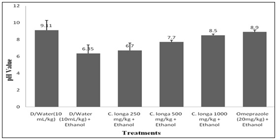
FIG. 1: EFFECT OF CURCUMA LONGA ON PH IN ETHANOL INDUCED GASTRIC TOXICITY AND TOTAL ACID SECRETION

FIG. 2: THE EFFECT OF CURCUMA LONGA ON TOTAL ACID SECRETION IN ETHANOL-INDUCED GASTRIC TOXICITY. The data displays the mean ± standard error of five mice. The percentages of inhibition; ###p<0.001; ***p<0.001 compared to D/water and D/water + Ethanol respectively, are displayed in parenthesis.
Curcuma longa Effects on Gastric Antioxidant Parameters in Rats: Ethanol administration significantly (p<0.001) increased MDA while reducing GSH, Catalase, and SOD, indicating oxidative stress and impaired antioxidant defense. Curcuma longa produced a dose-dependent improvement, with higher doses (500–1000 mg/kg) significantly lowering MDA and enhancing GSH, Catalase, and SOD levels, approaching the effects of omeprazole, which showed the strongest antioxidant activity Table 5.
TABLE 5: EFFECT OF CURCUMA LONGA ON GASTRIC ANTIOXIDANT PARAMETERS IN ETHANOL-INDUCED GASTRIC TOXICITY IN RAT
| Treatments | MDA (ղ moles/mg protein) | GSH (µmoles/mg protein) | Catalase (U/mg protein) | SOD (U/mg protein) |
| D/Water (10 ml/kg) | 3.39±0.21 | 14.97±0.91 | 10.75±0.12 | 1.59±1.41 |
| D/Water (10 ml/kg) + Ethanol | 5.75±1.12### | 8.42±1.42### | 9.29±1.15## | 1.51±0.23 |
| Curcuma longa 250 mg/kg + Ethanol | 5.73±0.31 | 13.22±1.02** | 13.46±0.32** | 1.69±0.03 |
| Curcuma longa 500 mg/kg + Ethanol | 4.29±1.32* | 21.19±0.13*** | 12.76±1.21* | 1.66±0.09 |
| Curcuma longa 1000 mg/kg + Ethanol | 3.82±0.23** | 20.26±1.22*** | 20.23±0.50*** | 2.39±1.21* |
| Ethanol + Omeprazole 20 mg/kg | 1.79±1.33*** | 25.96±0.13*** | 13.83±0.23** | 2.05±0.11* |
The data displays the mean ± standard error of five mice. ###p<0.001; ###p<0.001; *p<0.05; **p<0.01;***p<0.001 versus D/water (##; ###) and D/water + Ethanol (*; **; ***).
Effects Result of Curcuma longa Extract in Histological Studies in Ethanol-Induced Gastric Toxicity in Rat: Histological findings aligned with biochemical results, showing ethanol-induced severe mucosal injury.
Low-dose Curcuma longa (250 mg/kg) offered no protection, while higher doses (500–1000 mg/kg) reduced lesions in a dose-dependent manner, with near-complete protection at the highest dose. Omeprazole fully prevented damage, validating the model Fig. 3.
Histological sections showed no observable lesions in Plates A and F, while Plate B revealed severe erosion of neck cells with mucosal ulceration (yellow arrow). Severe atrophy of glandular cells was observed in Plate C (white arrow), moderate atrophy in Plate D (blue arrow), and mild atrophy in Plate E (green arrow).

FIG. 3: EFFECTS RESULT OF CURCUMA LONGA EXTRACT IN HISTOLOGICAL STUDIES IN ETHANOL-INDUCED GASTRIC TOXICITY IN RAT
DISCUSSION: This study demonstrates that Curcuma longa possesses significant phenolic content, antioxidant activity, and gastroprotective effects against ethanol-induced gastric ulceration. The observed increase in TPC and TAC with concentration supports earlier findings that polyphenolic compounds in turmeric contribute to its antioxidant potential by scavenging free radicals and strengthening endogenous defenses 22, 23.
In the ethanol ulcer model, untreated rats exhibited severe mucosal injury, elevated MDA, and reduced antioxidant enzymes, consistent with oxidative stress-driven pathogenesis 24. C. longa treatment restored gastric redox balance by lowering MDA and enhancing GSH, catalase, and SOD levels. These results are in line with previous studies by Vijayasteltar et al. 25, who reported protective effects of turmeric essential oils against oxidative stress. A noteworthy finding was the biphasic effect of Curcuma longa on gastric acidity. At 250 mg/kg, the extract lowered pH, enhancing acidity and aggravating injury. In contrast, higher doses (500–1000 mg/kg) restored gastric pH and reduced acid secretion, similar to omeprazole. This dual effect reflects the pharmacological complexity of curcumin, which has been reported to stimulate gastric secretion at low doses but suppress it at higher doses 26, 27.
Histological analysis provided further evidence. Ethanol-induced gastric mucosa showed severe atrophy and ulceration, while higher doses of C. longa significantly mitigated these changes. Near-complete mucosal protection at 1000 mg/kg suggests dose dependency, consistent with previous studies where turmeric extracts promoted epithelial regeneration and mucosal healing 28, 29. The protective effect observed with omeprazole validates the experimental model and emphasizes the therapeutic potential of Curcuma longa.
Overall, the findings demonstrate that Curcuma longa provides gastroprotection through several mechanisms, including phenolic-mediated antioxidant activity, inhibition of lipid peroxidation, restoration of endogenous antioxidant defenses, regulation of gastric acidity, and preservation of the mucosal lining. The dose-dependent pattern observed further indicates that higher doses are more likely to achieve meaningful protection. Supported by recent reports, these results highlight the promise of Curcuma longaas a natural anti-ulcer agent, with potential applications as either a complementary therapy or a starting point for drug discovery.
CONCLUSION: This study establishes that Curcuma longa exhibits dose-dependent gastroprotective effects against ethanol-induced gastric damage. Its protective action is largely attributed to its rich phenolic content, antioxidant properties, ability to reduce lipid peroxidation, and capacity to restore endogenous defense enzymes. Lower doses produced only modest effects, whereas higher doses (500–1000 mg/kg) offered significant protection, with the greatest effect at 1000 mg/kg, comparable to omeprazole. These findings suggest that Curcuma longa could serve as a valuable natural anti-ulcer remedy and a promising adjunct in the management of gastric ulcers.
ACKNOWLEDGEMENTS: The authors would like to thank all the Technical Staff of Department of Pharmacology and Toxicology, Olabisi Onabanjo University, Sagamu Campus Ogun State, Nigeria, for their assistance during bench work.
Ethical Approval: An ethical clearance request was submitted to the University of Lagos, (Lagos State, Nigeria), College of Medicine's Animal Care and Use Research Ethics Committee (CMUL/ACUREC), and was granted permission number CMUL/ACUREC/12/24/2089.
CONFLICTS OF INTERESTS: The authors have no relevant conflicts of interest to disclose.
REFERENCES:
- Ismail OI, El-Sayed M and El-Baz H: Curcumin ameliorates ethanol-induced gastric mucosal erosion in rats via alleviation of oxidative stress and regulation of pro-inflammatory cytokines and NF-κB activation. Scientific Reports 2022; 12: 19842.
- Gao J, Liu Y, Chen J, Tong C, Wang Q and Piao Y: Curcumin treatment attenuates chemically-induced gastric mucosal inflammation and apoptosis through inhibition of NF-κB and MAPK signalling. Toxicology Reports 2022; 9: 1557–1568.
- Ma G, Zhang J and Yang R: Therapeutic effects of curcumin and curcumin formulations on ethanol-induced gastric mucosal injury in rats: Comparative studies versus a proton pump inhibitor. Journal of Angiotherapy 2024; 8(1): 1–12.
- Wang Q, Yao C, Li Y, Luo L, Xie F, Xiong Q and Feng P: Effect of polyphenol compounds on Helicobacter pylori eradication: A systematic review with meta-analysis. BMJ Open 2023; 13(1): e062932.
- Deng R, Chen X, Zhao S, Zhang Q and Shi Y: The effects and mechanisms of natural products on Helicobacter pylori eradication. Frontiers in Cellular and Infection Microbiology 2024; 14: 1360852.
- Beiranvand M, Tashakori S and Habibi P: A review of the most common in-vivo models of stomach injury and their mechanisms. Journal of Ethnopharmacology 2022; 282: 114592.
- Xia YS, Zhang X and Li Q: Prevention of ethanol-induced gastric mucosal injury: Evidence that ethanol causes oxidative stress, apoptosis, and inflammatory activation in rat stomach. Frontiers in Nutrition 2021; 8: 769463.
- Asaad GF, Mostafa RE and El Sayed H: Lactoferrin mitigates ethanol-induced gastric ulcer via modulation of ROS/ICAM-1/Nrf2 signaling pathway in Wistar rats. Iranian Journal of Basic Medical Sciences 2022; 25(12): 1522–1527.
- Askgaard G, Christensen AI, Nordestgaard B, Grønbæk M and Tolstrup JS: Alcohol and risk of non-traumatic bleeding events requiring hospital care in the general population: A prospective study. Alcohol 2020; 87: 73–78.
- Antunes C, Copeland J and Rastegar V: Upper gastrointestinal bleeding. StatPearls 2024.
- Freedberg DE, Kim LS and Yang YX: The risks and benefits of long-term proton pump inhibitor therapy: A contemporary review. Gastroenterology 2020; 158(4): 706–715.
- World Health Organization: WHO traditional medicine strategy 2014–2023. World Health Organization 2013.
- Fuloria S, Singh M and Verma R: A comprehensive review on the therapeutic potential of curcumin: Mechanisms, delivery systems, and clinical evidence. Evidence-Based Complementary and Alternative Medicine 2022; 2022: 8990857.
- Ji J, Ma Z and Wang Y: Advancing gastrointestinal health: Curcumin’s efficacy and nanopreparations. Molecules 2024; 29(7): 1659.
- Ebrahimzadeh MA, Nabavi SM, Nabavi SF, Bahramian F and Bekhradnia AR: Antioxidant and free radical scavenging activity of H. officinalis L. var. angustifolius, V. odorata, B. hyrcana, and C. speciosum. Pakistan Journal of Pharmaceutical Sciences 2010; 23(1): 29–34.
- Phatak RS and Hendre AS: Total antioxidant capacity (TAC) of fresh leaves of Kalanchoe pinnata. Journal of Pharmacognosy and Phytochemistry 2014; 2(5): 32–35.
- Falcão HS, Mariath IR, Diniz MFFM, Batista LM and Barbosa-Filho JM: Plants of the American continent with antiulcer activity. Phytomedicine 2008; 15(1–2): 132–146.
- Agbaje OE and Okpara SC: Antiulcer activity of aqueous extract of fresh leaf of Brassica oleracea Linn. var. acephala (D.C.) Alef. International Research Journal of Pharmacy 2013; 4(8): 107–111.
- Bongu S and Vijayakumar S: Animal models in experimental gastric ulcer screening: A review. International Journal of Pharmacology Screening Methods 2012; 2(2): 82–87.
- Oyinloye EO, Murtala AA, Oladoja FA, Alabi AO, Aderinola AA, Okunye OL, Saka SA and Kasumu EO: Citrus aurantifolia (Christm.) Swingle peel extract attenuates nephrotoxicity induced by doxorubicin. Pharmacological Research - Modern Chinese Medicine 2024; 11: 100412.
- Mubarak S, Hamid SA, Farrag AR, Samir N and Hussein JS: Cardioprotective effect of date palm against doxorubicin-induced cardiotoxicity. Asian Journal of Pharmaceutical and Clinical Research 2018; 11: 141–146.
- Wu T, Zhou Y, Lu J, Zhang Q, Wu D, Zhang R and Wang X: Natural antioxidants in foods and their potential in preventing oxidative stress-related diseases: Molecular mechanisms and applications. Frontiers in Nutrition 2024; 11: 1344640.
- Swathi D and Prasad KVSRG: Antioxidant and antiulcer potential of ethanolic extract of bark of Myrica esculenta in pyloric ligation ulcer model. International Journal of Pharmacy and Pharmaceutical Sciences 2015; 7(11): 195–198.
- Matsuda H, Li Y, Murakami T, Yamahara J and Yoshikawa M: Protective effects of oleanolic acid oligoglycosides on ethanol- or indomethacin-induced gastric mucosal lesions in rats. Life Sciences 1998; 63(17): PL245–PL250.
- Vijayasteltar L, Nasir GP, Maliakel B, Kuttan R and Krishnakumar IM: Safety assessment of a standardized polyphenolic extract of clove buds (Clovinol): Oral acute and subchronic toxicity and mutagenicity studies. Toxicology Reports 2016; 3: 439–449.
- Kim YS, Kim TH and Choi CS: Effect of itopride, a new prokinetic agent, in patients with mild GERD: A pilot study. World Journal of Gastroenterology 2005; 11(27): 4210–4214.
- Goel RK, Sairam K and Rao CV: Role of gastric antioxidant and anti-Helicobacter pylori activities in antiulcerogenic activity of plantain banana (Musa sapientum var. paradisiaca). Indian Journal of Experimental Biology 2001; 39(7): 719–722.
- Savaringal JP and Sanalkumar KB: Gastroprotective effect of Curcuma longa Linn. against ethanol-induced gastric ulcer in rats. Journal of Evolution of Medical and Dental Sciences 2018; 7(3): 332–337.
- Savaringal JP and Sanalkumar KB: Anti-ulcer effect of extract of rhizome of Curcuma longa L. against aspirin-induced peptic ulcer in rats. National Journal of Physiology, Pharmacy and Pharmacology 2018; 8(5): 650–657.
How to cite this article:
Oyinloye EO, Murtala AA, Oladoja FA, Okonkwo VE and Adeneye QB: Phenolic-rich extract of Curcuma longa L. mitigates ethanol-induced gastric toxicity via antioxidant defense restoration. Int J Pharmacognosy 2026; 13(5): 436-44. doi link: http://dx.doi.org/10.13040/IJPSR.0975-8232.IJP.13(5).436-44.
This Journal licensed under a Creative Commons Attribution-Non-commercial-Share Alike 3.0 Unported License.
Article Information
7
436-444
1031 KB
116
English
IJP
Elijah Oladapo Oyinloye *, Abdullahi Akanji Murtala, Farouk Adedeji Oladoja, Victor Emmanuel Okonkwo and Qudus Babajide Adeneye
Department of Pharmacology and Toxicology, Faculty of Pharmacy, Olabisi Onabanjo University, Sagamu Campus, Ogun State, Nigeria.
oyinloye.oladapo@oouagoiwoye.edu.ng
15 April 2026
06 May 2026
12 May 2026
10.13040/IJPSR.0975-8232.IJP.13(5).436-44
15 May 2026