


PHARMACOGNOSY AND PHYTOCHEMICAL ANALYSIS OF LEAF GALLS OF MANGIFERA INDICA L
HTML Full TextPHARMACOGNOSY AND PHYTOCHEMICAL ANALYSIS OF LEAF GALLS OF MANGIFERA INDICA L
R. Anitha * and A. Suja
Department of Plant Biology and Plant Biotechnology, Ethiraj College for Women, Ethiraj Salai, Egmore, Chennai - 600008, Tamil Nadu, India.
ABSTRACT: The leaf galls of Mangifera indica L. are very characteristic and possess medicinal properties, due to their phytochemical profile. Hence, a pharmacognostic and phytochemical study was carried out. The microscopic evaluation of leaf gall powder of Mangifera indica revealed the presence of calcium oxalate crystals, cork cells with resin and sclerites. The anatomy of mature galls exhibited characteristic anomalous growth with an outer cork layer with cell differentiation. The young galls were only parenchymatous. They also showed the presence of tannins, alkaloids, terpenoids, and flavonoids. The flavanoid in the gall tissue was 60% more than the normal leaf tissue. The fluorescence study of the powder showed very characteristic bright yellow fluorescence indicating the presence of intense flavanoids. Water soluble and acid insoluble ash was found to be 24% and 10% respectively with a moisture content of 0.6%. Further, an in-vitro study on various therapeutic properties of leaf galls will be more beneficial.
| Keywords: |
Leaf galls, Mangifera indica, Powder analysis, Anatomical study, Phytochemical test, Physiochemical properties
INTRODUCTION: Galls are abnormal deformities commonly seen in young plant tissues. There are over 1500 species of gall producers. However, most galls are produced by plant mites, gall midges, and gall wasps. These galls are caused either by plant growth regulating chemicals induced by mechanical damage or salivary secretions or stimuli produced by an insect while feeding or during egg laying activity. Mangifera indica leaves play an important role in the Indian system of medicine. The leaves possess antimicrobial, antibacterial activity, antiulcerogenic action 1, hypoglycemic, antherogenic activity 2.
Leaves are used as a therapeutic agent in disease such as cancer. Mangifera is a super antioxidant. Mango leaf galls are produced by gall midge (Procontarinia matteiana) which triggers the hypertrophic activity and simultaneously increase enzymes (Protease, Chitinase, Cellulose, Amylase, Invertase, and Sugars) 3, 4 and antioxidants which is very useful for curing diabetes 5. Leaf galls contain substantial amounts of phytoconstituents such as phenolics, flavonoids and, tannins. The galls also are reported to be the sink of nutrients and contain starch, sugars, an amino acid such as proline and phenols 6.
The efficiency of herbal medicine depends on the quality and quantity of the important phytochemical constituent present in it. This is where pharmacognosy plays an important role not just in the identification of crude powder drug but also in standardizing the various physical parameters, morphology in terms of both macroscopic and microscopic characteristics 7. The pharmacognostic evaluation also helps in the detection of adulterants if any in the powder drug. The pharmacognostic study is mainly used for the standardization of crude drugs. Hence, this study was conducted to characterize the pharmacognostic properties of Mangifera indica leaf galls which possess enormous medicinal properties.
MATERIALS AND METHODS:
Organoleptic and Powder Analysis: Organoleptic characterization of dried gall and healthy leaf of Mangifera indica was carried out. The texture of the leaf, smell, color, taste was observed. The fine powder of gall and healthy leaves of Mangifera indica was placed on to a clean slide and observed under a microscope.
Anatomical Study: Freehand section of galls in leaves of young and mature Mangifera indica were taken, stained with safranin and mounted in glycerol and observed under light microscope and photographed.
Histochemical Study: The gall and healthy leaf sections were treated with various reagent such as Wagners reagent (potassium iodide and iodine) for detection of alkaloid, Orcinol in sulphuric acid for gums, Toluidine blue O for lignin, Ferric chloride in 1N Hydrochloric acid for tannin, Sulphuric acid for crystals, Methylene blue test for phenols., 10% Sodium hydroxide for flavonoids, Vanillin in acetic acid for terpenoids and Potassium iodide and iodine for starch.
Phytochemical Test: The gall and healthy leaves of Mangifera indica were washed thoroughly, shade dried and powdered. The powder was used for phytochemical detection. All phytochemical test was carried out following standard methods 8. The total flavonoid content was determined by according to 9 with rutin as standard.
Fluorescence Analysis: The dry powder was placed on a slide and treated with several drops of specified reagent like Hydrochloric acid, Sodium hydroxide, Nitric acid, Sulphuric acid, Ferric chloride, Iodine Acetic acid, HNO3 + Ammonia, Methanol, NaOH + Methanol 10. The slides were observed under UV 265 nm, and 365 nm and the emitted fluorescence was observed that helps in identifying the drug in the powdered sample.
Physicochemical Parameters: Determination of total ash, Insoluble acid ash, water-soluble ash, and moisture content was done according to Indian Pharmacopoeia 11.
RESULTS AND DISCUSSION:
Organoleptic and Powder Analysis: Organoleptic evaluation provide the simplest as well as quickest means to establish the identity and purity of a particular drug. Normal leaf was green in color, aromatic, astringent, coarse; Galls were yellowish brown in colour, aromatic, astringent and coarse. Microscopic study of the powder showed the presence of calcium oxalate crystals – rosettee and prismatic crystals, Macrosclerides, parenchyma cells, parenchyma with cork region, cork with resin, resin globules and fibres Plate 1. Microscopic characters can be used for standardization of drugs and also used for the preparation of plant monographs.
Anatomical Studies: The anatomical studies showed that the outermost layer of the galls was composed of 2-3 layers of sclerenchymatous cells in the case of young galls. However, the older galls had very characteristic cork formation. The cork cambium (phellogen) with phelloderm and phellem was evident. The cork was 3-4 layers in number and filled with resin. A vascular bundle was absent. Anomalous secondary growth was observed which is responsible for the formation of the cork layer. The outer parenchymatous cells differentiated into cork cambium and resulted in the cork formation Plate 2.
A central pith was absent. The entire cortex was parenchymatous. The central parenchyma cells were hexagonal whereas towards the periphery were elongated cells in the young gall; in the matured galls both central and periphery parenchyma cells were enormous. Tissue differentiation was absent. In most of the gall inhibition of development, differentiation, growth and tissue suppression along with activation of some gene is generally encountered 12.
The young galls showed calcium oxalate crystals which were rosette, prismatic and microspheroidal crystals were observed in the outer area of the cortex. The intercellular spaces were absent in the center whereas towards the periphery there were intercellular spaces. In galls induced Guarea macrophylla subsps tuberculata, the spongy parenchyma cells divide and become round with small intercellular spaces 13.
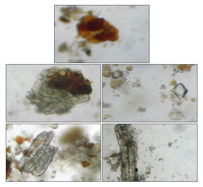
PLATE 1: POWDER ANALYSIS OF LEAF GALL OF MANGIFERA INDICA L. A- PARENCHYMA CELLS WITH RESIN B- CORK CELLS WITH RESIN C- PRISMATIC CALCIUM OXALATE CRYSTAL D- MACROSCLERIDES E- SEPTATE FIBRE
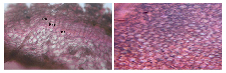
PLATE 2: ANATOMY OF LEAF GALL OF MANGIFERA INDICA L. A- T.S OF MATURE GALL SHOWING CORK REGION. NOTE THE DIFFERENTIATION OF PHELLEM (PH), PHELLOGEN (Phe) AND PHELLODERM (Pd). B- T.S OF YOUNG GALL SHOWING COMPACT PARENCHYMA CELLS IN THE CENTRE OF THE GALL TISSUE.
Histochemical Studies: Histochemical studies were carried out in young and mature galls. It was observed that alkaloid, tannin, flavonoid, terpenoids were present. Calcium oxalate was also present. However starch was absent. Generally, gall induced cells are reported to have elevated levels of phenols and tannins 14.
Phytochemical Test: The phytochemical tests revealed that the presence of tannin, alkaloid, resin, terpenoid, a flavonoid in galls infested Mangifera leaves; flavonoid and terpenoid in normal leaf Table 1. It is reported that various parts of Mangifera indica contain phenolic acids, phenolic esters, and flavonols. Flavonoid content in gall was highly elevated (90%) when compared to normal leaf (60%) in the present study. It is also known that three types of terpenes were found in Mangifera indica gall leaves 15.
TABLE 1: PHYTOCHEMICAL TEST IN NORMAL AND GALLED LEAF OF MANGIFERA INDICA
| S. no. | Phytochemicals | Test | Galls | Normal |
| 1 | Tannin | Fecl3 test | + | - |
| 2 | Alkaloid | Meyers test | + | - |
| 3 | Saponin | Frothing test | - | - |
| 4 | Resin | H2SO4 test | + | - |
| 5 | Gum | Ethanol test | - | - |
| 6 | Glycoside | NaOH test | - | - |
| 7 | Phlobatannin | HCl test | - | - |
| 8 | Flavonoid | Alkaline reagent | + | + |
| 9 | Terpenoid | H2SO4, chloroform test | + | + |
| 10 | Steroids | Libermann, Burchard test | - | - |
Fluorescence Analysis: With sodium hydroxide, the gall powder showed characteristic bright yellow fluorescence under UV Table 2. The ultraviolet light produces fluorescences in many natural products which do not visibly fluoresce in daylight 16. The characteristic yellow fluorescence indicates the presence of flavonoids. Hence, the crude drugs can be assessed qualitatively in this way, and it is an essential parameter for pharmacognostic evaluation.
TABLE 2: FLUORESCENCE ANALYSES-NORMAL AND GALLED LEAF OF MANGIFERA INDICA
| S. no. | Test | Normal tissue | Gall tissue | ||
| Visible Light | UV 265 nm | Visible Light | UV 265 nm | ||
| 1 | H2SO4 TEST | black | pink | black | brown |
| 2 | HCL | Greenish brown | brown | brown | Dark brown |
| 3 | Nitric acid | Reddish brown | pink | brown | No color |
| 4 | NaOH | Yellowish green | Fluorescent yellow | Yellowish brown | Intense fluorescent yellow |
| 5 | Ammonia | green | green | Dark brown | brown |
| 6 | Methanol | green | No color | black | yellow |
| 7 | Ethanol | green | No color | brown | No color |
Physicochemical Parameters: The physical evaluation of the drugs is an important parameter in detecting adulteration or improper handling of drugs 17. Ash values indicate the presence of various impurities like carbonate, oxalate and silicate. The water-insoluble ash is used to estimate the amount of inorganic compound present in the drug. Acid-insoluble ash consists of main silica and indicate contamination with earthly material 18. Total ash was found to be more in normal leaf. Water soluble and acid insoluble ash was found to be 24% and 10% in gall respectively Table 3. The moisture content of gall was found to be 0.6% and indicates that it generally not susceptible to bacterial growth.
TABLE 3: PHYSICOCHEMICAL PARAMETERS OF NORMAL AND GALL LEAF OF MANGIFERA INDICA
| S. no. | Physicochemical parameters | Normal | Gall |
| 1 | Total ash | 45% | 21.5% |
| 2 | Water soluble | 32% | 24% |
| 3 | Acid insoluble | 22% | 10% |
| 4 | Loss on drying | 0.6 | 0.4 |
The galls are not just mere abnormal structures, but these have elevated phytochemical profile since they act of food sink for the infesting insect. It is known that the leaves of Mangifera indica have medicinal values for treating various ailments.
The pharmacognostic study on these galls has shown that they can be effectively used as therapeutic agents due to its various phytochemical constituents. Further, an in-vivo study with the leaf galls can throw more light on its efficacy.
ACKNOWLEDGEMENT: The authors wish to thank The Principal and Head of the Department for providing the laboratory facility and constant encouragement.
CONFLICT OF INTEREST: Nil
REFERENCES:
- Severi JA, Lima ZP, kushima H, Brito ARM, Campaner dos santos L, Vilegas W and Lima AH: Polyphenols with antiulcerogenic action from an aqueous decoction of mango leaves (Mangifera indica L.). Molecules 2009; 14: 1098-11.
- Muruganandan S, Srinivasan K, Gupta S, Gupta PK and Lala J: Effect of Mangiferin on hyperglycemia and atherogenicity in Streptozotocin-diabetic rats. J Ethnopharmacol 2005; 97: 497-501
- Choudhary R and Kumar S: quantitative estimation of some metabolites and enzymes in insect induced leaf galls of Pongamia pinnata (L.) Journal of Chemical and Pharmaceutical Research 2012; 4(9): 4192-4197.
- Marmit, KS and Sharma SL: Quantitative estimation of some metabolites and enzymes in insect-induced leaf galls of Mangifera indica. Asian J Exp Sci 2008; 22(3): 343-346.
- Kamal SM, Vijay PM and Suman LS: Quantitative estimation of phenolics and related enzymes in insect induced leaf galls of Mangifera indica. Annals of Plant Protection Sciences 2008; 16(2): 306-308.
- Dsouza MR and Ravishankar BE: Nutritional sink formation in galls of Ficus glomerata roxb. (moraceae) by the insect Pauropsylla depressa (psyllidae, hemiptera). Tropical Ecology 2014; 55(1): 129-136.
- Chanda S: Importance of pharmacognostic study of medicinal plants. Journal of Pharmacognosy and Phytochemical 2014; 2(5): 69-73.
- Harborne JB: Methods of extraction and isolation. In: Phytochemical methods, 3rd ed, Chapman and Hall, London 1998: 60-66.
- ElFar MMM and Taie HAA: Antioxidant activities, total anthocyanins, phenolics and flavonoids contents of some sweet potato genotypes under stress of different concentrations of sucrose and sorbitol” Australian J Basic Applied Sci 2009: 3609-3616,
- Kokoshi CJ, Kokoshi RJ and Sharma FT: Fluorescence of powdered vegetable drugs under Ultraviolet radiation. J. Pharm. Asses 1958; 47: 715-717.
- Indian Pharmacopoeia, Vol.-II 4th Edition, Controller of Publications, Government of India, New Delhi 1996; A-47.
- Ananthakrishnan TA: Insect gall systems: Patterns, processes and adaptive diversity. Current Science 1998. 75: 672-680.
- Andreu GP, Delgado R, Velho J, Inada NM, Curti C and Vercesi AE: Mangifera Indica L. extract (Vimang) inhibits Fe2+-citrate-induced lipoperoxidation in isolated rat liver mitochondria. Pharmacol Res 2005; 51: 427-35.
- Upadhye AS and Rajopadhye AA: Pharmacognostic and phytochemical evaluation of leaf galls of Kakadshringi used in Indian system of medicine. Journal of scientific and industrial research 2010; 69: 700-704.
- Augustyn WA, Botha BM, Combrinck S and Du Plooy, W: Correlation of volatile profiles of twenty mango cultivars with their susceptibilities to mango gall fly infestation South African Journal of Botany 2010; 76(4): 710-716.
- Zhao Z, Liang Z and Guo P: Macroscopic identification of Chinese medicinal materials: Traditional experiences and modern understanding .J Ethnopharmacol 2011; 131: 556-561.
- Tatiya A, Surana S, Bhavsar S, Patil D and Patil Y: Pharmacognostic and preliminary phytochemical investigation of Eulophia herbacea Lindl. Tubers (Orchidaceae). Asian Pac J Trop Disease 2012; 2(1): S50-55.
- Vaghasiya Y, Nair R and Chanda S: Antibacterial and preliminary phytochemical and physic-chemical analysis of Eucalyptus citriodora HK leaf. Product Res 2008; 22: 51-54.
How to cite this article:
Anitha R and Suja A: Rhoifolin: Pharmacognosy and phytochemical analysis of leaf galls of Mangifera indica L. Int J Pharmacognosy 2015; 2(6): 301-05. doi: 10.13040/IJPSR.0975-8232.2(6).301-05.
This Journal licensed under a Creative Commons Attribution-Non-commercial-Share Alike 3.0 Unported License.
Article Information
5
301-305
593
2645
English
IJP
R. Anitha * and A. Suja
Department of Plant Biology and Plant Biotechnology, Ethiraj College for Women, Ethiraj salai, Egmore, Chennai, Tamil Nadu, India.
anitha.rajasekaran023@gmail.com
30 April 2015
23 June 2015
28 June 2015
10.13040/IJPSR.0975-8232.IJP.2(6).301-105
30 June 2015