


PHARMACOGNOSTIC EVALUATION OF ROOT OF BUTEA MONOSPERMA LAM. (FABACEAE)
HTML Full TextPHARMACOGNOSTIC EVALUATION OF ROOT OF BUTEA MONOSPERMA LAM. (FABACEAE)
Neetu Deshwal 1, Ajay Kumar Sharma 2, Pankaj Kumar Maurya * 3 and Saurabh Sharma 3
Department of Pharmacognosy 1, Department of Pharmacology 3, School of Pharm. Sciences and Health Care, CT University, Ludhiana - 142024, Punjab, India.
Department of Pharmacognosy 2, School of Pharmaceutical Science, Jaipur National University, Jaipur - 302025, Rajasthan, India.
ABSTRACT: All information about the herbal drug, including all its organoleptic characters, phytoconstituents, pharmacological actions, and its standardization in respect to various parameters, is required before developing an herbal drug formulation. Butea monosperma (Lam.) is commonly known as the Flame of the forest, belongs to the family Fabaceae. Commonly Butea monosperma is used as tonic, astringent, aphrodisiac, and diuretics. We are doing this work because it was thought worthwhile to explore this most functional plant based on this standardization parameter. The objective of the work is to study pharmacognostic and physicochemical characteristics of the root of Butea monosperma Fresh and dried root; root powder was studied for its morphology, microscopy, physicochemical, phytochemical characteristics, and other WHO-recommended methods for standardization. The root of Butea monosperma was observed to be brown in color and irregular in shape with a rough surface. The root has a characteristic odour and faint taste. Microscopy revealed the presence of cork cells; Medullary rays distinct parenchymatous cells narrow in the xylem region and wider in the phloem region. Xylem parenchymal contains calcium oxalate crystal. Pith contain Parenchymatous cells with intracellular space, Xylem Lignified, Xylem vessels thickened walls, lignified Phloem. Preliminary phytochemical screening revealed the presence of flavonoids and tannins & phenolic compounds, which is also confirmed by TLC results, so the total flavonoid and phenolic contents were also calculated in Butea monosperma to extract. It can be concluded that the pharmacognostic profile of Butea monosperma root is useful in standardization for quality, purity, and sample identification.
| Keywords: |
Butea monosperma, Pharmacognostic evaluation, Phytochemical characteristics, Standardization
INTRODUCTION: Butea monosperma (Lam.) is commonly known as flame of the forest, belongs to the family Fabaceae 1. It is locally called palas, Palash, mutthuga, bijasneha, dhak, khakara, chichra, Bastard Teak, Bengal Kino, Nourouc, and is common throughout India, Burma, and Ceylon except in very acrid parts.
Generally, it grows gregariously on open grasslands and scattered in mixed forest. Plantations can be raised both on irrigated and drylands. The pods should be collected and sown before the commencement of rains; root suckers are freely produced and help in vegetative propagation.
In India, palas ranks next to Kusum (schleicheratrijuga) as a host tree for lac insect 2, 3. It has proven to be a source of constitutive osteogenic agents belonging to isoflavonoid and pterocarpan groups. The genus Butea includes Butea monosperma, Butea parviflora, Buteaminor, and Butea superba widely distributed throughout India 4.
It holds an important place because of its medicinal and other miscellaneous uses of economic value. It is one of the most beautiful trees that has been put to some useful purpose. Butea monosperma is extensively used in Ayurveda, Unani and Homeopathic medicine and has become a cynosure of modern medicine. The plants of this genus are well known for their colouring matters. Commonly Butea monosperma is used as tonic, astringent, aphrodisiac, and diuretics 5.
It is an erect tree 12-15 m high with a crooked trunk and irregular branches, bark rough, ash-colored, young parts downy. Leaves are 3-foliate, petioles 10-15 cm long, stipules linear-lanceolate. Leaflets coriaceous (the terminal 10-20 cm long, broadly ovate from a cuneate base, the lateral smaller, 10-15 by 7.5 – 10 cm, obliquely rounded at the base, equilateral, the lower side the larger), all obtuse, glabrous above when old, finely silky and conspicuously reticulately veined beneath; petioles 6 mm long, stout-stipels subulate, deciduous. Flowers are large, in rigid racemes 15 cm long, 3 flowers together form the tumid nodes of the dark olive-green velvety rhachis, pedicels about twice as long as the calyx, densely brown-velvety: bracts and bracteoles small, deciduous. Calyx 13 mm long, dark olive-green, densely velvety outside, clothed with silky hairs within: teeth short, the 2 upper connate, the 3 lower equal, deltoid.
Corolla 3.8-5 cm long, clothed outside with silky, silvery hairs, orange or salmon colored: standard 2.5 cm broad: keel semicircular, beaked, veined. Pods stalked 12.5-20 by 2.5-5 cm, thickened at the sutures, reticulately veined argenteo - canescent: stalked 2 cm long 6.
MATERIALS AND METHODS: Fresh root bark of Butea monosperma collected in the month of August to September from the local region of Bhopal district of Madhya Pradesh, and fresh root bark of Butea monosperma were dried under the shade & powdered in a mixture grinder. The powdered root bark is packed in paper bags & stored in an airtight container until use.
Chemicals and Instruments: Methanol, hydrochloric acid, nitric acid, sodium hydroxide, ethanol, chloroform, HCl, conc. HNO3, NH4OH, FeCl3, potassium dichromate, all other solvents and/or reagents used in the study were of analytical grade and procured from Merck-India.
Instruments: Weighing balance, Refrigerator, Water bath, Vortex shaker, Ocular micrometer, Stage micrometer, Microscope, UV Spectro-photometer.
Authentication: The botanical identity was confirmed by the Department of botany Safia College of Science Bhopal (MP), herbarium, where voucher specimen number (149/BOT/SAFIA/2010) has been deposited for further reference.
Extraction: 250 gm dried powder plant material was taken and extracted with solvent Methanol: Water (1:1) by hot continuous extraction method at temp 40 to 50 ºC. After complete extraction, the extract was properly dried on the water bath and stored in an airtight container for further analysis. The yield of Butea monosperma hydroalcoholic extract was (BME) found to be 17.86% w/w.
RESULTS:
A. Morphological study: The morphological study includes color, odour, test, and shape were carried out on fresh root bark Fig. 1 Colour - Brown, Odour - Characteristic, Taste - Faint, Shape -Irregular, Surface – Rough 7.
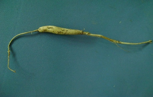
FIG. 1: BUTEA MONOSPERMA ROOT
B. Microscopical Study: Particle size analysis: Average length: 100.62µm, Average width: 38.54µm.
Particle size analysis was done at 5x eyepiece, and 40x objective lens with an ocular micrometer which was calibrated by using stage micrometer calibration factor was determine i.e. 1 division of ocular micrometer = 3.8µm, then 100 particle measure and their average was calculated for determination of average length and width.
Transverse section Fig. 2, 3, 4 cut a part of the root with the help of a sharp blade, and staining was done by safranin to impart a red colour to the lignified tissue. In the Transverse section of Butea monosperma root is circular in outline and shows the following regions: In young root, five to ten or more layer of cork cells. The outer layer contains dark brown matter, Medullary rays, Distinct parenchymatous cells narrow in the xylem region and wider in the phloem region. Parenchymal cells, Xylem parenchymal contain calcium oxalate crystal. Pith and Parenchymatous cells with intracellular space. Xylem Lignified, ensheathed by parenchyma. Xylem vessels thickened walls, lignified Phloem Lignified in the outer part containing parenchymal cells.
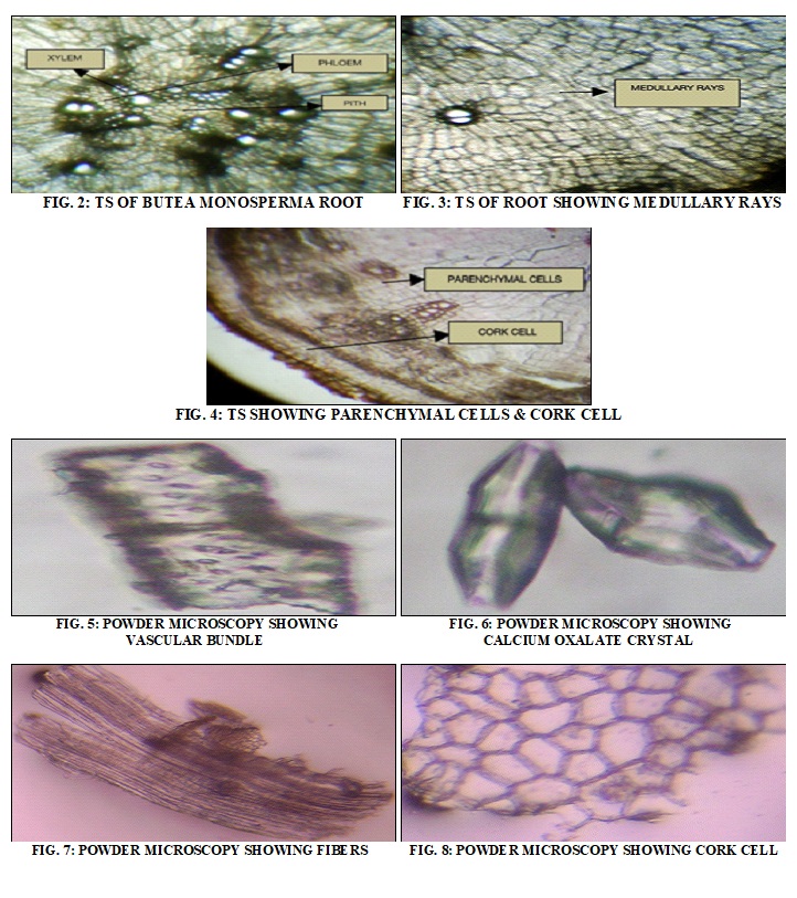
C. Powder Microscopical Study: Powder microscopy Fig. 5, 6, 7, 8: clear the powder with clearing reagent, make mount free from air bubbles, and use for microscopy. The root powder is slightly dark brown in color with a characteristic odour. The vascular elements with bordered pits are seen separately or found in association with fibers. Prism calcium oxalate is observed. Cork cells are also observed separately. Fibers are in seen in different shape and size 8.
Pharmacognostic Profile (Table 1):
A. Loss on Drying: 10 gm of the crude drug was weighed accurately in a tared evaporating dish. It was dried at 105 °C for 5 hours and weighed. The drying and weighing were continued at 1-hour interval until the difference between two successive weighings corresponds to not more than 0.25 percent constant weight were reached.
B. Total Ash Value: About 3 gm of coarse powder of dried root accurately weighed and incinerated in a silica dish at a temperature not exceeding 450 ºC until free from carbon. It was then cooled and weighed. The % w/w of ash with reference to the air-dried drug was calculated.
C. Acid Insoluble Ash: To the crucible containing the total ash was added 25 ml of hydrochloric acid. The crucible was then covered with a watch glass, and the mixture was boiled gently for 5 min. The watch glass was rinsed with 5 ml of hot water, and this liquid was added to the crucible. The insoluble matter was collected on an ashless filter paper and washed with hot water until the filtrate was neutral. The filter-paper contain the insoluble matter was transferred to the original crucible, dried on a hot-plate, and ignite to constant weight. The residue was allowed to cool in a desiccator for 30 min and then weighed.
D. Water Soluble Ash: To the crucible containing the total ash, was boiled for 5 min. with 25 ml of water. Insoluble matters were collected on ashless filter paper, washed with hot water, and ignite for 15 min. at a temperature not exceeding 450 °C. The weight of insoluble matter was subtracted from the weight of total ash.
E. Crude Fibre Content: 2 gm of the powdered crude drug was taken in a beaker and added 50 ml of 10% nitric acid. Heated to boil with constant stirring (till about 30 sec. after boiling start) strain through a fine cotton cloth on Buchner funnel. The residue was washed with boiling water, transfer the residue from the cloth to a beaker. 50 ml of 2.5% sodium hydroxide was added and heated to boil. Maintain at boiling point for 30 sec. with constant stirring. Strain and wash with hot water, as mentioned earlier. The residue was transferred in a crucible and weighed the residue, and the percentage of crude fiber was determined.
F. Water Soluble Extractive: Accurately weighed 4 gm of coarse powder of dried root was macerated with 100 ml of water in a closed flask for 24 h, shaken frequently during first 6 h, and allowed to stand for 18 h. It was then filtered rapidly, and 25 ml of the filtrate were evaporated to dryness in a tared flat-bottomed shallow dish and dried at 100°C to constant weight. The % w/w of water-soluble extractive value was calculated with reference to the air-dried drug.
G. Alcohol Soluble Extractive: Accurately weighed 4 gm of coarse powder of dried root was macerated with 100 ml of ethanol in a closed flask for 24 h, shaken frequently during the first 6 h, and allowed to stand for 18 h. It was then filtered rapidly, taking precautions against loss of the solvent, and 25 ml of the filtrate were evaporated to dryness in a tared flat-bottomed shallow dish and dried at 100 ºC to constant weight. The % w/w of alcohol-soluble extractive value was calculated with reference to the air-dried drug.
H. Acetone Soluble Extractive: Accurately weighed 4 gm of coarse powder of dried root was macerated with 100 ml of acetone in a closed flask for 24 h, shaken frequently during the first 6 hr, and allowed to stand for 18 hr. It was then filtered rapidly, taking precautions against loss of the solvent, and 25 ml of the filtrate were evaporated to dryness in a tared flat-bottomed shallow dish and dried at 100 ºC to constant weight. The % w/w of acetone soluble extractive value was calculated with reference to the air-dried drug
I. Chloroform Soluble Extractive: Accurately weighed 4 gm of coarse powder of dried leaves was macerated with 100 ml of chloroform in a closed flask for 24 h, shaken frequently during the first 6 h and allowed to stand for 18 h. It was then filtered rapidly, taking precautions against loss of the solvent, and 25 ml of the filtrate were evaporated to dryness in a tared flat-bottomed shallow dish and dried at 100 ºC to constant weight. The % w/w of chloroform soluble extractive value was calculated with reference to the air-dried drug.
J. Foaming Index: 1 gm coarse powdered crude drug was weighed accurately and transferred to a 500 ml conical flask containing 100 ml of water. It was maintained at moderate boiling for 30 min. It was cooled and filtered into a 100 ml volumetric flask, Volume was diluted by adding a sufficient amount of water. The decoction was poured into ten stoppered test tubes in a successive portion of 1 ml, 2 ml, 3 ml -- up to 10 ml, and the volume of liquid in each test tube was adjusted to 10 ml with water. The tubes were stoppered, and they were shaken in a lengthwise motion for 15 sec. two shakes per sec were allowed to stand for 15 min, and the height of foam was measured.
TABLE 1: STANDARDIZATION PARAMETERS
| S. no. | Evaluation Parameter | Value (%w/w) |
| 1 | Moisture Contains | 12.89 |
| 2 | Total AshValue | 7.33 |
| 3 | Acid Insoluble Value | 1.5 |
| 4 | Water Soluble Value | 2 |
| Crude Fiber Analysis | 14.85 | |
| 6. | Extractive Values
1. Water Extractive Value 2. Alcoholic Extractive Value 3. Acetone Extractive Values 4. Chloroform Extractive Values |
4.5 1.75 0.75 0.75
|
K. Swelling Index: It is the volume in ml taken up by the swelling of 1gm of plant material under specified conditions. 1 gm fine powdered drug was weighed accurately and transferred into specified 25ml stoppered measuring cylinder. Add 25ml of water & shake the mixture thoroughly every 10 min for 1hr. Allowed to stand for 3 h at room temperature, measure the volume in ml occupied by the plant material, including any sticky mucilage. Calculate the mean value of the individual determination, related to 1 gm of plant material Table 2 9.
TABLE 2: OBSERVATION OF SWELLING INDEX
| S. no. | Volume Occupied By Drug In ML After 3h |
| 1 | 7.0 |
| 2 | 7.0 |
| 3 | 7.2 |
| Mean | App 7.0 |
Phytochemical Studies: The chemical tests were performed for testing different chemical groups present in extracts Table 3.
A. Tests for Alkaloids: Evaporate the aq., alcoholic & chloroform extract separately. To the residue, add dil. HCl shake well & filter with filtrate, perform the following tests: To the 2-3 ml filtrate, add few drops of following 4 reagents;
- Dragendorff’s Reagent: Orange Brown ppt
- Mayer’s Reagent: Creamish ppt
- Hager’s Reagent: Yellow ppt
- Wagner’s Reagent: Reddish-brown ppt
- Murexide Test for Purine Alkaloids: to 3-4 ml test solution, add 3-4 drops of conc. HNO3 evaporate to dryness cool & add 2 drops of NH4OH. Purple color is observed.
B. Tests for Tannins & Phenolic Compounds: To 2-3 ml of aq. or alcoholic extract, add few drops of the following reagents: -
- 5% FeCl3 solution: deep blue-black color
- lead acetate solution: white ppt
- Bromine water: decoloration of bromine water
- Acetic acid: red color solution
- Gelatin solution: white ppt
- Potassium dichromate: red ppt
- Dil. Iodine solution: transient red color
- Dil. HNO3: reddish to yellow color
- Dil. NH4OH & potassium ferricyanide solution: red color solution
- Dil. Potassium permanganate solution: decoloration
C. Test for Flavonoids:
- Shinoda Test: to the dry powder or extract, add 5ml 95% ethanol, a few drops conc. HCl & 0.5g magnesium turnings. the pink color observed.
- to a small quantity of residue, add lead acetate solution yellow-colored ppt is formed
- Addition of an increasing amount of NaOH to the residue shows yellow coloration, which decolorizes after addition of acid.
D. Test for Steroid:
- Salkowski Reaction: to 2ml of extract, add 2ml chloroform & 2ml conc. H2SO4. Shake well. chloroform layer appears red & acid layer shows greenish-yellow fluorescence.
- Liebermann–Burchard Reaction: mix 2ml extract with chloroform. add 1-2 ml acetic anhydride & 2 drops conc. H2SO4 form side of the test tube. first red then blue & finally green color appears.
- Liebermann’s Reaction: mix 3ml extract with 3ml acetic anhydride. Heat & cool, add a few drops conc. H2SO4. The blue color appears.
E. Test for Glycosides:
F. Cardiac Glycoside:
- Baljet’s Test:-a thick section shows yellow to orange color with sodium picrate.
- Legal’ test (test for cardenolides): to the aq. or alcoholic extract, add 1ml pyridine & 1ml sodium nitroprusside. Pink to red color appears.
- Keller Killiani’s Test (for deoxysugars): to 2ml extract, add glacial acetic acid, one drop 5% FeCl3 & conc. H2SO4 the reddish-brown color appears at the junction of the 2 layers & the upper layer appears bluish-green.
- Liebermann’s Test (for bufadienolides): see in steroids
G. Anthraquinone Glycosides:
- Borntrager’ test: to 3ml extract, add dil. H2SO4. boil & filter. To the cold filtrate, add equal volume benzene or chloroform. shake well. Separate the organic solvent add ammonia. ammonical layer turns pink or red.
- Modified Borntrager’ test: to 5ml extract, add 5ml 5% FeCl3 & 5ml dil. HCl, heat for 5min. in the boiling water bath. Cool & add benzene or any organic solvent. Shake well. The separate organic layer, add equal volume dil. Ammonia. Ammonical layer shows pinkish-red color.
H. Saponin Glycosides:
- Foam Test: shake the drug extract or dry powder vigorously with water. The persistent foam was observed.
- Hemolytic Test: add drugs or dry powder to one drop of blood placed on a glass slide. Hemolytic zone appears 8.
TABLE 3: PHYTOCHEMICAL INVESTIGATION
| Extract | Hydro-alcoholic |
| Alkaloids | - |
| Carbohydrate | - |
| Taninand Phenolic Compound | + |
| Glycoside | - |
| Flavanoid | + |
| Saponin | - |
| Steroid | - |
Total Flavonoids and Phenol Determination Table 4: Aluminum chloride colorimetric method was used for flavonoids determination 10. Each plant extracts (0.5 ml of 1:10 g ml-1) in methanol were separately mixed with 1.5 ml of methanol, 0.1 ml of 10% aluminum chloride, 0.1 ml of 1 M potassium acetate and 2.8 ml of distilled water. It remained at room temperature for 30 min; the absorbance of the reaction mixture was measured at 415 nm with a double beam spectrophotometer. The calibration curve was prepared by preparing quercetin solutions at concentrations 12.5 to 100g ml-1 in methanol.
Total phenols were determined by the FolinCiocalteu reagent 11. A dilute extract of each plant extract (0.5 ml of 1:10 g ml-1) or gallic acid (standard phenolic compound) was mixed with FolinCiocalteu reagent (5 ml, 1:10 diluted with distilled water) and aqueous Na2CO3 (4 ml, 1 M). The mixtures were allowed to stand for 15 min, and the total phenols were determined by spectrophoto-meter at 765 nm. The standard curve was prepared using 0, 50, 100, 150, 200, 250 mg L-1 solutions of gallic acid in methanol: water (50:50, v/v). Total phenol values are expressed in terms of gallic acid equivalent (mg g–1 of dry mass), which is a common reference compound Table 4.
TABLE 4: FLAVONOID AND PHENOL CONTENTS IN BME
| Plant Species | Flavonoids mg/g | Phenol mg/g |
| Butea monosperma | 100.96 ± 0.60 | 30.89 ± 0.01 |
Thin Layer Chromatography: An aliquot of hydroalcoholic extract of Butea monosperma was dissolved in 1 mL of an appropriate solvent. For TLC, we used silica gel sheets (silufol 60 F254, aluminum support; Merck) in the appropriate solvent system: for flavonoids; n-Butanol: Acetic acid: water (4:1:5), Chloroform: Acetone: Formic acid (75:16.5:8.5), Ethyl acetate: Formic acid: Glacial acetic acid (100:11:11:26), Chloroform: Ethyl acetate (60:40). Rf (distance traveled by solute from origin line/distance traveled by solvent from origin line) was calculated for every constituent Table 5 12, 13.
TABLE 5: THIN LAYER CHROMATOGRAPHY
| S. no. | Solvent System | Rf | Colour in UV | Conclusion |
| 1 | n-Butanol: Acetic acid:Water
(4:1:5) |
AL=0.95 | White | Flavones, Flavonols
Isoflavones, Flavonones |
| 2 | CHCl3: Acetone:Formic acid (75:16.5:8.5) | AL=0.91
AJ=0.97 |
White
|
Flavanolignans |
| 3 | Ethylacetate: Formicacid: Glacial acetic acid (100:11:11:26) | AL=0.97 | White | Flavonoids
Glycosides |
| 4 | CHCl3: Ethylacetate
(60:40) |
AL=0.97,0.94 | White | Flavonoids
Aglycones |
AJ =Sample dissolve in water, AL =Sample dissolve in alcohol
DISCUSSION: The root of Butea monosperma was observed to be brown in color and irregular in shape with a rough surface. The root has a characteristic odour and faint taste. Microscopically evaluation is an indispensable tool for the identification of medicinal herbs and is one of the essential parameters in modern monograph. In this regard, the important microscopic features of the various parts of the plant have been documented.
It was found that the transverse section of Butea monosperma root is circular in outline and shows the following Regions: cork cells, medullary rays, distinct parenchymatous cells narrow in the xylem region and wider in the phloem region.
Parenchymal cells, xylem parenchymal contains calcium oxalate crystal. Pith consist of paren-chymatous cells with intracellular space. Xylem was Lignified, ensheathed by parenchyma, Xylem vessels thickened walls. Phloem was also Lignified in the outer part containing parenchymal cells. The quantitative determination of some pharmaco-gnostic parameters is useful for setting standards for crude drugs. The constant physical evaluation of the drugs is an important parameter in detecting adulteration or improper handling of drugs. The moisture content of the drug was found not high to be for bacterial, fungal or yeast growth.
Ash value is also a significant parameter for the evaluation of crude drugs. The total ash is significant in evaluating the purity of drugs, i.e., the presence or absence of foreign inorganic matter such as metallic salts and/or silica. Since the plant Butea monosperma is useful in traditional medicine for the treatment of some ailments, so its standardization was required.
It has been recognized that flavonoids show antioxidant activity, and their effects on human nutrition and health are considerable. The mechanisms of action of flavonoids are through the scavenging or chelating process 14, 15. Phenolic compounds are a class of antioxidant agents that act as free radical terminators 16. The flavonoid content of the extract in terms of quercetin equivalent (the standard curve equation: y = 0.0067x + 0.0132, r2 = 0.999) was found to be 100.96 ± 0.60 mg/g in the extract. The contents of total phenolic content was measured by FolinCiocalteu reagent in terms of gallic acid equivalent (standard curve equation: y = 0.05x + 0.0545, r2= 0.9873). The total phenolic content was found to be 30.89 ± 0.01 mg/g in the extract.
CONCLUSION: Since the plant Butea monosperma is useful in traditional medicine for treating some ailments, it is important to standardize it for use as a drug with various evaluation parameters. Also, the diagnostic microscopic features reported in this work could be useful for the compilation of a suitable monograph for its proper identification. Today sophisticated modern research tools are available for the evaluation of the plant drug, but the microscopic method is still one of the simplest and cheapest methods to start for establishing the correct identity of the source material. The macroscopic and microscopic description of a medicinal plant is the first step towards establishing the identity and purity of the drug. The macromorphology study gives the important sensory characteristics of the drug, which are useful for initial identification. Microscopical evaluation is important in the identification of drugs as well as small fragments of crude or powdered drug by characteristic tissue features. Every plant possesses a characteristic tissue structure that is observed when properly mounted in stains and reagents. Determination of moisture content is important because high moisture content may cause the decomposition of plant drugs. The presence or absence of inorganic matter such as metallic salts and/ or silica can be determined by performing the total ash. This includes both ‘physiological-ash’ which is derived from the plant tissue itself, and ‘non-physiological ash’, which is the residue of extraneous matter adhering to the plant surface. Water-soluble ash is the water-soluble portion of total ash. Acid-insoluble ash indicates the non-physiological ash due to adherence of inorganic dust, dirt to the crude drug. The ash values of the crude drug signify the presence or absence of adulteration. The extractive values in different solvents indicate the nature of phytoconstituents from the crude drug and their solubility in a given solvent. Normally alcohol and water are used as solvents to determine extractive value as per pharmacopeias. The plant material was subjected to preliminary phytochemical screening by different chemical tests for the qualitative determination of phytoconstituents present in plant drug, and this data was useful for the selection of solvents for extraction purposes.
ACKNOWLEDGEMENT: The author, Neetu Deshwal, Research scholar and Assistant Professor is thankful to Dr. Saurabh Sharma, head of pharmaceutical sciences, CT University Ludhiana, Punjab, India, for constant encouragement, support, and providing the laboratory facilities to carry out this work.
CONFLICTS OF INTEREST: Conflict of interest declared none.
REFERENCES:
- Patil MV, Pawar S and Patil DA: Ethnobotany of Butea monosperma (Lam.) Kuntze in North Maharashtra, India. Nat Prod Rad 2006; 5(4): 323-25.
- Kirtikar KR and Basu BD: Indian medicinal plants. Lalit mohan Basu Allahabad (India), 2nd edition 1935
- Kapoor LD: Handbook of Ayurvedic Medicinal Plants. Herbal Reference Library Edition, Replica Press Pvt. Ltd., 2005.
- The Wealth of India. A dictionary of India raw material and Industrial products, Publication and Information Directorate, CSIR, New Delhi 1988: 341-46.
- Nadkarni’s KM: Indian Materia Medica. Bombay Popular Prakashan, Vol. I, 2002: 223-25.
- Burlia DA and Khadeb AB: A Comprehensive review on Butea monosperma (Lam.) Kuntze. Pharmacognosy Reviews. 2007; 1(2): 333-37.
- Mukharjee PK: Quality control of herbal drugs. Business Horizons, Pharmaceutical Publishers 2002.
- Khandelwal KR: Practical Pharmacognosy. Nirali Prakashan Pune, 5th ed, vol 1. 2005: 149-53.
- World health Organisation. Quality control methods for medicinal plant material, Switzerland 53.
- Chang CC, Yang MH, Wen HM and Chern JC: Estimation of total flavonoid content in propolis by two complementary colorimetric methods. Journal of Food and Drug Analysis 2002; 10(3): 178-82.
- McDonald S, Prenzler PD, Antolovich M and Robards K: Phenolic content and antioxidant activity of olive extracts. Food Chemistry 2001; 73(1): 73-84.
- Harborne JB: Phytochemical methods. Published by Springer (India) private limited, 3rd ed, vol. 2, 2005: 60-68.
- Wager H and Bladt S: Plant drug analysis. A thin layer chromatography, 2nd ed, Atlas 2002.
- Kessler M, Ubeaud G and Jung L: Anti‐and pro‐oxidant activity of rutin and quercetin derivatives. Journal of Pharmacy and Pharmacology 2003; 55(1): 131-42.
- Cook NC Journal of Pharmacy and Pharmacology Samman S: Flavonoids-chemistry, metabolism, cardio-protective effects, and dietary sources. The Journal of Nutritional Biochemistry 1996; 7(2): 66-76.
- Shahidi F, Janitha PK Journal of Pharmacy and Pharmacology Wanasundara PD: Phenolic antioxidants. Critical Reviews in Food Science & Nutrition 1992; 32(1): 67-103.
How to cite this article:
Deshwal N, Sharma AK, Maurya PK and Sharma S: Pharmacognostic evaluation of root of Butea monosperma Lam. (Fabaceae). Int J Pharmacognosy 2021; 8(1): 33-40. doi link: http://dx.doi.org/10.13040/IJPSR.0975-8232.IJP.8(1).33-40.
This Journal licensed under a Creative Commons Attribution-Non-commercial-Share Alike 3.0 Unported License.
Article Information
5
33-40
673
1234
English
IJP
N. Deshwal, A. K. Sharma, P. K. Maurya * and S. Sharma
Department of Pharmacology, School of Pharm. Sciences and Health Care, CT University, Ludhiana, Punjab, India.
pankajkmr374@gmail.com
18 September 2020
27 January 2021
29 January 2021
10.13040/IJPSR.0975-8232.IJP.8(1).33-40
31 January 2021