


PHARMACOGNOSTIC AND PHYTOCHEMICAL SCREENING OF DESMODIUM TRIFLORUM LINN.
HTML Full TextPHARMACOGNOSTIC AND PHYTOCHEMICAL SCREENING OF DESMODIUM TRIFLORUM LINN.
Namrata Singh * 1, Mukul Tailang 2 and S. C. Mehta1
Department of Pharmacology 1, G. R. Medical College, Gwalior - 474009, Madhya Pradesh, India.
SOS in Pharmaceutical Sciences 2, Jiwaji University, Gwalior - 474001, Madhya Pradesh, India.
ABSTRACT: The plant of Desmodium triflorum Linn (Fabaceae) is reported to have great medicinal value. The aim of this study to evaluate pharmacognostic evaluation including examination of morphological characters, ash value, powder analysis, and extractive values was carried out. Phytochemical screening including chemical examination and chromatographic study were also carried out. This would help to scientifically justify its pharmacological activities of particular chemical constituents in different extracts.
| Keywords: |
Desmodium triflorum, Flavonoids, Alkaloids
INTRODUCTION: Botanical Description: Desmodium triflorum is an ornamental plant commonly known as Jangali Methi, belonging to family Fabaceae. A small perennial much branched prostrate tailing herb with slender stems rooting at the nodes; leaves trifoliate, leaflet membranous, obovate, cuneate, truncate, or emarginate or rarely rounded; flowers pink or white, 1-5 fascicled in the exils of the leaves; fruits pods, with straight upper edge and indented lower edge reticulately veined 1.
Chemical Constituents: The leaf contains alkaloids (0.01-0.15%), beta-phenylethylamine, tyramine, hypaphorine, and flavonoids. The root contains (0.01-0.02%) alkaloids. Ursolic acid, vitexin, genistin (Chio and Huang et al., 1995), fucosterol, 2-O-β-xylosylvitexin (Sreenivasan et al., 1984) and a rare diholosylflavone, 2-O-glucosylvitexin had been isolated from DF.
The alkaloids of DF include hypaphorine, N, N-methyltryptophan, betaine, choline, β-phenethyl amine (a minor constituent), N, and N-dimethyl tryptamine oxide 2.
Ethnobotanical Uses: The plant is acrid, sweet, cooling, expectorant, and galactagogue, and used in the vitiated condition of pitta, cough, bronchitis, wounds, abscess, sores, pruritus, dysentery, flatulence and burning sensation1. This plant also used in ache (stomach), dermatosis, dysentery, abscess, diarrhea, ophthalmia, rheumatism, sore, tonic, diuretic, and tumor 3.
Reported Activities: Antioxidant and antiproliferative activity 4, analgesic and anti-inflammatory activity 5, anthelmintic action 6, anticonvulsant activity 7, antibacterial Activity 8, antinociceptive Activity 9.
Classification of Desmodium triflorum Plant:
Kingdom : Plantae
Subkingdom : Tracheobionta
Superdivision : Spermatophyta
Division : Mognoliophyta
Class : Magnoliopsida
Order : Fabales
Family : Fabaceae
Genus : Desmodium
Species : D. triflorum L
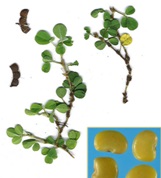
FIG. 1: DESMODIUM TRIFLORUM PLANT
MATERIAL AND METHODS: The present section deals with the detailed description of various methods and techniques employed for carrying out different studies categorized into the following heading.
Collection and Authentication of the Plant Leaves: The leaves of Desmodium triflorum were collected from outfield medicinal garden near to Gwalior (Madhya Pradesh) that show the green color with the rough surface. The plant leaves were washed thoroughly in tap water, dried in the shade, finely powdered and used for extraction. The plant was identified, and herbarium specimen was submitted in the Department of Pharmacognosy for future references.
Extraction of Plant Material: Extraction is the separation of medicinally active portions of plant tissues using selective solvents through standard procedures. The extraction was done by following the general procedure. Powdered material (leaves) was packed in soxhlet apparatus. The drug was defatted with petroleum ether (60-80 °C) for about 30-35 complete cycles. Defatted material was subjected to further extraction process by dichloromethane, methanol, and water. All the extracts were concentrated under vacuum. After completion of the total, the extracted powder was discarded and the extracts so obtained were further processed. The excess solvent in the extracts was removed by distillation and the concentrated extracts so obtained were also dried at a temperature not exceeding 40 ºC in the water bath. The extracts were then collected kept in Petri dish and stored in desiccators at room temperature. The yield values and other physical properties were observed 10.
The % Yield of the Petroleum ether, dichloromethane, Methanol, & Aqueous extract was calculated by using the following formula.
% Yield = Net weight of powder in gram after extraction × 100 / Total weight of leaf powder in gram taken for extraction
Determination of Physico-Chemical Parameters: Various physicochemical parameters were analyzed for the confirmation of identity & purity. The extractive values with alcohol and water were also determined.
Moisture Content: Moisture is an inevitable component of crude drugs, which must be eliminated. The moisture content should be minimized to prevent decomposition of crude drugs either due to chemical changes or microbial contamination.
The powdered sample of H. rosa-sinensis and C. gigantea was weighed accurately and kept in IR moisture balance. The loss in weight was recorded as percentage moisture concerning an air-dried sample of crude drug 10-11.
% Moisture = (Fw - Pw) × 100 / W
Where:
Fw = Final constant weight of drug along with the container
Pw = Weight of empty container
W = Total weight of drug taken
Ash Values:
I. Total Ash Value: For the determination of total ash, 2 gm of the air-dried crude drug was weighed in the tarred silica dish and incinerated at a temperature 450°C until free from carbon in Muffle furnace and then was cooled and weighed. The residue was collected on an ashless filter paper and then incinerated until the residue is white or nearly so. The percentage of ash was calculated concerning the air-dried drug.
II. Acid-Insoluble Ash Value: The ash obtained from the previous process was boiled with 25 ml of 2M HCI for 5 min. and the insoluble matter was collected on ash-less filter paper and was washed with hot water, ignited, cooled in a desiccator and weighed. Percentage of insoluble acid ash was calculated concerning the air-dried drug.
III. Water Soluble Ash: The ash was boiled with 25ml of water for 5 min. and the insoluble matter was collected on ash-less filter paper and was washed with hot water, ignited for 15 min. at a temperature not exceeding 450 °C. The weight of the insoluble matter was subtracted from the weight of the ash, and this represents the water-soluble ash. Percentage of water-soluble ash was calculated with reference to the air-dried drug 10-13.
Phytochemical Screening: Preliminary phyto-chemical screening was performed for all the extracts 14-15.
Detection of Carbohydrate: 500 mg of extract was dissolved in 5 ml of distilled water and filtered. The filtrate was used to test the presence of carbohydrates.
Molisch’s Test: To 1 ml of filtrate, 2 drops of Molisch’s reagent was added in a test tube, and 2 ml of concentrated sulphuric acid added carefully along the side of the test tube. Formation of a violet ring at the junction indicates the presence of carbohydrate.
Fehling’s Test: To 1 ml of filtrate, 4 ml of Fehling’s solution was added in a test tube and heated for 10 min in a water bath. Formation of red precipitate indicates the presence of reducing sugar.
Detection of Glycosides: 0.5 gm of the extract was hydrolyzed with 20 ml of dilute hydrochloric acid (0.1 N) and filtered. The filtrate was used to test the presence of glycosides.
Modified Borntrager’s Test: To 01 ml of filtrate, 02 ml of 1% ferric chloride solution was added in a test tube and heated for 10 minutes in a boiling water bath. The mixture was cooled and shaken with an equal volume of benzene. The benzene layer was separated and treated with half of its volume of ammonia solution. Formation of rose pink or cherry color in the ammonia layer indicates the presence of glycoside.
Killer Killian Test: A small portion from the respective extracts was shaken with 1 ml glacial acetic acid containing a trace of ferric chloride. 1 ml of conc. Sulphuric acid (H2SO4) was added carefully by the sides of the test tube. A blue color in the acetic acid layer and red color at the junction of the two liquids indicate the presence of glycosides.
Detection of Alkaloids: 0.5 gm of the extract was dissolved in 10 ml of dilute hydrochloric acid (0.1 N) and filtered. The filtrate was used to test the presence of alkaloids.
Mayer’s Test: Filtrates were treated with Mayer’s reagent; formation of yellow cream colored precipitate indicates the presence of alkaloids.
Dragendorff’s Test: Filtrates were treated with Dragendroff’s reagent; formation of red colored precipitate indicates the presence of alkaloids.
Hager’s Test: Filtrates were treated with Hager’s reagent; formation of yellow colored precipitate indicates the presence of alkaloids.
Detection of Phytosterols and Triterpenoids: 0.5 gm of the extract was treated with 10 ml chloroform and filtered. The filtrate was used to test the presence of phytosterols and triterpenoids.
Salkowaski Test: To the test, extract solution added few drops of conc. H2SO4 shaken and allowed to stand, the lower layer turns reddish brown or golden yellow indicating the presence of triterpenes.
Detection of Protein and Amino Acid: 100 mg of each extract was taken in 10 ml of water and filtered. The filtrate was used to test the presence of protein and amino acids.
Millon’s Test: 2 ml of filtrate was treated with 2 ml of Million’s reagent in a test tube and heated in a water bath for 5 min, cooled and added a few drops of Sodium Nitrate solution. Formation of white precipitate, which turns to red upon heating, indicates the presence of proteins and amino acid.
Ninhydrin Test: To 2 ml of filtrate, 0.25% Ninhydrin reagent was added in a test tube and boiled for 2 minutes. Formation of a blue color indicates the presence of amino acids.
Biuret Test: 2 ml of filtrate was treated with 2 ml of 10% sodium hydroxide solution in a test tube and heated for 10 min. A drop of 7% copper sulphate solution was added in the above mixture. Formation of purplish violet color indicates the presence of proteins.
Detection of Fixed Oils and Fats:
Oily Spot Test: One drop of each extract was placed on filter paper, and the solvent was allowed to evaporate. An oily stain on filter paper indicates the presence of fixed oil.
Detection of Phenolics and Tannins: 100 mg of each extract was boiled with 1 ml of distilled water and filtered. The filtrate was used for the following tests.
Ferric Chloride Test: To 2 ml of filtrate, 2 ml of 1% ferric chloride solution was added in a test tube. Formation of bluish black color indicates the presence of the phenolic nucleus.
Lead Acetate Test: To 2 ml of filtrate, few drops lead acetate solution was added in a test tube. Formation of yellow precipitate indicates the presence of tannins.
Detection of Flavonoids:
Alkaline Reagent Test: To 100 mg of extract, few drops of sodium hydroxide solution were added in a test tube. Formation of intense yellow color that becomes colorless on the addition of a few drops of dilute acid (HCl) indicates the presence of Flavonoids.
Detection of Saponin:
Foam Test: Extracts were diluted with distilled water to 20 ml and Shaken in a graduated cylinder for 15 min. Formation of one cm layer of foam indicates the presence of Saponin.
Detection of Mucilage: 10 ml of the aqueous extract was tested for mucilage; the extract was added with 25 ml of 95% alcohol with constant stirring. The so formed precipitate was centrifuged and washed with alcohol, the dissolved in water (10 ml) and reprecipitated. After washing the precipitate was collected & dried in desiccators. On addition of a drop of water and allowed to stand for some time, it swelled to give a viscous mass which indicated the presence of mucilage.
Chromatographic Study:
TLC of Methanolic Extract of Desmodium triflorum: TLC for the separation of various bioactive compounds from bioactive extract, the methanolic extract was developed to find out the probable number of compounds present in them. On the pre-coated TLC plate, test samples (after dissolving in respective solvents) were applied in the form of spots with the help of fine capillary. Spots were marked on the top of the plate for their identification. Rectangular glass chambers were used for chromatography. To avoid insufficient chamber saturation and undesirable edge effect, a smooth sheet of filter paper was placed in the TLC chamber and was allowed to be in the developing solvent. A number of developing solvent systems were tried during the study. Each time plate was sprayed with Vanillin sulphuric acid and heated at 115 ºC for 5 min. The solvent system in which there is a satisfactory resolution was taken as a final solvent system. Solvent systems; Ethyl acetate: methanol: water (76:20:4) was found to be the most satisfactory solvent system. After the development of plates, they were air-dried and a number of spots, color and Rf values were recorded 10.
Rf value = Distance traveled by solute/Distance travelled by solvent
RESULTS AND DISCUSSION: Successive solvent extraction values in various organic solvent were observed as n-hexane, dichloromethane, methanol, water as shown in Table 1. The proximate analysis revealed the moisture content, total ash, insoluble acid ash, & water soluble ash values were observed to be 11.5%, 3.1%, 9.2%, 5.7%, respectively as shown in Table 2. The preliminary phytochemical analysis revealed that different active constituent present in different extracts such as carbohydrates, proteins, amino acids, fat, oils, steroids, terpenoids, glycosides, alkaloids, tannins and other phenolic compounds as shown in Table 3.
TABLE 1: DIFFERENT EXTRACTS WITH THEIR APPEARANCE AND % YIELD (IN gm)
| Extracts | The color of dried extracts | The consistency of dried extracts | % Yield (w/w) |
| n-hexane extract of Desmodium triflorum | Dark Brown | Sticky | 12 % |
| Dichloromethane extract of Desmodium triflorum | Dark Green | Dried Powder | 18 % |
| Methanolic extract of Desmodium triflorum | Dark Orange | Sticky | 10 % |
| Water extract of Desmodium triflorum | Dark Brown | Resinous | 4 % |
TABLE 2: EVALUATION OF PHYSICOCHEMICAL PARAMETER
| S. no. | Parameters | Values obtained (%w/w) dry weight basis |
| 1 | Moisture content | 3.9 |
| 2 | Total ash | 9.7 |
| 3 | Acid-insoluble ash | 3.4 |
| 4 | Water-soluble extractive | 5.2 |
TABLE 3: QUALITATIVE CHEMICAL ANALYSIS OF DESMODIUM TRIFLORUM BY CHEMICAL TESTS
| S. no. | Test | n-hexane | Dichloromethane | Methanolic | Aqueous |
| 1 | Carbohydrate
Molish test Felling test |
- - |
- - |
- - |
+ + |
| 2 | Glycosides
Bronteger test |
- |
- |
+ |
+ |
| 3 | Alkaloid
Mayer test Hager test |
- - |
+ + |
+ + |
- - |
| 4 | Phytosterol + Triterpenoids
Salkowaski test |
- |
+++ |
- |
- |
| 5 | Protein + Amino acid
Biuret test Ninhydrin test |
- - |
- - |
- - |
- - |
| 6 | Phenolic test
Ferric test Lead acetate test |
- - |
- - |
+ + |
+ + |
| 7 | Flavonoids
Alkaline test |
- |
- |
+ |
+ |
| 8 | Saponin
Foam test |
- |
- |
- |
- |
Note: + ve indicates a positive result, whereas – ve indicates a negative result
TLC of Methanolic Extract of Desmodium triflorum: TLC study had shown the presence of different components present in the methanolic extract of Desmodium triflorum when the extracts were run in a specific solvent system. Before reaching to the most optimum solvent system, a number of systems were employed as shown in Table 4, Fig. 2.
TABLE 4: SUMMARY OF TLC
| S. no. | Fractions | Solvent systems | Detecting reagents | Color | No. of spots | Rf value of
Spots |
| 1 | Butanolic | Ethyl acetate: methanol: water (76:20:4) | Vanillin sulphuric acid, heated at 110 ºC for 5 Min. | Green & yellow | 6 | 0.20, 0.30, 0.53, 0.58, 0.68, 0.72 |
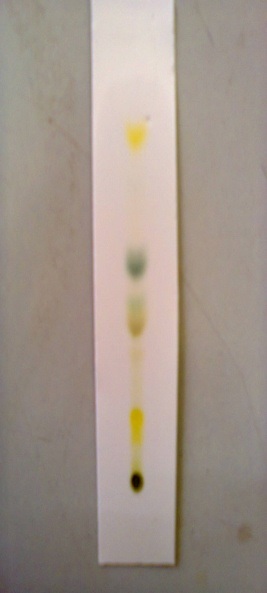
FIG. 2: TLC OF METHANOLIC EXTRACTS
CONCLUSION: The preliminary pharma-cognostic and phytochemical analysis revealed the successive solvent extraction value in different solvents, moisture content, total ash, acid insoluble ash, sulfated ash, & water soluble ash, different active constituent present in different extracts such as carbohydrates, proteins, amino acids, fat, oils, steroids, terpenoids, glycosides, alkaloids, tannins and other phenolic compounds, and TLC study of methanolic extract has shown the different components present in extract.
ACKNOWLEDGEMENT: The work was supported by the Department of Pharmacology, G R Medical College, Gwalior M.P. India. I would like to thanks Dr. S.C. Mehta for providing a platform to carry out this work.
CONFLICT OF INTEREST: Nil
REFERENCES:
- Kirtikar KR and Basu BD: Indian medicinal plants, National book distributors, Dehradun 1995; 1: 335-336.
- Ghosal S, Srivastava RS, Banerjee PK and Dutta SK: Alkaloids of Desmodium triflorum. Phytochemistry 1971; 10(12): 3312-3313.
- Khare CP: Indian medicinal plant: An Illustrated Dictionary, Springer-Verlag Heidelberg publication 2007;
- Lai SC, Ho YL, Huang SC, Huang TH, Lai ZR, Wu CR, Lian KY and Chang YS: Am J Chin Med 2010; 38(2): 329-42.
- Lai SC, Peng WH, Huang SC, Ho YL, Huang TH, Lai ZR and Chang YS: Am J Chin Med 2009; 37(3): 573-88.
- Raj RK: Indian J Physiol Pharmacol 1975; 19(1).
- Bhosle V: Rev Bras Farmacogn Braz J Pharmacogn 2013; 23(24).
- Ethnobotanical Leaflet 2008; 12: 227-230.
- Daya RW:International Research Journal of Pharmacy 2011; 2(7): 120-123.
- Mukherjee PK: “Quality Control of Herbal Drugs”, Business Horizons Pharmaceutical Publishers, 1st, 2002: 186-189, 193, 256-370.
- Khandelwal SK: “Practical Pharmacognosy”, Vth Edition, Nirali Prakashan 2003: 149-155, 157-8, 159.
- “Quality Control methods for medicinal plant materials,” World health organization, Genera 1998: 28-9, 30.
- “Indian Pharmacopoeia,” IV Edition, Vol-II, Controller of Publications, Govt. of India, New Delhi 1996: A-54.
- Kokate CK: Practical Pharmacognosy. Delhi, Vallabh Prakashan 1996: 107-111.
- Khandelwal KR: Practical Pharmacognosy. Pune, Nirali Prakashan 2006: 149-155, 157- 159.
How to cite this article:
Singh N, Tailang M and Mehta SC: Pharmacognostic and Phytochemical Screening of Desmodium Triflorum Linn. Int J Pharmacognosy 2016; 3(1): 43-49. doi: 10.13040/IJPSR.0975-8232.3(1).43-49.
This Journal licensed under a Creative Commons Attribution-Non-commercial-Share Alike 3.0 Unported License.
Article Information
7
43-49
664
2429
English
IJP
N. Singh*, M. Tailang and S. C. Mehta
Department of Pharmacology, G R Medical College, Gwalior, Madhya Pradesh, India.
namrata.singhms@gmail.com
20 October 2015
28 December 2015
24 January 2016
10.13040/IJPSR.0975-8232.IJP.3(1).43-49
31 January 2016