


OCCURRENCE OF CALCIUM OXALATE CRYSTALS IN THE LEAVES OF MEDICINAL PLANTS
HTML Full TextOCCURRENCE OF CALCIUM OXALATE CRYSTALS IN THE LEAVES OF MEDICINAL PLANTS
R. Anitha * and T. Sandhiya
Department of Plant Biology and Plant Biotechnology, Ethiraj College for Women, Ethiraj Salai, Egmore, Chennai - 600008, Tamil Nadu, India.
ABSTRACT: Calcium oxalate crystals have a definite biological function in medicinal plants and act as a good diagnostic tool for the identification and detection of adulterants in crude drugs. The occurrence of calcium oxalate crystals was evident in Moringa pterygesperma, Mentha arvensis, Cissus quadrangularis, Murraya koenigii and Amaranthus gangiticus leaves. Rosette, druse, raphides, acicular and prismatic crystals were recorded. The presence of calcium oxalate crystals was confirmed; further Cissus quadrangularis leaves had both druse and raphides belonging to type-II. Druse in Amaranthus gangiticus was the largest, measuring about 12.98-18.82 µm in length, 9.74-14.28 µm in breadth in the light microscope and 22.1 µm Scanning Electron microscopy. Mentha arvensis leaves showed acicular crystals as well as druse in the Scanning Electron Microscope observation.
| Keywords: |
Calcium oxalate crystals, Raphides, Druse, Acicular, Rosette, Medicinal plants
INTRODUCTION: Calcium oxalate is the most abundantly present biomineral in higher plants. The crystals of calcium oxalate are found in a variety of defined shapes in higher plants such as raphide druse, styloid, prismatic and sand crystals 1. Calcium oxalate occurs in different plants tissue including leaves, stems, roots, seeds 2, ovaries, anther and petals 3. The only place where the crystals have not been seen is the pollen. Calcium oxalate crystals are widely distributed in the plant kingdom and found in over 215 families 4. The type of crystal is genetically determined 5; however, the quantity of crystals is determined by several environmental factors like light density 6,
Herbivory and Ca existence 4. Hence, the distribution and the shape of these crystals are characteristic of each plant family 4. They seem to play an important role in the protection against herbivory 4, storage of calcium and oxalic acid, regulation of calcium levels in plant tissues 5, influence the photosynthetic process 6, give mechanical support 7 and also detoxification of heavy metals 1 in the plant tissue. Recent studies on calcium oxalate crystals in biological functions revealed that they are stored during the depositions for the secondary metabolites or metabolic ions.
Calcium oxalate crystals have diagnostic value, the presence and absence of crystals; their dimensions are of great importance in the identification of the crude drugs. It also helps in the detection of adulterants in herbal drug and in taxonomical identification. Since, the availability of literature on calcium oxalate crystals in medicinally important leaves is not abundant, present work was undertaken.
MATERIALS AND METHODS:
Plant Selection: Twenty different medicinal plants were randomly selected and screened for the presence of calcium oxalate crystals. They were Amaranthus gangeticus (Amaranthaceae), Mentha arvensis (Lamiaceae), P. niruri (Euphorbiaceae), Murraya koenigii (Rutaceae), Ocimum sanctum (Lamiaceae), Lawsonia inermis (Lythraceae), Solanum nigrum (Solanaceae), Mirablis jalapa (Nyctaginaceae), Tridax procumbens (Asteraceae), Leucas aspera (Lamiaceae), Stenolobium stanza (Bignoniaceae), Hibiscus kannabinus (Malvaceae), Coleus aromaticus (Lamiaceae), Hydrocotyle asiatica (Apiaceae), Alternanthera sessilis (Amaranthaceae), Solanum trilobatum (Solanaceae), Moringa ptergesperma (Moringaceae) Cissus quandrangularis (Vitaceae), Coriandrum sativum (Apiaceae) and Clitoria ternatea (Fabaceae).
Powder Analysis: The leaves of the selected plants were taken, washed thoroughly and shade dried for few days. The dried leaves were grounded using a mortar and pestle. The powder thus produced was stored in an airtight container. A small quantity of powder was taken in a glass of slide and observed under a light microscope for the presence of calcium oxalate crystals. The shape of the crystal was observed. All the selected plant materials were observed for the presence of calcium crystals.
The Conformation of Calcium Oxalate Crystals: The shape and appearance of the crystals were noted. The dried powder of the selected plant was to treated with the concentrated sulphuric acid and observed for the formation of calcium sulphate needles. Those plants which showed the presence of calcium oxalate crystals alone was chosen for the further study.
Transverse Section of the Leaf: The leaves of the medicinal plants which were positive for the calcium oxalate test were taken, then washed properly and cut into small bits. A transverse section of the leaf was treated with sodium hypochlorite for bleaching, stained with safranin and observed for the presence of calcium oxalate crystals and photographed.
The Dimension of the Calcium Oxalate Crystals: The light microscope was calibrated at10X using an Ocular and Stage micrometry.
Value of 1 division = Stage Division ×10 / Ocular division. Divisions that coincided were taken and used for calibrating of the microscope. The section of the selected medicinal plants was observed in the microscope using an ocular. The length and breadth of the crystals are measured, and an average was taken.
Scanning Electron Microscopy of Calcium Oxalate Crystals: The leaf section of Murraya koenigii, Amaranthus gangeticus, Cissus quadrangularis and Mentha arvensis, was mounted on a stub and observed in Hitachi S340010. Scanning electron microscope and photograph.
RESULTS AND DISCUSSION:
Powder Analysis: Screening the dry powder of the 20 medicinal plants revealed that only 5 plants were positive for the presence of calcium oxalate crystals. They were Murraya koenigii, Moringa pterygesperma, Cissus quadrangularis, Mentha arvensis and Amaranthus gangeticus. The occurrence of calcium oxalate crystals in these medicinal plants has a definite role in its medicinal activity. It is reported that the presence of calcium oxalate crystals increases the antioxidant property of medicinal plants apart from being useful in treatment of fractures and bone ailments.
The Conformation of Calcium Oxalate Crystals: Upon adding concentrated sulphuric acid, the calcium oxalate crystals dissolve and reforms crystals of calcium sulphate. This principle was utilized in the conformation of calcium oxalate crystals. Needles of calcium sulphate were observed. Cissus quadrangularis showed fine needles, Murraya koengii had flat, broad crystals, whereas Moringa pterygesperma showed stellate crystals, Mentha arvensis showed a tuff of fine needles as a bundle and Amaranthus gangeticus had forked, flattened needles.
Transverse Section of the Leaf: The transverse section of the leaf of the selected medicinal plant revealed the distribution of calcium oxalate crystals. Murraya koenigii leaf section showed hexagonal prismatic crystal in the parenchyma cells below and above the lower and upper epidermis, and it was evident near the vascular bundle as well. Moringa pterygesperma had a large number of small druse in idioblast cells.
Druses were grey and abundantly present in the lower epidermis. Cissus quadrangularis leaf had abundant raphides and druse. The raphides and druse were dull greys in color. The section of the plant was observed, and the plant had raphides in idioblast the lower epidermis of the plant. Druse was evident near the vascular bundle. Larger druses were evident in Amaranthus gangeticus along the mesophyll cells. Druse was predominantly in the central vascular bundle. Many small druses were seen in the lower epidermis in Mentha arvensis Fig. 1.
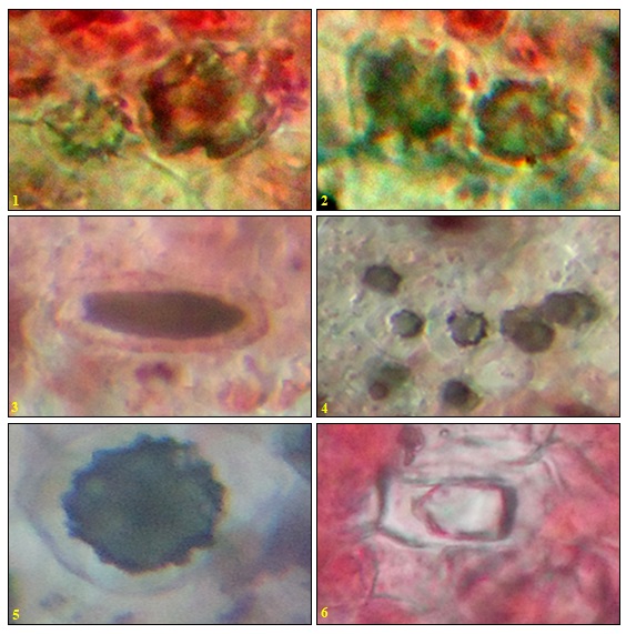
FIG. 1: T. S. OF LEAF SHOWING CALCIUM OXALATE CRYSTALS IN LIGHT MICROSCOPY AT 100X. 1-2; MORING PTERGESPERMA, 3-4; CISSUS QUADRANGULARIS, 5- AMARANTHUS GANGETICUS, 6-MURRAYA KOENIGII
The Dimension of the Calcium Oxalate Crystals: The calcium oxalate crystal observed in the transverse section of the leaf was measured in ocular and stage micrometry. It was found that Murraya koenigii leaf had hexagonal crystals of length 6.49-10.38 µm and breadth 6.492-8.44 µm. Mentha arvensis leaf showed many rosette whos length was 6.49-11.48 µm and breadth 6.49-11.68 µm. Amaranthus gangeticus leaf had druse measuring 12.98-18.82 µm in length and breadth of 9.74-14.28 µm. Cissus quarangularis leaf had raphides of length 32.47-37.66 µm and breadth of 6.43-12.33 µm. Moringa pterygesperma leaf also had rosette length was 12.98-18.17 µm and breadth 6.49-9.73 µm.
Scanning Electron Microscopy of Calcium Oxalate Crystals: The Scanning Electron Micrographs revealed that Amaranthus leaf had a large druse measuring 22.1 µm and Murraya exhibited rosette of 16.8 µm; while Cissus quadrangularis had a rosette of 25 µm, druse of 15.4 µm and raphides of 69.7 µm. Mentha arvensis had acicular crystals and hexagonal crystals measuring 30-50 µm. The acicular crystals had a pointed end and a forked opposite end. This is characteristic of Type-II raphides that are commonly seen in Vitaceae family Fig. 2.
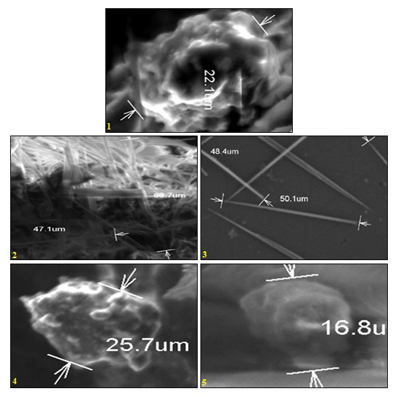
FIG. 2: SCANNING ELECTRON MICROGRAPHS OF CALCIUM OXALATE CRYSTALS. 1 & 2- DRUSE AND RAPHIDES IN CISSUS QUADRANGULARIS, 3- ACICULAR CRYSTALS IN MENTHA ARVENSIS, 4- DRUSE IN AMARANTHUS GIGANTECUS, 5- ROSETTE IN MURRAYA KOENIGII
DISCUSSION: Distribution of calcium oxalate crystals is comparatively more common in the leaves than in stems. Earlier it is reported in species of Moraceae 8, Prunus sp. 9 Crataegus 10 and Aster squamates 11. They are generally distributed in the epidermis, mesophyll, and vascular tissue. In the present study it was evident that druse was present in specialized idioblast cells in mesophyll region in Moringa pterygesperma, Cissus quadrangularis, Amaranthus gangeticus and in Mentha arvensis, it was found above the lower epidermis.
A similar observation was made in mesophyll cells of A. squamates. They occurred both in epidermis and mesophyll cells of Conyza spp. of the Asteraceae tribe 12. Druse provides mechanical strength to the leaf tissue and moreover they also regulate the calcium levels in the tissue. This is an important function in the medicinal leaves since the medicinal property could be attributed to the presence of these crystals. Rosette crystals were reported Munronia pinnata and needle in Andrographis paniculata 13.
In the present study, Moringa pterygesperma had rosette crystals in the lower epidermis region. The raphides are crystal formed in vacuoles of the specialized cells or idioblast that are scattered in otherwise homogeneous cell types such as the mesophyll cells in the leaves. Generally, idioblast contains only one crystal type raphide or druse. The raphide crystal idioblast is concentrated at the lower portion of the leaf in tissue partitions between aerenchyma air spaces whereas the druse crystal idioblast is usually found at the upper portion of the leaf in compact chlorenchyma above the aerenchyma. In Cissus quadrangularis both druse and raphides were observed; however, they appeared in separate idioblast cells distributed in mesophyll cells and above the lower epidermis respectively. Rosette, acicular, raphides and prismatic crystals of calcium oxalate are very common in Cissus quadrangularis Linn., Vitis trifolia Linn., Cissus repanda & Vitis vinifera Linn. 14 Panchvalkala is an ayurvedic preparation containing plant species belonging to Malvaceae and Moraceae. The powder analysis of these showed the presence of rosette & prismatic crystals 15. Presence or absence of different crystal types in the plant may, therefore, represent a useful taxonomic character in some groups.
CONCLUSION: The occurrence of calcium oxalate crystals among the selected twenty medicinal plants was only 25% belonging to Amaranthaceae, Lauraceae, Vitaceae, Rutaceae, and Moringaceae. It is concluded that the occurrence of calcium oxalate crystals in these medicinal plants serves as a diagnostic tool for taxonomic identification and purity of the plant sample.
ACKNOWLEDGEMENT: The authors wish to thank the principal and The Head of the Department for their laboratory facility and encouragement.
CONFLICT OF INTEREST: Nil
REFERENCES:
- Nakata PA: Advances in our understanding of calcium oxalate crystal formation and function in plants. Plant Sci 2003; 164(6): 901-909.
- Ilarslan H, Palmer RG and Horner HT: Calcium oxalate crystals in developing seeds of soybean. Ann Bot 2001; 88: 243-257
- Meric C and Dane F: Calcium oxalate crystals in floral organs of Helianthus annuus and H. tuberosus L. (Asteraceae). Acta Biol Szeged 2004; 48(1-4): 19-23.
- Molano-Flores B: Herbivory and calcium concentrations affect calcium oxalate crystal formation in leaves of Sida (Malvaceae). Ann Bot 2001; 88(3): 387-391.
- Franceschi VR and Nakata PA: Calcium oxalate in plants: formation and function. Annual Rev Pl Biol 2005; 56(1): 41-71.
- Kuo-Huang LL, Ku MSB and Franceschi VR: Correlations between calcium oxalate crystals and photosynthetic activities in palisade cells of shade-adapted Peperomia glabella. Bot Stud 2007; 48(2): 155-164.
- Franceschi VR and Horner HJ: Calcium oxalate crystals in plants. Bot Rev 1980; 46: 361-427.
- Wu CC and Kuo-Huang LL: Calcium crystals in the leaves of some species of Moraceae. Bot Bull Acad Sin 1997; 38(2): 97-104.
- Lersten NR and Horner HT: Crystal macro pattern development in Prunus serotina (Rosaceae, Prunoideae) leaves. Ann Bot 2006; 97(5): 723-729.
- Demiray H: Calcium oxalate crystals of some Crataegus (Rosaceae) species growing in Aegean region. Biologia 2007; 62(1): 46-50.
- Meric C: Calcium oxalate crystals in Aster squamatus and Bellis perennis (Asteraceae: Astereae) Phytologia Balcanica 2009; 15(2): 255-259.
- Meric C: Calcium oxalate crystals in Conyza canadensis (L.) Cronq. and Conyza bonariensis (L.) Cronq. (Asteraceae: Astereae). Acta Biol Szeged 2008; 52(2): 295-299.
- Dharmadasa RM, Samarasinghe K, Adhihetty P and Hettiarachchi PL: Comparative pharmacognostic evaluation of Munronia pinnata (Wall.) Theob. (Meliaceae) and its substitute Andrographis paniculata (Burm.f.) Wall. Ex Nees (Acanthaceae). World Journal of Agricultural Research 2013; 1(5): 77-81.
- Hemant and Harisha CR: Importance of calcium oxalate in family Vitaceae. Int. Journal of Comprehensive Pharmacy 2012; 3(2): 1-3.
- Vyas P and Prajapati H: Importance of calcium oxalate crystals in panchavalkala. Int J Pharmacognosy and Phytochemical Res 2012; 4(3): 112-116.
How to cite this article:
Anitha R and Sandhiya T: Occurrence of calcium oxalate crystals in the leaves of medicinal plants. Int J Pharmacognosy 2014; 1(6): 389-93. doi link: http://dx.doi.org/10.13040/IJPSR.0975-8232.IJP.1(6).389-93.
This Journal licensed under a Creative Commons Attribution-Non-commercial-Share Alike 3.0 Unported License.
Article Information
6
389-393
473
4821
English
IJP
R. Anitha * and T. Sandhiya
Department of Plant Biology and Plant Biotechnology, Ethiraj college for women, Ethiraj salai, Egmore, Chennai, Tamil Nadu, India.
anitha.rajasekaran023@gmail.com
29 April 2014
19 May 2014
29 May 2014
http://dx.doi.org/10.13040/IJPSR.0975-8232.1(6).389-93
01 June 2014