


NEUROPROTECTIVE ACTIVITY OF ASCOPHYLLUM NODOSUM ON EXPERIMENTLLY INDUCED GLOBAL CEREBRAL ISCHEMIA/REPERFUSION INJURY IN MICE
HTML Full TextNEUROPROTECTIVE ACTIVITY OF ASCOPHYLLUM NODOSUM ON EXPERIMENTALLY INDUCED GLOBAL CEREBRAL ISCHEMIA/REPERFUSION INJURY IN MICE
Mohan Pankaj Dhyani *, Arun Kumar, Preeti Kothiyal and Alka N. Choudhary
Shri Guru Ram Rai Institute of Technology, Patel Nagar, Dehradun - 248121, Uttarakhand, India.
ABSTRACT: Background: Ascophyllum nodosum has been used in oriental medicine to treat a variety of remedies, including some neurological disorders. Ascophyllum nodosum rich in phenolic antioxidant (rich in polyphenol) have been identified as major active component exhibiting antioxidant, anti-inflammatory and neuroprotective effects. The present study is to evaluate the neuroprotective effect of Ascophyllum nodosum on experimentally induced Global Cerebral Ischemia/Reperfusion Injury in mice. Material and Methods: Neuroprotective activity was carried out by global cerebral ischemia on Swiss albino mice by carotid artery occlusion for 17 min followed by 24 h reperfusion. Then after 24 h blood sample was collected by retro-orbital route and after that mouse was sacrificed by cervical dislocation under light area, the brain was removed for various biochemical analyses. Results: Ascophyllum nodosum hydroalcholic extract showed dose-dependent neuroprotective activity by a significant decrease in lipid peroxidation (LPO), lactate dehydrogenase (LDH) and serum nitrite level in extract treated the group as compared to the ischemia/reperfusion group. Cerebral infraction area was significantly reduced in extract treated groups as compared to ischemia/reperfusion group. Conclusion: In the present investigation, a polyphenolic fraction of A. nodosum has shown promise as a Cerebral Ischemia/Reperfusion Injury curing, as well as enhancing agents in mice in the entire laboratory model employed. Furthermore, the polyphenolic extract of A. nodosum was found more effective than against global Cerebral Ischemia/Reperfusion Injury.
| Keywords: |
A. nodosum, Polyphenol, Lipid peroxidation, Lactate dehydrogenase
INTRODUCTION: Stroke is a term used to describe an abrupt-onset focal neurological deficit that lasts 24 h and is of presumed vascular origin 1. Cerebral ischemia is caused by a reduction in blood flow that lasts longer than several seconds.
Neurological symptoms are manifest within seconds because neurons lack glycogen, so energy failure is rapid. If the cessation flow lasts for more than a few min, infarction or death of brain tissue results 2. Stroke can be either ischemic or hemorrhagic (80% and 12%) respectively,
1. Ischemic strokes are due either to local thrombus formation or to the embolic phenomenon, resulting in occlusion of the cerebral artery.
2. Hemorrhagic strokes: these types of hemorrhages very often are associated with uncontrolled high blood pressure and sometimes antithrombotic or thrombolytic therapy 1. Stroke is a major health problem in India. The average annual incidence rate of strokes in a recent study from India was 145 per 100,000 populations, which compares well with the developed countries. Stroke burden has been rising in India as compared to the developed countries where it has reached a plateau or decreased 3. Natural product (especially the medicinal plants) probably represents an ideal source to develop safe and effective agents for the management of stroke and deserve scientific probe.
Oxidative stress is believed to be one of the mechanisms taking part in the neuronal damage of stroke 4. Oxidative stress is a state of imbalance between free radical production, in particular, reactive oxygen species (ROS), and the ability of the organism to defend against them, leading to progressive oxidative damage. It is assumed that oxidative stress contributes to the initiation and development of stroke via different interrelated mechanisms: excitotoxicity resulting in cellular enzyme activation and ROS generation; inflammation leading to leukocyte priming and activation and accompanied by an excessive radical production; activation and oxidative damage of endothelium resulting in reduced bioavailability of nitric oxide (NOS); free radical-mediated hyperhomocysteinemia; lipid peroxidation of plasma and cellular components including those in the arterial vessel wall and macrophages, processes each one of which may exacerbate oxidative damage through mechanisms of positive feedback4.
Polyphenol is natural substances with variable phenolic structures and is elevated in vegetables, fruits, grains, bark, roots, tea, and wine. In addition, to their well-known antioxidant effects, select polyphenol also have insulin-potentiating, anti-inflammatory, anti-carcinogenic, anti-viral, anti-ulcer, and anti-apoptotic properties 5. A significant interest on the protective effects of polyphenol has principally been because of their antioxidant properties 6, 7. Phenolic antioxidants have been shown to inhibit the oxidation of lipids and other molecules and protect against free radicals 8.
Ascophyllum nodosum was commonly known as seaweed. The seaweed is enriched in the polyphenolic compound. The extract of A. nodosum comprises at least about 20% by weight of the polyphenolic compound. The highly polymeric phenolic components from Ascophyllum nodosum demonstrated antioxidant and ant diabetic properties 9. Resveratrol is a natural polyphenol found in grapes and wine and has been associated with protective effects against cardiovascular diseases. Resveratrol protects the heart, brain, and kidney from ischemia-reperfusion injury 10.
The purpose of the present study is to know the safe and potent neuroprotective effect of Ascophyllum nodosum against global cerebral ischemia/reperfusion injury in mice.
MATERIAL AND METHODS:
Drugs and Chemicals: Trichloroacetic acid (TCA), 2-thiobarbituric acid (TBA), Triphenyl-tetrazolium chloride (TTC) etc. available from Sigma Aldrich and resveratrol was procured from Zenith nutrition Pvt. Ltd., Bangalore.
Plant Material: In the present study, the ex-gratia of seaweed was provided by Shri Vinayaka trading company, Hosur, Tamil Nadu. Before extraction of a polyphenolic fraction of A. nodosum, the lipophilic component was removed by extracting powdered drug with petroleum ether at room temperature for five days. A crude extract of A. nodosum enriched in the polyphenolic compound was prepared by extracting residue with aqueous ethanol (90:10) solution at room temperature for extraction time of seven days. The crude extract was then concentrated in a rotatory evaporator. The percent yield from (90:10) hydroalcoholic solvent was 18.96%.
The presence of polyphenol was confirmed by ferric chloride test for tannins. Addition of ferric chloride solution to A. nodosum extract gave bluish-black color, confirming the presence of polyphenol (tannins) in the extract.
Animals: Swiss albino mice of either sex weighing 20 ± 5 g maintained on the standard laboratory diet and water ad libitum were employed in the present study. They were housed in the departmental animal house and exposed to 24 h of the natural light/ dark cycle.
Institutional Animal Ethical Committee (IAEC) approved the experimental protocol vide M.PH/IAEC/01/2012/ECC-9 and care of animals was as per guidelines of Committee for Control and Supervision of Experiments on Animals (CPCSEA) (Reg No.-264/CPCSEA).
Experimental Protocol: Eight groups of an animal comprising twelve animals were used.
Group 1: Control: Normal animals.
Group 2: Sham Control: Carotid artery will be exposed for 17 min.
Group 3: Negative control group: Vehicle will be administered for 7 days, then BCA will be occluded for 17 min, followed by reperfusion for 24 h.
Group 4: Disease (I/R Injury): Bilateral carotid artery occlusion (BCA) for 17 min followed by reperfusion for 24 h.
Group 5: Ascophyllum nodosum extract was administered for 7 days (Low dose 50 mg/kg orally), then BCA was occluded for 17 min, followed by reperfusion for 24 h.
Group 6: Ascophyllum nodosum extract was administered for 7 days (medium dose 100 mg/kg orally), then BCA was occluded for 17 min, followed by reperfusion for 24 h.
Group 7: Ascophyllum nodosum extract was administered for 7 days (high dose 200 mg/kg orally), then BCA was occluded for 17 min, followed by reperfusion for 24 h.
Group 8: Resveratrol was administered for 7 days (20mg/kg orally), then BCA was occluded for 17 min, followed by reperfusion for 24 h.
Induction of Global Cerebral Ischemia: Mice were anesthetized by using chloral hydrate (400 mg/kg, i.p). A midline ventral incision was made in the neck to expose the right and left common carotid arteries, which were isolated from surrounding tissue and vagus nerve. A cotton thread was passed below both the carotid arteries. Global cerebral ischemia was induced by occluding the carotid arteries. After 17 min of global cerebral ischemia, reperfusion was allowed for 24 h. The incision was sutured back in layers. The sutured area was cleaned with 70% ethanol and was sprayed with antiseptic dusting powder.
The animals were shifted individually to their home cage and were allowed to recover overnight. During surgery, the animals were kept on a heating pad to maintain the body temperature, to avoid the effect of temperature variations on the final results11.
Preparation of Post-Mitochondrial Supernatant: Following decapitation, the brain was removed and washed in cooled 0.9% saline, kept on the ice and subsequently blotted on filter paper, then weight and homogenized as 10% (w/v) in cold phosphate buffer (0.05M, pH 7.4). The homogenates were centrifuged at 10,000rpm for 20 min at 4 °C, and post-mitochondrial supernatant (PMS) was kept on ice until assayed 12.
Biochemical Estimation:
Lactate Dehydrogenase (LDH): It was estimated using a commercially available kit from Aspen laboratories.
Lipid Peroxidation (LPO): 13 The most common method of measuring MDA is based on its reaction with TBA. The TBA reactive substances (TBARs) assay is a colorimetric method used for the detection of lipid peroxidation in biological materials. MDA reacts with TBA at high temperature (90-100 °C) in acidic conditions. The reaction yields a pink MDA-TBA adduct. This colored complex can be measured by spectro-photometrically at 532 nm. 2.0 ml of the tissue homogenate (supernatant) was added to 2.0 ml of freshly prepared 10% w/v trichloroacetic acid (TCA), and the mixture was allowed to stand in an ice bath for 15 min.
After 15 min, the precipitate was separated by centrifugation and 2.0 ml of the clear supernatant solution was mixed with 2.0 ml of freshly prepared thiobarbituric acid (TBA). The resulting solution was heated in a boiling water bath for 10 min. It was then immediately cooled in an ice bath for 5 min. The color developed was measured at 532nm against reagent blank. Different concentrations (0-23nM) of standard malondialdehyde (1, 1, 3, 3-tetraethoxypropane) were taken and processed as above for standard graph. The values were expressed as nM of MDA/mg protein.
Serum Nitrite Concentration: 14, 15 Estimation of nitrite and nitrate, the stable end products of nitric oxide oxidation, is a common indirect method used to monitor nitric oxide levels in various body fluids. 100 μl of serum or standards was added to a test tube to which 400 μl of carbonate buffer was added. The tubes were then incubated at room temperature for 1 h with thorough shaking. The reaction was stopped by the addition of 100 μl of 0.35 M sodium hydroxide, followed by 400 μl of 120 mM zinc sulfate solution under vortex and the solution was allowed to stand for 10 min. The tubes were centrifuged at 4000g for 10 min. Aliquots (500 μl) of the clear supernatant were transferred to another test tube, and 250 μl 1.0% sulfanilamide (prepared in 3N HCl) and 250 μl 0.1% N-naphthylethylenediamine (prepared in water) was added with shaking. After 10 min the absorbance was measured at 545 nm against a blank containing the same concentration of ingredients but no biological sample.
Assessment of Cerebral Infarct Size: At the end of 24 h of reperfusion after global cerebral ischemia, the animals were sacrificed by decapitation, and the brain was removed. Brain samples were immediately sliced into uniform coronal sections of about 1mm thickness. The slices were incubated with 1% triphenyl-tetrazoliumchloride (TTC) at 37 °C in 0.2M Tris buffer (pH7.4) for 20 min. TTC is converted to red form zone pigment by NAD and acetate dehydrogenase and thus stained the viable cells deep red. The infracted cells lost the enzyme as well as a cofactor and thus remain unstained dull yellow. The brain slices were placed over a glass plate. A transparent plastic grid with 100 squares in 1 cm2 was placed over it. The average area of each brain slice was calculated by counting the number of squares on either side. Similarly, the number of squares falling over the non-stained dull yellow area was also counted. Infracted area was expressed as a percentage of the total brain volume. Whole brain slices were weighed. Infracted dull yellow part was dissected out and weighed. Infarct size was expressed as a percentage of the total wet weight of the brain 11.
Statistical Analysis: The result were expressed as the mean ± standard error means Statistical analysis for all result was done using a one-way ANOVA followed by Tukey’s multiple comparison tests. A value of P<0.05 was considered to be statistically significant.
RESULTS:
Effect of Ascophyllum nodosum on Cerebral Infarction Size: Global cerebral ischemia of 17 min followed by reperfusion for 24 h produced a significant increase in the cerebral infarct size in ischemia-reperfusion (I/R) injury group. A. nodosum extract (100, 200 mg/kg, p.o) and resveratrol (20 mg/kg, p.o) administered 7 days before I/R significantly decreased the I/R- induced increase in the cerebral infarct size in by volume and weight method Fig. 1.
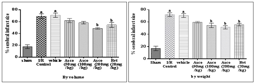
FIG. 1: EFFECT OF ASCOPHYLLUM NODOSUM ON GLOBAL CEREBRAL I/R INJURY INDUCED CEREBRAL INFARCT SIZE IN MICE
Each group (n=12) represents mean ± standard errors of means. a=p<0.001 vs. sham group, b=p< 0.01 vs. I/R and vehicle.
Effect of Ascophyllum nodosum on Global Cerebral I/R Injury Increases Lactate Dehydrogenase (LDH): Global cerebral ischemia of 17 min followed by reperfusion for 24 h produced a significant increase in LDH level in I/R injury group. Administration of Ascophyllum nodosum extract (100, 200 mg/kg, p.o) and resveratrol (20 mg/kg, p.o) for 7 days prior to I/R significantly decreased the I/R-induced increase in the LDH level in blood and Asco (200 mg/kg, p.o) was effective in reducing the LDH level and when we compared the vehicle-treated group to I/R injury group there is no significant difference, so it shows that vehicle does not have any effect Fig. 2.
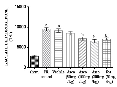
FIG. 2: EFFECT OF ASCOPHYLLUM NODOSUM ON GLOBAL CEREBRAL I/R INJURY INCREASES LACTATE DEHYDROGENASE (LDH) CONTENT
Each group (n=12) represents mean ± standard errors of means. a=p<0.001 vs. sham group, b=p< 0.01 vs. I/R and vehicle.
Effect of Global Cerebral I/R Induced Increase in Thiobarbituric Acid Reactive Substances (TBARS): Global cerebral ischemia of 17 min followed by reperfusion for 24 h produced a significant increase in TBARS level in I/R injury group. Administration of Ascophyllum nodosum extract (100, 200 mg/kg, p.o) and resveratrol (20 mg/kg, p.o) for 7 days prior to global cerebral ischemia significantly decreased the I/R-induced increase in the TBARS level in blood and Asco (200 mg/kg, p.o) was effective in reducing the TBARS level and when we compared the vehicle-treated group to I/R injury group there is no significant difference, so it shows that vehicle does not have any effect Fig. 3.
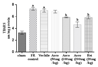
FIG. 3: AFFECT OF ASCOPHYLLUM NODOSUM ON GLOBAL CEREBRAL ISCHEMIA I/R INJURY INDUCED INCREASES IN THIOBARBITURIC ACID REACTIVE SUBSTANCES (TBARS)
Each group (n=12) represents mean ± standard errors of means. a=p<0.001 vs. sham group, b=p< 0.01 vs. I/R and vehicle.
Effect of Ascophyllum nodosum on Global Cerebral Ischemia-Induced Increases in Serum Nitrite Level: Global cerebral ischemia of 17 min followed by reperfusion for 24 h produced a significant increase in level serum nitrite level in I/R injury group. Administration for Ascophyllum nodosum extract (200 mg/kg, p.o) and resveratrol (20 mg/kg, p.o) for 7 days before I/R significantly decrease in serum nitrite level compared to the I/R injury group, and when we compared the vehicle-treated group to I/R injury group there is no significant difference so it shows that vehicle does not have any effect Fig. 4.
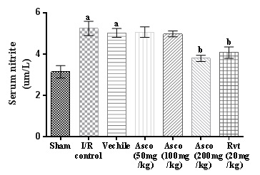
FIG. 4: GLOBAL CEREBRAL ISCHEMIA INDUCED INCREASES IN SERUM NITRITE LEVEL
Each group (n=12) represents mean ± standard errors of means. a=p<0.001 vs. sham group, b=p< 0.01 vs. I/R and vehicle.
Cerebral Infarction Area: The cerebral infarction area revealed a significant decrease in Ascophyllum nodosum extract and Rvt treated the group as compared to I/R injury group especially in Caudal and rostral side Fig. 5.
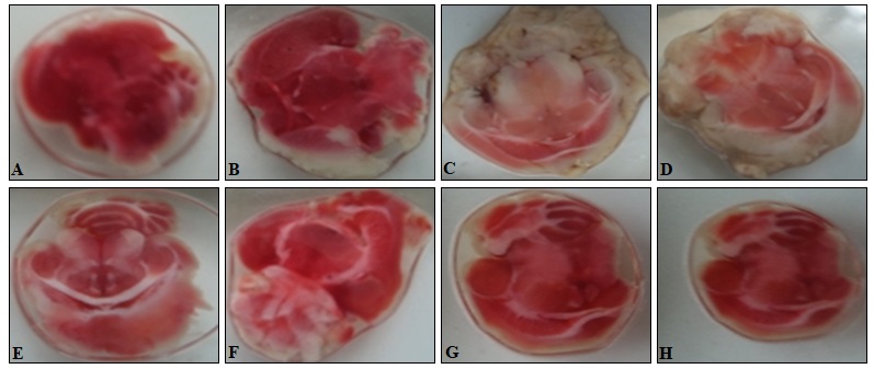
FIG. 5: NEUROPROTECTIVE EFFECT OF ASCOPHYLLUM NODOSUM EXTRACT AGAINST GLOBAL CEREBRAL ISCHEMIA / REPERFUSION DAMAGE IN MICE EVALUATED BY 2, 3, 5-TRIPHENYLTERTRAZOLIUM CHLORIDE (TTC) STAINING. BRAIN CORONAL SECTIONS WERE PREPARED AND THEN EACH SECTION WAS STAINED WITH TTC: (A) Control: normal animal, (B) Sham control: Carotid artery will be exposed for 17 min,(C) Negative control: distilled water + ischemia for 17 min, (D) I/R control: ischemia for 17 min, (E) Asco (50 mg/kg, p.o), (F) Asco (100 mg/kg, p.o), (G) Asco (200 mg/kg, p.o), (H) Rvt (20 mg/kg, p.o). The infarction was markedly reduced in the mice brain treated with 100 mg/kg (F), 200 mg/kg (G), 20 mg/kg (H); n=12
DISCUSSION: In the present study neuro-protective activity of Ascophyllum nodosum was evaluated by using global cerebral ischemia /reperfusion injury. It is well-known that ischemia/reperfusion induces neuronal injuries through several pathophysiological mechanisms, including excitotoxicity, lipid peroxidation and free radical production, etc., which finally triggers cerebral ischemic injury 16. LDH is a very important metabolic enzyme in neuron and is released into bloodstream by injured neuron.
Thus, increasing activity of LDH released from cerebral cells into blood serum may reflect the damage of neurons 17. MDA, the product of lipid peroxidation of the cell membrane, is another index to evaluate the cerebral ischemic injury. In this study, mice were subjected to carotid artery occlusion for 17 min and reperfusion for 24 h, as indicated by the formation of cerebral infarction, increases in serum LDH activity, serum nitrite level, and MDA level. The pre-treatment with Ascophyllum nodosum for 7 days prior to the ischemic insult significantly inhibited the increases of serum LDH activity, serum nitrite level and MDA level and reduced cerebral infarction size indicating a delayed protective effect of Ascophyllum nodosum against global cerebral I/R injury. Free radicals are highly reactive molecules with one or more unpaired electrons.
Free radicals can react with DNA, proteins, and lipids, causing varying degrees of damage and dysfunction. Free radicals, involved in stroke induced brain injury; include superoxide anion radical, hydroxyl radical and nitric oxide (NO). The damaging effects of free radicals are normally prevented or reduced by antioxidant enzymes and free radical scavengers. The primary source of oxygen-derived free radicals (often referred to as ‘reactive oxygen species’) during ischemic-stroke injury is the mitochondria, which produce superoxide anion radicals during the electron transport process 16. Previous study suggests that Mango peel extract (Mangifera indica L.) protect against peroxide-induced oxidative damage in rats due to its antioxidant activity. Mango peel contains various bioactive compounds like polyphenol, carotenoid etc. in it. The mango peel extract showed protection against lipid peroxidation caused by H2O2 18.
It has been reported that Cyanidin-3-glucoside fraction extracted from mulberry (Morus alba L.) rich in anthocyanin (polyphenol) are protective against oxidative stress in ischemia-reperfusion injury model of the brain 19. Quercetin represents the preponderant flavones in dried apple peel polyphenol (DAPP) and, according to previous studies; it has exhibited antioxidant & anti-inflammatory activities 20. Magnolia (Magnolol) is isomer polyphenolic compounds from the bark of Magnolia officinalis & has been identified as major active components exhibiting anti-oxidative, anti-inflammatory and neuroprotective effect 21.
Cyanidin-3-rutinoside (natural polyphenolic antioxidant) treatment resulted in reactive oxygen species (ROS) dependent activation of p-38 MAPK and JNK, which contribute to cell death by activating the mitochondrial pathway and causes antioxidant activity reduces the level of reactive oxygen species 22. These studies suggest that polyphenol play a vital role in decreases oxidative stress. Mice subjected to I/R exhibited a significant elevation of LDH. LDH is very important metabolic enzyme in neuron and is released into blood stream by injured neurons. The extract of Ascophyllum nodosum comprises at least about 20% to 100% by weight polyphenolic extract 9. It is showed that phenolic antioxidant had been shown to inhibit the oxidation of lipid and other molecules and protect against free radicals 8. Overproduction of ROS during ischemia /reperfusion can damage lipids, proteins, and nucleic acids, thereby inducing apoptosis or necrosis. Increasing evidence supports the hypothesis that plant polyphenol provides protection against neurodegenerative changes associated with cerebral ischemia 7.
The increasing activity of LDH released from cerebral cells into blood serum may reflect the damage of neurons. The pre-treatment with Ascophyllum nodosum (200 mg/kg p.o) for 7 days before ischemia significantly decrease the serum LDH activity. Reactive oxygen species (ROS) produces malondialdehyde (MDA), an end product of lipid peroxidation. MDA reacts with thiobarbituric acid and is thus estimated as thiobarbituric acid reactive substance (TBARS) 23. Therefore in the present study MDA was estimated using TBARS assay to estimate extent of ROS formation. The pre-treatment with A. nodosum (200 mg/kg p.o) for 7 days before ischemia significantly normalized the TBARS.
In the present study, I/R injury may cause an elevation in blood serum nitrite level. The pre-treatment with Ascophyllum nodosum (200 mg/kg p.o) for 7 days before ischemia significantly decreases the serum nitrite level in blood. In the present study, we observed that the Ascophyllum nodosum significantly improved cerebral ischemia. Ascophyllum nodosum also reduces Infract size, TBARS, serum nitrite concentration and LDH level which were increased by ischemia-reperfusion injury in mice.
CONCLUSION: In the present investigation, a polyphenolic fraction of A. nodosum has shown promise as a Cerebral Ischemia /Reperfusion Injury curing, as well as enhancing agents in mice in all the laboratory model employed. Furthermore, the polyphenolic extract of A. nodosum was found more effective than against global Cerebral Ischemia/Reperfusion Injury. In conclusion, the present results constitute the first evidence for the therapeutic potential of A. nodosum in Global Cerebral Ischemia/Reperfusion Injury. The study supports an important concept that onset of neurodegenerative disease may be delayed or mitigated with the use of dietary antioxidants that protect against oxidative stress and neurodegeneration.
ACKNOWLEDGEMENT: Nil
CONFLICT OF INTEREST: Nil
REFERENCES:
- Fagan SC and Hess DC: Stroke. Pharmacotherapy- A pathophysiologic approach. New Delhi. McGraw Hill Medical Publishing Division; Edition 6th, 2005.
- Smith SW, Johnston CS and Easton DJ: Cerebrovascular disease. Harrison principle of internal medicine. McGraw-Hill Companies; America, Edition 16th, 2005.
- Khadilkar SV: Neurology: The Scenario in India. JAPI 2012; 60: 42-44.
- Panickar KS and Anderson RA: Effect of polyphenols on oxidative stress and mitochondrial dysfunction in neuronal death and brain edema in cerebral ischemia. International Journal of Molecular Sciences 2011; 12: 8181-8207.
- Alexandrova ML and Bochev PG: Oxidative stress during the chronic phase after stroke. Free Radical Biology & Medicine 2005; 39: 297-316.
- Urquiaga I and Leighton F: Plant polyphenol antioxidants and oxidative stress. Biol Res 2000; 33: 55-64.
- Dajas F, Rivera-Megret F, Blasina F, Arredondo F, Abin-Carriquiry JA, Costa G, Echeverry C, Lafon L, Heizen H and Ferreira M: Neuroprotection by flavonoids. Braz J Med Biol Res 2003; 36: 1613-1620.
- Halliwell B: Are polyphenols antioxidants or pro-oxidants? What do we learn from cell culture and in-vivo studies? Arch Biochem Biophys 2008; 476: 107-112.
- Zhang J, Ewart HS, Shen JK and Barrow CJ: Ascophyllum composition and methods. United States patent application publication. US 2008; 2008/0280994A1.
- David DM, Kunjan RD, Anthony D, Ying C B, Ami PR, and Miguel A: Perez-Pinzon. Resveratrol pretreatment protects rat brain from cerebral ischemic damage via a SIRT1-UCP2 pathway. Neurosci 2009; 159(3): 993 02.
- Rehni AK, Singh TG, Jaggi AS and Singh N: Pharmacological preconditioning of the brain: a possible interplay between opioid and calcitonin gene-related peptide transduction systems. Pharmacological Reports 2008; 60: 904-913.
- Poonam K, Sita A and Viswanathan P: Oxidative changes in the brain of aniline exposed rats. Arch Environ Con Tox 1992; 23: 307-309.
- Slater TF and sawyer BC: The stimulatory effects of carbon tetrachloride and other halogenoalkanes on peroxidative reactions in rat liver fractions in-vitro. Biochem J 1971; 123: 805-814.
- Cortas NK and WakId NW: Determination of inorganic nitrate in serum and urine by a kinetic cadmium-reduction method. Clin Chem 1990; 36: 1440-1443.
- Sastry KVH, Moudgal RP, Mohan J, Tyagi JS and Rao GS: Spectrophotometric determination of serum nitrite and nitrate by copper-cadmium alloy. Analytical Biochemistry 2002; 306: 79-82.
- Woodruff MT, Thundyil J and Tang SC and Arumugam TV: Pathophysiology, treatment, and animal and cellular model of human ischemic stroke. Molecular Degeneration 2011; 6: 2-5.
- Shang YZ, Miao H, Cheng JJ and Qi JM: Effects of amelioration of total flavonoids from stems and leaves of Scutellaria baicalensis Georgi on cognitive deficits. Neuronal damage and free radicals disorder induced by cerebral ischemia in rats. Biol Pharm Bull 2006; 4: 805-10.
- Berardini N, Carle R and Schieber A: Characterization of gallotannins and benzophenone derivatives from mango (Mangifera indica cv. Tommy Atkins) peels, pulp and kernels by High-Performance Liquid Chromatography/ Electrospray Ionization Mass Spectrometry. Rapid Communications in Mass Spectrometry 2004; 18: 2208-2216.
- Kang TH, Hur JY, Kim HB, Ryu JH and Kim SY: Neuroprotective effects of the cyanidin-3-O-beta-d-glucopyranoside isolated from mulberry fruit against cerebral ischemia. Neurosci Lett 2006; 391: 122-126.
- Moon SK, Cho GO, Jung SY, Gal SW and Kwon TK: Quercetin exerts multiple inhibitory effects on vascular smooth muscle cells: the role of ERK1/2, cell cycle regulation, and matrix metalloproteinase-9. Biochem Biophys Res Commun 2003; 301: 1069-1078.
- Dennis YC, Ming HC, Yijia Z, Wenwen S, Yan H, Jing HJ, Agnes S, Zezong G, Kevin LF, Jiankun C, James CL, William RF, Dennis BL, Albert YS and Grace YS: Magnolia polyphenol attenuate oxidative and inflammatory responses in neurons and microglial cells. Biomed central. Journal of Neuroinflammation 2013; 10: 1-15.
- Rentian F, Hong MN, Shiow YW, Irina LT, Michael RS, Hisashi H and Xiao MY: Cyanidin-3-rutinoside, a natural polyphenol antioxidant selectively kills leukemic cells by induction of oxidative stress. The Journal of Biological Chemistry 2007; 282(18): 13468-13476.
- Bora KS, Arora S and Shri R: Role of Ocimum basilicum L. in the prevention of ischemia and reperfusion-induced cerebral damage, and motor dysfunctions in mice brain. Journal of Ethnopharmacology 2011; 137: 1360-1365.
How to cite this article:
Dhyani MP, Kumar A, Kothiyal P and Choudhary AN: Neuroprotective activity of Ascophyllum nodosum on experimentally induced Global Cerebral Ischemia/Reperfusion Injury in mice. Int J Pharmacognosy 2014; 1(2): 153-60. doi link: http://dx.doi.org/10.13040/ IJPSR.0975-8232.IJP.1(2).153-60.
This Journal licensed under a Creative Commons Attribution-Non-commercial-Share Alike 3.0 Unported License.
Article Information
10
153-160
684
2309
English
IJP
M. P. Dhyani *, A. Kumar, P. Kothiyal and A. N. Choudhary
Shri Guru Ram Rai Institute of Technology, Patel nagar, Dehradun, Uttarakhand, India
pankaj.dhyani93@gmail.com
01 November 2013
14 January 2014
26 January 2014
http://dx.doi.org/10.13040/IJPSR.0975-8232.1(2).153-60
01 February 2014