


IDENTIFICATION OF CURCUMA LONGA RHIZOMES BY PHYSICOCHEMICAL AND TLC FINGERPRINT ANALYSIS
HTML Full TextIDENTIFICATION OF CURCUMA LONGA RHIZOMES BY PHYSICOCHEMICAL AND TLC FINGERPRINT ANALYSIS
M. Duraisankar * and A. David Ravindran
Department of Biology, Gandhigram Rural University, Gandhigram, Dindigul - 624005, Tamil Nadu, India.
ABSTRACT: Curcuma longa L. rhizomes are widely used by traditional medical practitioners for curing various diseases. The rhizomes of C. longa Linn. is commonly known as Haldi or Turmeric. Haldi is of particular importance to a human with the discovery that its rhizome powder, when added to various food preparations, preserves their freshness and imparts a characteristic flavor. The Indian system of medicine comprises of Ayurveda, Siddha, and Unani. In these systems, many medicines are made up of C. longa rhizomes. In Ayurveda, turmeric has been used internally as a stomachic, tonic, blood purifier and externally in the prevention and treatment of skin diseases. The World Health Organization (WHO) in 1999 had given a detailed protocol for quality control of single herbal drugs. We have developed a simple and rapid identification method for authentication of C. longa rhizome. Physicochemical parameters and TLC fingerprint analysis of C. longa rhizomes were also carried out. The study revealed different analytical parameters of the crude drug which will be useful in the identification of certain drug and control of adulterations.
| Keywords: |
Curcuma longa, Secondary metabolites, Standardization, Phytochemical analysis
INTRODUCTION: Plants have an almost unlimited ability to synthesize diverse secondary metabolites that have a wide range biological functions that are useful to human Curcuma longa Linn., Turmeric (Curcuma longa) and several other species of the Curcuma genus grow wild in the forests of Southern Asia including India, Indonesia, Indochina, nearby Asian countries, and some Pacific Islands including Hawaii. All of these areas have traditional culinary and medicinal uses going back to pre-history 1.
In the Indian Ayurveda system of herbal medicine, turmeric is known as strengthening and warming to the whole body. Traditional uses in India include to improve digestion, to improve intestinal flora, to eliminate worms, to relieve gas, to cleanse and strengthen the liver and gallbladder, to normalize menstruation, for relief of arthritis and swelling, as a blood purifier, to warm and promote proper metabolism correcting both excesses and deficiencies, for local application on sprains, burns, cuts, bruises, insect bites and itches, for soothing action in cough and asthma, as antibacterial and anti-fungus, and in any condition of weakness or debility.
According to Michael Moriarty, “The ancient Hawaiians used this herb for many things, including the prevention and treatment of sinus infections (it is very astringent and appears to pull mucus out), ear infections (swimmers ear) and gastrointestinal ulcers 2. Turmeric is eaten as food both raw and cooked throughout Asia. While turmeric root looks much like ginger root, it is less fibrous and is more chewable, crunchy, and succulent 3. The fresh root (not the powder) has a somewhat sweet and nutty flavor mixed with its bitter flavor. As a result, it is not unpleasant to eat and not difficult to chew. It is sometimes chewed plain or chopped up and put in salads raw. Traditional use includes mashing / grinding it in a mortar to make a paste to mix with other spices for flavoring in curries. In modern times, most common use is of the dried root powder as the base of most curries in India and other nearby countries 4.
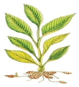
FIG. 1: CURCUMA LONGA
The Curcuma longa, the yellow root, has been cultivated in India, Southern China and other tropical and subtropical countries. It is conjectured that the yellow root originates from East India, but this is not certain because the plant has never been found in the wild 5. The plant’s flower and growth pattern look very similar to that of ginger, and it is also in the same family as ginger (Zingiberaceae). The yellow root can grow to a height of approximately 1 meter. A bundle of leaves and flower stalk with a 20-cm-long flower rises from the rootstock 6. Similarly, to the ginger plant, side shoots with corm-like swellings develop underground. The yellow root grows best in damp and warm areas 7.
The underground parts of the plants are dug up when the flowers wilt in December and January. The corms and the rootstock are separated from the side shoots 15-17. These are then submerged in boiling water and placed in the sun to dry. Through drying in the sunlight, the underground parts become yellow: the heat from the sun causes the pigment from the glandular cells to be distributed over the rootstock and corms. Once they are dry, the rootstocks are ground to form a powder which is called “curcumin” or “curcuma.” The powder is then sold under the name “fumeric.”
MATERIALS AND METHODS: Fresh samples of turmeric rhizomes were purchased and authenticated. These are processed prepared and dried and evaluated for pharmacognostic and phytochemical studies were evaluated.
In this study, the identity of the drug has been taken into consideration from the plant (genuine) after due taxonomical identification and preparing the rhizomes after collection as done in the case of the commercial sample and the characters compared with the market sample both macroscopically and microscopically to detect any changes or adulterants 8.
Microscopy: Fresh rhizomes of Curcuma longa were subjected for the microscopical studies. Then the sections were cut by freehand sectioning. The numerous temporary and permanent mounts of the microscopical section of the specimen were made and examined microscopically.
Powder Characteristics: Preliminary examination and behavior of the powder with different chemical reagents were carried out as per the reported method.
Quantitative Microscopy: Fresh turmeric showed abundant individual starch grain. They were ovate, simple, with a small beak at one end where the hilum was situated but it was altered during curing due to gelatinization.
Micrometry: Quantitative microscopy of the transverse sections and rhizome powder were performed to determine the size and dimension of tissues, cell and cell content.
Physicochemical Evaluation: Physicochemical parameters such as foreign organic matter, moisture content, ash values, extractive values, pH, etc were determined as per procedures mentioned by WHO guidelines.
Preliminary Phytochemical Screening: The chemical evaluation includes qualitative chemical tests which are used for identification of various phytoconstituents present in the powdered crude drug.
Fluorescence Analysis: Dried rhizomes were powdered and observed under visible light, short ultraviolet light, long ultraviolet light after treatment with different reagents like chloroform, ethyl acetate, methanol, petroleum ether (60-80°C), 50% sulphuric acid, 50% hydrochloric acid, 50% nitric acid, 10% sodium hydroxide, etc.
Chromatographic Study:
Thin Layer Chromatography:
Alcoholic Extract: Alcoholic extract of Curcuma which has shown the hypoglycaemic effect was subjected to thin layer chromatography.
| Stationary phase | Silica gel |
| Mobile phase | Benzene: Chloroform: Alcohol
(45: 45: 10) |
| Detection | Boric acid in methanol |
Method: Alcoholic extract of turmeric was spotted separately on a TLC plate and developed in a solvent system as stated above. Then the plate was dried and sprayed with the diagnostic agent. The Rf value of the separate components was calculated.
Rf value =Distance travelled by solute/Distance travelled by solvent
Separation of Curcuminoids by Column Chromatography:
| Absorbent | Silica gel
(column chromatography grade) |
| Solvent | Benzene |
| Sample | Benzene extract |
The column was packed with slurry (silica gel in benzene). It was run for 1-2 hours with benzene. About 1 gram of the benzene extract from the marc obtained after the extraction with pet ether to remove fixed and volatile oils was passed down the column and when the solution has just sunk into the column, fresh benzene equilibrated with water was added for the development of the chromatogram. There is a gradual separation of bands on the column, and the complete chromatogram will show 3 distinct zones and a few minor zones development was continued with benzene. Any fixed or volatile oil if turmeric which was not removed completely by petroleum ether passes down the column first as a pale yellow fraction. Evaporation of the solvent leaves a yellow oily residue having the characteristic odor of turmeric. The different zones were collected as liquid chromatogram in separate tubes as each band got washed down the column. The eluates from the interzones were nearly colorless or only faintly colored were rejected. The different fractions collected were concentrated by distillation under reduced pressure and further purified by re-chromatography on smaller columns using benzene9.
Determination of Curcumin Content as Curcuminoids by Spectroscopy method: As per the literature cited the following method has been adopted using the standard value 1 percent at 430nm - 1560. About 200 mg of turmeric powder was weighed accurately and extracted with alcohol and made up to 100 ml. From this 5 ml was pipette out into the 50 ml standard flask and made up to the volume. Absorbance was studied at 430 nm.
RESULTS AND DISCUSSION:
Macromorphological Description: The central or primary rhizomes are ovate, irregularly ovoid, cylindrical or fusiform, curved, sometimes slightly branched into a Y-shape, 1.1-10.3 cm long, 5-30 mm in diameter to, rough, with wrinkled striations, distinct cyclic nodes, and rounded scars of root branches and rootlets. The organoleptic evaluation of the rhizomes revealed that the rhizomes were Yellowish to yellowish-brown in color, with characteristic and aromatic odor and slightly bitter and pungent in taste. The results of morphological characters are mentioned in Table 1.
T.S. of rhizome Description: The transverse section of the rhizome shows cork as an outer layer followed by epidermis, cortex, and endodermis and ground tissue. Cork composed of thin-walled brown cells which are large and polygonal in shape. The epidermis is consisting of thin-walled cubical cells of various dimension. The cortex consists of thin-walled rounded parenchymatous cells and having oleoresin cells. These cells are filled with gelatinized starch grains and yellow coloring matter. The ground tissue is parenchymatous and cells filled with gelatinized starch grains and yellow pigment. Fibrovascular bundle and oil cells scattered throughout ground tissue 10.
Organoleptic Characters:
Prepared Sample Powder:
- Colour: Dark yellow
- Odor: Dromatic
- Taste: Spicy, bitter
- Texture: Fine
Powder Characters:
- Fragments of cork cell both sectional view and surface view was seen.
- Group of rounded parenchyma cells with dark-brown pigment cells were seen.
- Altered pasty masses of starch, colored yellow (in unstained preparation) were present.
- Parenchymatous cells with unlignified vessels were seen.
- The vessels were found to have spiral, reticulate and annular thickenings.
- Calcium oxalate crystals were absent.
Power of market sample showed similar characters as above. Hence, from the above power microscopical characters of the powered prepared and market samples of the drug, the genuinely of the market sample was determined.
Quantitative Microscopy: The measurement of starch grains was made in a fresh sample. It was found to conform to standard values specified in brackets. The results were expressed in Table 2.
TABLE 1: MORPHOLOGICAL CHARACTERS
| Fresh sample | Prepared sample | Market sample | |
| Colour | Pale brown | Yellow-yellowish brown | Yellow-yellowing brown |
| Taste | Spicy bitter | Initially spicy and later bitter | Spicy bitter |
| Odor | Aromatic | Aromatic | Aromatic |
| Shape | Cylindrical | Cylindrical | Cylindrical |
| Size | 3-5cm and 1.8cm | 3-5cm and 1.5cm | 3-5cm and 1.8cmrs |
| Fracture | Short | Short | Short |
| Fractured surface | Yellowish-orange | reddish-brown | reddish-brown |
| Surface
|
Notes leave visible | a few scales leaves and root, branchlet, scars seen | Scale leaves, scars of the root, branchlet, seen |
| Bud | Apical bud present | bud scars are seen | bud scars are seen |
TABLE 2: QUANTITATIVE MICROSCOPY
| Minimum | Average | Maximum | ||
| Fresh sample | Length | 29µ | 47.7µ | 66.5µ (30-60µ) |
| Starch grain | width | 12.8µ | 23.8µ | 30.8µ (25-35µ) |
By comparing the above-observed value with that of standard value, the genuinely of the fresh sample was confirmed
Fluorescence Analysis: Fluorescence analysis of the powder treated with different solvents and reagents is exhibited in Table 4. Fluorescence is the phenomenon exhibited by numerous phytoconstituents present in the plant material.
In fluorescence, the fluorescence light is always of greater wavelength than the exciting light. Light rich in short wavelength is very active in producing fluorescence, and for this reason, ultraviolet light produces fluorescence in many substances which does not visibly fluoresce in daylight. The results were expressed in Table 3 and 4.
Physicochemical Evaluation: The results of the physicochemical constants of raw material within the limit which is mentioned in Table 5. This signifies that the quality and purity of raw material was good enough; the results of foreign organic matter denote presence of any organism, part or product of an organism other than that named in the specification and description of the herbal material concerned which was found to be 0.23 ± 0.015% w/w, it indicates that there may be present of part or product of an organism in less amount. Insufficient drying favors spoilage by molds and bacteria and makes possible the enzymatic destruction of active principles. Not only the ultimate dryness of the drug is essential equally important is the rate at which the moisture is removed and the condition under which it is removed thus the determination of moisture content also provides the method of preparation of drug, and it is observed that the moisture content of the drug was found to be 7.42 ± 0.021% w/w which signifies that the drug is appropriately dried and properly stored.
TABLE 3: FLUORESCENCE ANALYSIS OF EXTRACTS
| Material | Extracts | Daylight | UV light |
| Turmeric
|
Petroleum ether
Benzene Alcohol Chloroform Water |
Yellow
Yellow yellowish green yellowish green Nil |
Light yellowish green
yellowish green yellowish green yellowish green Nil |
TABLE 4: FLUORESCENCE ANALYSIS OF TURMERIC
| Material | Reagents | Daylight | UV light |
|
Turmeric |
Powder
Powder + 1N NaOH( aqu) Powder +1N NaOH ( Alc) Powder + 50% H2SO4 |
Yellow
Red Reddish orange yellowish |
yellowish green
yellowish green yellowish green yellowish green |
TABLE 5: PHYSICOCHEMICAL PARAMETERS OF CURCUMA LONGA (RHIZOME)
| S. no. | Parameters | Analysis % w/w | Remark |
| 1
2 3 4 5 |
Foreign matter
Total ash Acid-insoluble ash Alcohol soluble extractive value Water-soluble extractive value |
Nil
4.5±0.03 0.86±0.61% 9.42±0.57% 18.26±0.01% |
NMT -2.0%
NMT-9% NMT-1% NLT-8% NLT-12% |
The total ash is particularly important in the evaluation of purity of drugs, i.e., the presence or absence of foreign matter such as metallic salts or silica. An analytical result for total ash was found to be 4.5 ± 0.03% w/w.
The amount of acid-insoluble siliceous matter present was 0.86 ± 0.61% w/w; as the ash values of the crude drugs lies within the reasonable limit which signifies its quality and purity and gives an idea about the total inorganic content. The water-soluble extractive value indicated the presence of sugar, acids and inorganic compounds the water-soluble extractive value found to be 18.28 ± 0.01% w/w and alcohol soluble extractive values indicated the presence of polar constituents like phenols, alkaloids, steroids, glycosides, flavonoids. The alcohol soluble extractive value was found to be 9.42 ± 0.57% w/w which signifies the nature of the phytoconstituents present in the plant.
Preliminary Phytochemical Screening: 500g of the coarse powder of Curcuma (market sample) was subjected to extraction by cold percolation petroleum ether (40-60 °C), benzene, chloroform, ethanol (70%) and chloroform water. Percentage yield of various extracts was mentioned in Table 6.
TABLE 6: PERCENTAGE YIELD OF VARIOUS EXTRACTS
| Extracts | Turmeric | |||
| Yield in % w/w | Colour | Consistency | Yield in w/w | |
| Petroleum ether (40-60 °C) | 2.1 | Yellow | Viscous | 2.2 |
| Benzene | 10.3 | Brown | Solid | 2.8 |
| Chloroform | 6.8 | Reddish | Solid | 3.8 |
| Ethanol | 10.2 | Reddish | Solid | 9.8 |
| Water | 2.8 | Pale Yellow | Solid | 15.8 |
The preliminary phytochemical Investigations of Aqueous extract, acetone extract, ethanolic extract and methanolic extract of Curcuma longa rhizome were performed which reveals the presence of phenolic compound, tannins, alkaloids, terpenes, saponin type of major secondary metabolites which revealed their potential therapeutic activity. The results were expressed in Table 7.
TABLE 7: QUALITATIVE CHEMICAL EXAMINATION OF VARIOUS EXTRACTS
| Plant constituent
test |
Turmeric | ||||
| P.E | BEN | CHE | ALC | AQ | |
| Alkaloids
Mayers’s reagents |
|
+ | |||
| Carbohydrates and glycosides
Molisch reagent |
|||||
| Phytosterols
Liebermann’s test |
|||||
| Fixed oil and fats
Spot test |
+ | ||||
| Saponins
Foam test |
|||||
| Phenolic
Ferric chloride |
+ | + | + | ||
| Proteins
Millons’s |
+ | ||||
| Resin
With water |
+ | + | + | ||
| Volatile oil
Test for aroma |
+ | ||||
| Vitamins
Ascorbic acid |
|||||
Chromatographic Study:
Thin Layer Chromatography: A chromato-graphic study of curcuminoids present in Curcuma longa was undertaken since during the screening for the bioactive substances, it was found, that the curcumin affects bringing down blood sugar level in normal fasting rabbits. Hence, it was thought that curcumin might be having a major role to play in the fall of blood-sugar level 11, 12, 13. A standard sample of curcumin was not available for comparison. Hence, isolation was taken up of curcumin. Through, curcumin is highly soluble in alcohol, since tannins also may interfere, it was considered curcumin be isolated from benzene extract, taking into account the past work done of a chromatographic study of curcuminoids in Curcuma longa. The results were expressed in Table 8.
TABLE 8: THIN LAYER CHROMATOGRAPHY
| Spots | Rf value | Colour | ||
| As per Ref | As per TLC | As per Ref | As per TLC | |
| Spot I | 0.25-0.35 | 0.26 | Yellow | Yellow |
| Spot II (Curcumin derivative) | 0.35-0.45 | 0.39 | Orange | Light orange |
| Spot III (Curcumin) | 0.40-0.45 | 0.42 | Orange | Dark orange |
Column Chromatography:
Fraction 1: This is the eluate containing the orange-yellow zone. As the benzene solution is evaporated, curcumin is thrown out of solution in the form of orange-red crystals. Recrystallization from acetone gave curcumin in the form of needles of crystals having a melting point of 180 °C. The yield was 50 mg. This was confirmed using a chemical test. This was used as standard curcumin which is adsorbed lowest on the column and forms by far major constituent.
Spectroscopy Method: The average weight of the one tablet was found out from 20 ml tablets. It was extracted with alcohol and made up to 100ml. From this 5ml was pipette out into the 50ml standard flask and made up to volume. Measured at 430 nm using (Shimadzu – 1201 – spectrophotometer)
Calculation:
For 1560 Absorbance - 1 gram
For 0581 Absorbance - x
The content of curcumin as curcuminoids in each tablet of average weight has been calculated after incorporating appropriate dilution factor. The percentage of curcumin content = 0.756%.
CONCLUSION: Summary the standardization, qualitative and quantitative determination of phytochemicals and TLC profiling were accomplished. Furthermore, the result may be useful in developing potential phytopharmaceutical products based on C. langa. However, there is a need to further identify specifically the flavonoid, saponin and alkaloid compounds that are present in the extracts.
ACKNOWLEDGEMENT: Nil
CONFLICT OF INTEREST: Nil
REFERENCES:
- Vaidya yoga ratnavali (formulary of Ayurvedic medicines) - impcops, Madras-41.
- Trease and Evans: pharmacognosy, English language, Edition 12th, 1980: 261.
- Atal CK and Kapur BM: Cultivation and utilization of Aromatic plants, CSIR publications jammu- Tawi 1982: 209-210.
- Tyler E and Brady R: Pharmacognosy leans febiger, Edition 8th, 1981: 499.
- The wealth of India: Council of Scientific and Industrial Research, New Delhi.
- Nadkarni KM: Indian Materia medica popular Prakashan Pvt. Ltd., Bombay, Vol. I, 1982: 418.
- Medicinal plants of India: Indian Council and Medical Research, New Delhi, Vol. 1, 1976: 313.
- Heber W and Youngken HW: Textbook of Pharma-cognosy, McGraw- Hill Book Co., New Yerk, Edition 6th, 1950: 230.
- Kokate CK, Purohit AP and Gokhale SB: Textbook of Pharmacognosy, Nirali Prakashan, 1990.
- Wallis TE: Analytical microscopy J & A Churchill Ltd., Edition 3rd, 1965: 162-163.
- Dictionary of medicinal plants of India Central Institute of medicinal and aromatic plants, 1992: 340.
- Iyer N and Kolammal: Pharmacognosy of ayurvedic drugs, Dept. of pharmacognosy, University of Kerala Trivandrum Vol. 8, 1964.
- Gamble JS: Flora of the Presidency of madras, Bi-Shen singh Mahendra pal singh Vol. II, 1979: 1295.
- Kirithikar and Basu: International book distributors, Book Sellers & Publishers, Vol. III, 1987: 2221.
- Goel A, Kunnumakkara AB and Aggarwal BB: Curcumin as “Curcumin”: From kitchen to clinic. Biochem Pharmacol 2008, 75 (4), 787-809.Price LC and Buescher RW: Decomposition of turmeric curcuminoids as affected by light, solvent and oxygen. J Food Chem 2007; 20: 125-133.
- Paramasivam M, Poi R, Banerjee H and Bandyopadhyay A: High-Performance Thin Layer Chromatographic method for quantitative determination of curcuminoids in Curcuma longa Food Chem 2009; 113: 640-644.
- Herebian D, Choi JH, Abd El-Aty AM, Shim JH and Spiteller M: Metabolite analysis in Curcuma domestica using various GC-MS and LC-MS separation and detection techniques. Biomed Chromatogr 2009; 23: 951-965.
How to cite this article:
Duraisankar M and Ravindran AD: Identification of Curcuma longa rhizomes by physicochemical and TLC fingerprint analysis. Int J Pharmacognosy 2015; 2(9): 459-65. doi link: http://dx.doi.org/10.13040/IJPSR.0975-8232.IJP.2(9).459-65.
This Journal licensed under a Creative Commons Attribution-Non-commercial-Share Alike 3.0 Unported License.
Article Information
7
459-465
556
2174
English
IJP
M. Duraisankar * and A. D. Ravindran
Department of Biology, Gandhigram Rural University, Gandhigram, Dindigul, Tamil Nadu, India.
dsankar2012@gmail.com
31 July 2015
04 September 2015
17 September 2015
10.13040/IJPSR.0975-8232.IJP.2(9).459-65
30 September 2015