


ANTISECRETORY AND ANTIOXIDANT ACTIVITIES OF THE LEAVES AQUEOUS EXTRACT OF BOCSIA ANGUSTIFOLIA (CAPPARACEAE) ON GASTRIC ULCERS IN WISTAR RATS
HTML Full TextANTISECRETORY AND ANTIOXIDANT ACTIVITIES OF THE LEAVES AQUEOUS EXTRACT OF BOCSIA ANGUSTIFOLIA (CAPPARACEAE) ON GASTRIC ULCERS IN WISTAR RATS
André Perfusion Amang * 1, Jonas Walantini 1, Gustave Lebeau Otto Ndji 2, Gael Tchokomeni Siwe 3, Luc Vandza Vandi 1 and Paul Vernyuy Tan 3
Department of Biological Sciences 1, Faculty of Science, University of Maroua, P.O. Box 814, Maroua, Cameroon.
Department of Life Science 2, Higher Teachers’ Training College, University of Ngaoundéré, P.O. Box 652, Bertoua, Cameroon.
Department of Animal Biology and Physiology 3, Faculty of Science, University of Yaoundé I, P.O. Box 812, Yaoundé, Cameroon.
ABSTRACT: The aim of this study was to evaluate antisecretory and antioxidant activities of the leaves aqueous extract of Boscia angustifolia (LAEBA) on gastric ulcers induced in male rats. LAEBA at the doses of 125, 250, and 500 mg/kg were tested on three models of gastric ulcers, namely: pylorus ligation, pylorus ligation + histamine, and pylorus ligation + carbamylcholine. Some parameters of oxidative stress (malondialdehyde (MDA), superoxide dismutase (SOD), catalase (CAT), and reduced glutathione (GSH)) were evaluated in stomach’s homogenates. The qualitative phytochemical screening of LAEBA was carried out. Antisecretory screening showed that LAEBA at 500 mg/kg significantly decreased (p<0.01) gastric acidity (8.06 mEq/l) compared to the negative control (14.07 mEq/l), with a significant reduction (p<0.001) of gastric volume (1.30 ml) compared to the negative control (5.12 ml), as well as a reduction of pepsin activity (64.64%). These variations were similar to pylorus ligation + histamine and pylorus ligation + carbamylcholine models. LAEBA at all the doses induced a significant reduction (p< 0.05) of ulcerated surface correlated with an increase of mucus secretion (16.89 to 187.50%) compared to the negative control. It also induced a significant reduction of MDA level and a significant increase in catalase and GSH. LAEBA would protect the gastric mucous membrane by a mechanism which would involve the cholinergic and histaminic ways, the reinforcement of mucus secretion and antioxidant status, via the presence of flavonoids, saponins and tannins in LAEBA.
| Keywords: |
LAEBA, Phytoconstituents, Gastric Ulcer, Antisecretory, Antioxidant, Wistar rats
INTRODUCTION: The stomach is the organ of the digestive tract that digests food and acts as a temporary reservoir. It is located in the upper left quadrant of the abdominal cavity 1. It is daily exposed to aggression factors.
Gastric acid, Helicobacter pylori 2, tobacco, alcohol and non-steroidal anti-inflammatory drugs 3, psychological stress 4 and defense factors anti-oxidants, estrogens and mucus.
Imbalance in favor of aggression factors can lead to the deterioration of the gastric wall and gastro-intestinal disorders, among which, gastric ulcers. Gastric ulcer is the principal pathology of stomach 3. It represents a loss of substances resulting from the rupture of the mucosa and sub-mucosa layers of the stomach, which can be associated with vascular lesions 5.
Prevalence of gastroduodenal ulcers is estimated at approximately 10% of the world population 6, with approximately 5.1% in South Africa and 17.1% in Cameroun, men being preferentially concerned 7. The incidence of gastric ulcers increases with aging as the peak of patients is around 65 years 8. The most frequent symptoms of gastric ulcers described are: stomach burns and/or distending, unexplained weight loss, presence of blood in vomiting 9. Gastric ulcers can lead to complications such as digestive hemorrhages and perforation 9. The treatment of gastric ulcers consists of a tri-therapy which associates: antibiotics, antisecretory, and antacids 10. However, the adverse effects of these drugs in addition to their prohibitive cost (according to a low-income population) have convinced the majority of developing countries population to use medicinal plants as an alternative for the treatment of gastric ulcers. Thus, several plants are used for this purpose: Eremomastax speciosa 11, Ocimum suave 12, Alstonia boonei 13. Bocsia angustifolia is a plant growing in Sahelian areas and belonging to the family of Capparaceae 14. Ethnobotanical studies led in Togo 15 and Burkina Faso 16 reported the use of this plant for the management of articular pains, bilharzias, edemas, jaundice, Guinea worm, and impotence. Hassan et al. 17 showed the hepatoprotective effect of the chloroform extract of B. angustifolia roots, which would be due to the presence of saponins and alkaloids in that extract.
Arbonnier 14 also reported the use of B. angustifolia for the treatment of gastric ulcers in Burkina Faso. Hypersecretion of acid being a crucial cause of gastric ulcers occurrence18, its control represents a cornerstone in the treatment process. Thus, this study was carried out to evaluate the antisecretory and antioxidant properties of LAEBA on gastric ulcers induced in male rats.
MATERIALS AND METHOD:
Plant Material: Fresh leaves of Bocsia angustifolia were collected in May 2019, in Edoum-hai village (Far-North region of Cameroun) (10°47’4.4088” N and 14°6’12.942” E). The plant was identified by Pr. TCHOPSALA (Botanist) and authenticated at the Herbarium of the Wildlife School of Garoua by Dr. DAMBOYA Emmanuel by comparison with the existing specimen registered under the number HEFG/558.
The leaves were shade dried at room temperature for two weeks. Dry leaves were reduced into powder using a mortar. The powder obtained was used for extract preparation.
Animals: Male Wistar rats (13 ± 1 weeks old) weighing between 150 to 180 g were used. They were raised in the animal house of the Laboratory of Animal Physiology, Faculty of Science, University of Yaoundé I. Animals were fed with standard laboratory diet composed as follows: maize flour maize (50%), soya flour (20%), fish flour (15%), bone flour (4%), vitamins (0.1%), cotton oil cake (10%), palm oil (0.1%), cooking salt (0.8%), with free access to tap water. These animals were acclimatized for two weeks at the University of Maroua, before any experimentation.
Extract Preparation: Three hundred grams (300 g) of powder were boiled in 2 liters of distilled water for 15 min. The solution was then filtered using coffee filter paper number 4. The filtrate was evaporated in an oven at 50 °C for 24 h, to obtain 36.6 g of extract (12.20% yield).
Antisecretory Screening: The antisecretory scree-ning was realized using pylorus ligation, according to the method described by Shay et al. 19. Twenty-five (25) rats were divided in 5 groups of 5 animals each, as follows: negative control, positive control and 3 test groups. Animals were fasted, with free access to water, during forty-eight hours (48 h) before their respective treatments. Negative and positive control groups received, respectively, distilled water (1 ml/ 200 g) and ranitidine (100 mg/kg), and the three extracts treated groups received LAEBA at the respective doses of 125, 250, and 500 mg/kg. One hour later, animals underwent laparotomy, under anesthesia to ethyl ether, the pylorus of each rat was tightened using a wire, and the abdomen was then stitched up. All the rats were sacrificed under deep anesthesia to ethyl ether 8 h after pylorus ligation. The gastric contents of each rat were collected in dry tubes and centrifuged at 2000 rpm for 10 min. The supernatant of each tube was collected, volume, pH, as well as acidity were measured, and pepsin activity was evaluated. Ulcers formed on the glandular area of stomachs were measured, and scores were attributed according to the method described by Tan et al. 20
The mucus of each animal was carefully scraped using a glass slide and weighed using a sensitive electronic balance. Homogenates of stomachs were prepared for the measurement of some oxidative stress parameters.
Evaluation of Antihistaminic and Anticholinergic Effects of LAEBA: The antihistaminic and anti-cholinergic activities of LAEBA were evaluated according to the protocol described by Vela et al. 21 For each experiment, twenty (20) rats were fasted during 48 h, with free access to water, and then distributed in 4 groups of 5 animals each. Negative and positive control groups received, respectively distilled water (1 ml/200 g) and ranitidine or cimetidine (100 mg/kg) and two groups received LAEBA at 250 and 500 mg/kg, respectively.
One hour after pylorus ligation, histamine (2.5 mg/kg, s.c.) or carbachol (1 mg/kg, s.c.) was administered to animals, and 4 h later, animals were sacrificed, and the remaining procedure was same as described in antisecretory screening.
pH Measurement and Acidity Titration: The gastric acidity of each rat was titrated according to the protocol described by Syed et al. 22, and pH was measured as described by Amang et al. .23. One milliliter of centrifuged gastric juice was diluted (1/10 v) with distilled water, and pH was measured using a pH-meter (JENWAY, PSAC12R-120, made in China). The solution was then titrated with NaOH (0.01 N) in phenolphthalein until obtaining pink color. The final volume of NaOH used was noted and total acidity calculated as per the following formula:
Acidity = Volume of NaOH × Normality of NaOH × 100 / 0.1
Determination of Pepsin Activity: To determine the pepsin activity, a solution of Bovine Serum Albumin (BSA) (50 mg/ml) was incubated with gastric juice at 37 °C during 10 min.
The quantity of proteins hydrolyzed in each tube was determined following the method of Biuret 24.
Evaluation of In-vivo Antioxidant Parameters: MDA, SOD, catalase, and reduced glutathione levels were determined according to the methods described by Wilbur et al. 25, Misra et al. 26, Sinha et al. 27 and Ellman 28, respectively.
Statistical Analysis: Results were expressed as mean ± standard error (SEM). They were analyzed (GraphPad PRISM 5) using a one-way analysis of variance (ANOVA), followed by Newman Keuls post-test. Values of p<0.05 were considered significant.
RESULTS:
Results of Phytochemical Tests: Phytochemical analysis of LAEBA revealed the presence of flavonoids, coumarins, saponins, and tannins Table 1.
TABLE 1: PHYTOCHEMICAL SCREENING OF LAEBA
| Classes of Compounds | Observations |
| Alkaloids | - |
| Coumarins | + |
| Flavonoids | + |
| Quinones | - |
| Saponins | + |
| Steroids | - |
| Tannins | + |
Présence +, Absence
Effects of LAEBA on Gastric Ulcers Induced by Pylorus Ligation: Table 2 represents the effects of LAEBA on gastric ulcers induced by pylorus ligation. Extract, at all the doses, induced a dose-dependent decrease (p< 0.001) of ulcerated surface (15.60, 8.40, and 2.0 mm2, respectively) compared to the negative control (52.40 mm2), which corresponded to percentages of inhibitions (38.15, 45.17 and 64.91%, respectively). It was also noticed a dose-dependent increase of mucus in extract treated groups (24.60, 36.00, and 41.40 mg) by comparison to the negative control (29.60 mg).
TABLE 2: EFFECTS OF LAEBA ON GASTRIC ULCERS INDUCED BY PYLORUS LIGATION
| Treatment | N | Doses (mg/kg) | US (mm2) | UI | % I | WM (mg) | % MI |
| Negative control | 5 | - | 52.40 ± 2.20 | 4.56 ± 0.27 | - | 29.60 ± 2.16 | - |
| Ranitidine | 5 | 100 | 8.20 ± 1.93*** | 2.45 ± 0.20*** | 46.27 | 45.60 ± 5.30 | 35.08 |
| LAEBA | 5 | 125 | 15.60 ± 3.09*** | 2.82 ± 0.20*** | 38.15 | 24.60 ± 2.02 | 16.89 |
| LAEBA | 5 | 250 | 8.40 ± 1.69*** | 2.50 ± 0.16*** | 45.17 | 36.00 ± 2.10 | 21.62 |
| LAEBA | 5 | 500 | 2.40 ± 1.93*** | 1.60 ± 0.40*** | 64.91 | 41.40 ± 1.97 | 39.86 |
N: number of animals per group, US: ulcerated surface, UI: ulcer index, % I: percentage of inhibition, WM: weight of mucus, % MI: percentage of mucus increase, *** p <0.001: significant difference compared to the negative control. Values represent means ± SEM.
Effects of LAEBA on Gastric Secretion Induced by Pylorus Ligation: Table 3 shows the effect of LAEBA on gastric secretion induced by pylorus ligation. Extract induced a significant decrease of gastric acidity at all the doses (12.05, 10.47, and 8.06 mEq/l, respectively) compared to the negative control (14.07 mEq/l). This reduction was correlated with a significant increase (p<0.01) of gastric juice pH in extract-treated groups (4.10, 4.98, and 5.99) compared to the negative control (3.33), as well as a significant decrease (p < 0.001) of gastric juice volume (2.42, 1.15 and 1.30 ml) compared to the negative control (5.12 ml). The extract also induced a significant (p<0.01) increase of hydrolyzed proteins percentage at the doses of 250 and 500 mg/kg (56.36 and 64.64%, respectively), compared to the negative control.
TABLE 3: EFFECTS OF LAEBA ON GASTRIC SECRETION INDUCED BY PYLORUS LIGATION
| Treatment | N | Doses (mg/kg) | VGJ (ml) | pH | Gastric acidity (mEq/l) | PA |
| Negative control | 5 | - | 5.12 ± 0.25 | 3.33 ± 0.09 | 14.07 ± 1.07 | 3.30±0.60 |
| Ranitidine | 5 | 100 | 4.39 ± 0.48 | 5.63 ± 0.47 *** | 9.90 ± 1.10 * | 3.63±0.20 |
| LAEBA | 5 | 125 | 2.42 ± 0.45 *** | 4.10 ± 0.32 | 12.05 ± 0.97* | 18.18±0.23* |
| LAEBA | 5 | 250 | 1.15 ± 0.18 *** | 4.97 ± 0.26 ** | 10.47 ± 1.13* | 56.36±0.24** |
| LAEBA | 5 | 500 | 1.30 ± 0.05 *** | 5.99 ± 0.32 *** | 8.06 ± 0.62 ** | 64.64±0.19** |
N: number of animals per group, VGJ: volume of gastric juice, PA: pepsin activity in the percentage of hydrolyzed protein, *p< 0.05, ** p< 0.01, *** p < 0.001: significant difference compared to the negative control, Values represent means ± SEM.
Effects of LAEBA on Gastric Ulcers induced by a Combination of Pylorus Ligation and Histamine: Table 4 shows the effect of LAEBA on gastric ulcers induced by a combination of pylorus ligation and histamine. LAEBA induced a significant reduction (p<0.01) of ulcerated surface in extract treated groups (18.60 and 17.00 mm2) compared to the negative control (83.80 mm²). The mucus secretion increased significantly (p<0.05) in extract- treated groups (90.00 and 92.00 mg) compared to the negative control (32.00 mg).
TABLE 4: EFFECTS OF LAEBA ON GASTRIC ULCERS INDUCED BY COMBINATION OF PYLORUS LIGATION AND HISTAMINE
| Treatment | N | Doses (mg/kg) | US (mm2) | UI | % I | WM (mg) | % MI |
| Negative control | 5 | - | 83.80 ± 1.95 | 5.60 ± 0.10 | - | 32± 1.69 | - |
| Ranitidine | 5 | 100 | 17.20 ± 2.18** | 3.07 ± 0.31*** | 45.51 | 90.60 ± 2.92* | 184.24 |
| LAEBA | 5 | 250 | 18.60 ± 2.38** | 3.42 ± 0.17*** | 38.92 | 90.00 ± 2.94* | 181.25 |
| LAEBA | 5 | 500 | 17.00 ± 3.89** | 3.32 ± 0.13*** | 40.71 | 92.00 ± 3.41* | 187.50 |
N: number of animals per group, US: ulcerated surface, UI: ulcer index, % I: percentage of inhibition, WM: weight of mucus, % MI: percentage of mucus increase, *p < 0.05, **p < 0.01 and ***p < 0.001: significant difference compared to negative control. Values represent means ± SEM.
Effects of LAEBA on Gastric Hypersecretion Induced by Histamine: Table 5 shows the effect of LAEBA on gastric hypersecretion induced by histamine.
It is observed a significant decrease (p <0.001) of gastric acid secretion in LAEBA treated-group (4.15 and 3.13 mEq/l) compared to the negative control (8.90 mEq/l).
This decrease was correlated with a significant increase (p<0.01) of pH (5.07 and 5.62) compared to the negative control (4.24).
The percentage of hydrolyzed protein increased in a dose-dependent manner in the groups treated with the extract (27.99 and 48.08%), reflecting a significant decrease in pepsin activity.
TABLE 5: EFFECTS LAEBA ON HYPERSECRETION INDUCED BY HISTAMINE
| Treatment | N | Doses (mg/kg) | VGJ (ml) | pH | Gastric Acidity (mEq/l) | PA |
| Negative control | 5 | - | 13.54±1.68 | 4.24±0.10 | 8.90±1.11 | 4.18±0.54 |
| Ranitidine | 5 | 100 | 10.65±0.56 | 5.06±0.20* | 3.09±0.61*** | 30.86±O.30** |
| LAEBA | 5 | 250 | 11.83±0.94 | 5.07±0.20* | 4.15±0.80*** | 27.99±0.12** |
| LAEBA | 5 | 500 | 10.60±0.95 | 5.62±0.34** | 3.13±0.31*** | 48.08±0.37*** |
N: number of animals per group, VGJ: volume of gastric juice, PA: pepsin activity in the percentage of hydrolyzed protein, *p < 0.05, **p< 0.01, ***p < 0.001: significant difference compared to the negative control, Values represent means ± SEM.
Effects of LAEBA on Gastric ulcers Induced by Combination of Pylorus Ligation and Carbachol: Table 6 presents the results of the effect of LAEBA on gastric ulcers induced by the combination of pylorus ligation and carbachol (carbamylcholine). LAEBA at 250 and 500 mg/kg prevented significantly (p<0.001) and dose-dependently the development of gastric lesions, corresponding to inhibition percentages of 31.04 and 51.53, respectively. Mucus production also increased in extract-treated groups in a dose-dependent manner.
TABLE 6: EFFECTS OF LAEBA ON GASTRIC ULCERS INDUCED BY THE COMBINATION OF PYLORUS LIGATION AND CARBACHOL
| Treatment | N | Doses (mg/kg) | US (mm2) | UI | % I | WM (mg) | % MI |
| Negative control | 5 | - | 37.49 ± 16.77 | 5.29 ± 0.51 | - | 30 ± 4.47 | - |
| Cimetidine | 5 | 100 | 3.70 ± 1.60*** | 3.13 ± 0.08** | 40.86 | 44 ± 5.09 | 46.66 |
| LAEBA | 5 | 250 | 9.91 ± 4.43*** | 3.65 ± 0.32*: | 31.04 | 38 ± 6.63 | 26.66 |
| LAEBA | 5 | 500 | 9.53 ± 4.76*** | 2.57 ± 0.64** | 51.53 | 40 ± 5.47 | 33.33 |
N: number of animals per group, US: ulcerated surface, UI: ulcer index, % I: percentage of inhibition, WM: weight of mucus, % MI: percentage of mucus increase, *p < 0.05, **p < 0.01 and ***p < 0.001: significant difference compared to negative control. Values represent means ± SEM.
Effects of LAEBA on Gastric Hypersecretion Induced by Carbachol: Table 7 presents the effect of LAEBA on gastric hypersecretion induced by carbachol. Extract induced a significant and dose-dependent reduction (p<0.001) of gastric acidity at 250 and 500 mg/kg (5.17 and 2.35 mEq/l, respectively) compared to the negative control (8.75 mEq/l). The percentage of hydrolyzed protein increased significantly (p< 0.01) in the extract-treated groups compared to the negative control.
TABLE 7: EFFECTS OF LAEBA ON GASTRIC HYPERSECRETION INDUCED BY CARBACHOL
| Treatment | N | Doses (mg/kg) | VGJ (ml) | pH | Gastric Acidity (mEq/l) | PA |
| Negative control | 5 | - | 3.28 ± 0.23 | 8.75 ± 0.88 | 3.81 ± 0.17 | 4.08±0.31 |
| Cimetidine | 5 | 100 | 1.96 ± 0.43 | 3.10 ± 0.71*** | 5.06 ± 0.59 | 36.29±0.26** |
| LAEBA | 5 | 250 | 2.49 ± 0.56 | 5.17 ± 0.89** | 4.35 ± 0.21 | 55.06±0.02** |
| LAEBA | 5 | 500 | 1.70 ± 0.20 | 2.35 ± 0.49*** | 5.45 ± 0.59 | 33.33±0.43** |
N: number of animals per group, VGJ: volume of gastric juice, PA: pepsin activity in the percentage of hydrolyzed protein, ** P < 0.01, *** p < 0.001: significant difference compared to the negative control, Values represent means ± SEM.
Effects of LAEBA on Some Oxidative Stress Parameters: Fig. 1 shows the effect of LAEBA on some in-vivo antioxidant parameters in the three models studied. Extract provoked a significant (p<0.05) decrease of MDA level at the dose of 500 mg/kg (pylorus ligation and pylorus ligation + carbachol models) compared to their respective negative control. Extract also induced a dose-dependent increase of catalase and glutathione in extract-treated groups by comparison to negative controls.
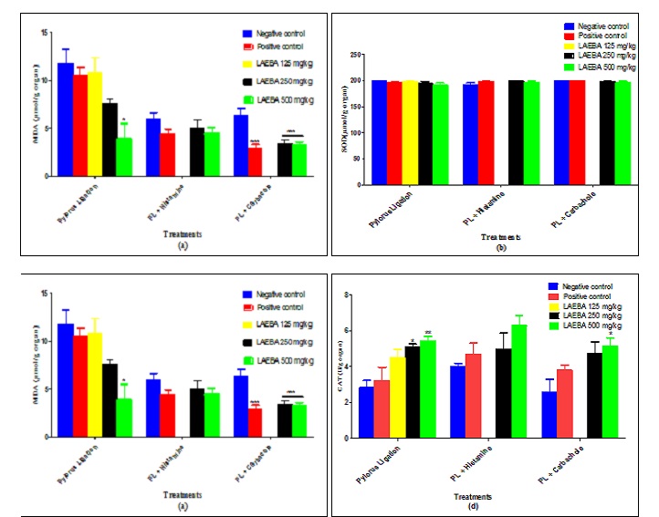
FIG. 1: EFFECTS OF LAEBA ON SOME OXIDATIVE STRESS PARAMETERS A) MALONDIALDEHYDE; B) SUPEROXIDE DISMUTASE; C) CATALASE; D) GLUTATHION; N= NUMBER OF RATS PER GROUP, PL: PYLORUS LIGATION; VALUES REPRESENT MEANS ± SEM; *P<0.05, ***P<0.001: SIGNIFICANT DIFFERENCE COMPARED TO NEGATIVE CONTROL”.
DISCUSSION: The objective of this study was to evaluate the antisecretory and antioxidants effects of LAEBA on gastric ulcers induced in rats. Three models of induction were realized, namely: pylorus ligation (to evaluate antisecretory activity) and pylorus ligation potentialized by histamine/ carbachol (to investigate the antisecretory mecha-nism of this extract). Ulcers formed by the pylorus ligation model are due to the accumulation of gastric juice in the stomach. Indeed, gastric acid contained in this juice, in addition to its corrosive action on the glandular epithelium of the stomach, provides an optimal pH (1.6 - 3.2) necessary to convert pepsinogen into pepsin 19. Pepsin possesses a proteolytic capacity and contributes to release of free radicals by digesting membrane proteins.
Thus, HCl and pepsin are the two elements responsible of gastric ulcer formation in pylorus ligation model. In this model, extract (125, 250 and 500 mg/kg) induced a dose-dependent decrease (p < 0.001) of ulcer index (2.82, 2.50 and 1.60) compared to negative control (4.56).
It was accompanied by a significant reduction of gastric acidity (12.05, 10.47, and 8.06 mEq/l) compared to the negative control (14.07 mEq/l) and correlated with a significant increase of pH compared to the negative control. These results are similar to those obtained by Manowar et al. 29, who reported that reduction of ulcer index, after induction of gastric ulcers by pylorus ligation, was due to antisecretory activity of the extract.
The physiopathological mechanisms of gastric ulcers induced with acetylcholine and histamine are linked. Indeed, acetylcholine synthesized by postganglionary neurons of the parasympathetic system binds on M3 muscarinic receptors coupled with Gq protein to stimulate gastric secretion of acid by direct action on parietal cells and indirect action through induction of gastrin and histamine secretion. Acetylcholine also stimulates the secretion of pepsinogen by principal cells via the increase of Ca2+ following the opening of calcium-channels, which will allow its entry in cells through IP3 and DAG action 30. Histamine stimulates surrounding parietal cells by paracrine pathway via H2 receptors coupled with adenyl cyclase, which transforms ATP in AMPc 31.
It was observed an increase of gastric juice volume in negative control was treated with histamine (13.54 ml) compared to negative control used for pylorus ligation (5.12 ml). LAEBA (250 and 500 mg/kg) induced a reduction of gastric juice volume in pylorus ligation potentialized with histamine (11.83 and 10.60 ml) or carbachol (2.49 and 1.70 ml) compared to their negative controls (13.54 and 3.28 ml, respectively). These results are similar to those obtained by Amang et al. 11 with Eremomastax speciosa (Acanthaceae), which concluded that the antisecretory mechanism could imply both antihistaminergic and anticholinergic pathways. pH interval obtained in test groups, with the three experimental methods, was comprised between 3.81 and 5.99, while the optimal interval to inactivate pepsin is 4-6 32. The extract induced an increase of hydrolyzed protein percentage (18.18, to 64.64%), indicating a reduction of pepsin activity in extract-treated groups compared to the negative control. Mezui et al., 13 found similar results while evaluating antiulcer activity of Corchorus olitorius and suggested that the studied extract could inhibit protein digestion by inactivating pepsin, which could lead to constipation as an adverse effect. Extract globally induced an increase of mucus secretion compared to negative controls. It is established that mucus plays a significant role in the protection of mucous membranes against aggressive agents (hydrochloric acid and pepsin). Its protective effects depend on the quality and quantity or thickness, which covers mucous surface 33, and its secretion is stimulated by endogenous prostaglandins 34. These results those of Amang et al. 35 who suggested that mucus secretion would be implied in protecting mucous membrane against aggressions.
Damages caused by reactive oxygen (ROS) species can be revealed by lipidic peroxidation, which generates the formation of MDA 13. Indeed, tissue lesions start with the formation of superoxideanion (O-2), peroxide hydrogen (H2O2), and hydroxyl radical (OH-) by increasing lipids peroxidation of cellular membranes 36. Thus, ulcer formation can be revealed by an increase of MDA 37. The extract induced a dose-dependent decrease of MDA level in extract-treated groups compared to the negative control. Similar results were observed by Amang et al.11, who suggested that reduction of ulcer index was due to LAEBA through reduction of lipidic peroxidation. SOD converts free radicals into hydrogen peroxide H2O2, which is then transformed in H2O by catalase 38.
The extract induced a dose-¶dependent increase of catalase and GSH levels compared to the negative control. This suggests that the cytoprotective effect of LAEBA could be related to its ability to reinforce antioxidant status of rats.
Qualitative phytochemical screening of LAEBA revealed the presence of coumarins, flavonoids, saponins, and tannins. Antioxidant activities of these classes of compounds are well-known 39; this suggests that these classes of bioactive compounds could be responsible for antioxidant properties of LAEBA.
CONCLUSION: The leaves aqueous extract of B. angustifolia protected gastric mucous membrane by promoting mucus secretion, reinforcing antioxidant status, and reducing gastric acid secretion through mechanisms which would imply both anti-cholinergic and antihistaminergic pathways. These results could justify the use of B. angustifolia in ethnomedicine for the symptomatic treatment of gastric ulcers.
ACKNOWLEDGEMENT: The present work has been financed by the author’s personal funds.
CONFLICTS OF INTEREST: Authors declare no conflicts of interest.
REFERENCES:
- Marieb EN and Hoehn K: Anatomie et physiologie humaine, Québec: Pearson. 8ème Edition 2010; 985.
- Francoeur C, Tremblay-Coutu E, Desroches J, Poitras P, Beaulieu P and Havsteen HB: The biochemistry and medical significance of the flavonoids, Pharmacology and Therapeutics Journal 2002; 96 :67-202.
- Perlemuter G, Perlemuter L, Pitard L and Quevauvilliers J: Anti-inflammatoires stéroïdiens non stéroïdiens. In Pharmacologie Thérapeutiques Italie Elsevier Masson 2011; 133-34.
- Kuipers EJ and Blaser MJ: Ulcères gastroduodénaux, In Goldman L and Schafer AI: Gold man’s ce cil Médicine maladies gastro-intestinales. Elsevier Masson 24 èmeédition 2013; 97-110.
- Aziz K, Bonnet D and Foppa B: Hépato-gastro-entérologie: chirurgie digestive. Elsevier-Masson, 2ème Edition 2012; 441-67.
- Adiaratou T, Korotimi K, Adama D, Mahamane H, Rokia S and Drissa D: Effet protecteur des feuilles de Opilia celtidifolia contre l’ulcère induit par l’éthanol chez le rat. International Formulae Group 2014; 2416-23.
- Eloumou B, Louma NH, Noa NN and Manga A: Facteurs des risques associés aux lésions gastroduodénales dans un hôpital de référence à Douala Cameroun. Médecine Santé Tropicale 2016; 26(1):104-109.
- Drive C: gastro entérologie clinique, Œsophage-estomac. Capelle et Bordeaux 1986; 139.
- Brown LF and Wilson DE: Gastroduodenal ulcers: Causes, diagnostic, prevention and treatment. Comprehensive Therapy 1999; 25(1): 418-22.
- Balian A: Ulcères gastrique et duodénal, In: Hépato-gastroentérologie. Paris Elsevier Masson 2ème Edition 2011; 79-83.
- Amang AP, Tan PV, Patamaken SA and Mefe MN: Cytoprotective and Antioxidant Effects of the Methanol Extract of Eremomastax speciosa in Rats. African Journal Traditional Complement Alternative Medicine 2014; 11(1): 165-71.
- Tan PV, Mezui C, Enow-Orock GE and Agbor G: Antioxidant Capacity, Cytoprotection, and Healing Actions of the Leaf Aqueous Extract of Ocimum suave in Rats Subjected to Chronic and Cold-Restraint Stress Ulcers Ulcers 2013; 2013: 1-9.
- Mezui C, Amang AP, Nkenfou C, Sando Z, Betou D, Moulioum H and Tan PV: Anti-ulcer and antioxidant activities of the leaf aqueous extract of Corchorus olitorius (Tiliaceae) in rats. International Journal of Phyto-pharmacology 2016; 7(1): 17-28.
- Arbonnier M and Ligneux du Sahel: Montpellier, Museum national d’histoire naturelle. Paris CIRAD 2008; 1: 574.
- Ould MH and Abdallahi E: Contribution à l’étude des plantes médicinales de Mauritanie. Série Scien 2009; 9-27.
- Oumar S, Amy B, Sékouna D and Léonarde A: L’arbre en milieu soudano-sahélien dans le bassin arachidier (Centre-Sénégal). J of Applied Biosciences 2013; 61: 4515 -29.
- Hassan Z, Ghanem DS, Foda BM and Ibrahim: Evaluation of the anti-ulcer effect of Aerva javanica aerial parts against ethanol induced gastric lesions in albino rats. Journal of Innovations in Pharmaceutical and Biological Sciences 2017; 2349-59.
- Grossman ED: Peptic Ulcer: A guide for the practicing, physician. Year book Medical Chicago Ill USA 1981.
- Shay JP, Komarov SA, Fels SS, Meranze D, Grunstein M and Simpler H: A simple method for the uniform production of gastric ulceration in the rat. Journal of Gastroenterology 1945; 5: 43-61.
- Tan PV, Nditafon GN, Yewah MP, Ayafor JF and Dimo T: Eremomastax speciosa: Effect on the leaf aqueous extract on ulcer formation and gastric secretion in rats. Journal of Ethnopharmacology 1996; 54(3):139-42.
- Vela S, Souccar TR, Lima L and Manand AJ: Lapa“Inhibition of gastric acid secretion by the aqueous extract and purifed extracts of Stachytarpheta cayennensis, Planta Medica 1997; 63(1): 36-39.
- Syed A and Arshaduddin A: Anti-ulcer activity of Nardostachys jatamansi against Pylorus ligation induced gastric ulcer. Scholars Journal of Applied Medical Sciences 2016; 4(8): 3048-53.
- Amang AP, Tan PV, Nkwengoua E and Nyasse B: Antisecretory Action of the Extract of the Aerial Parts of Eremomastax speciosa (Acanthaceae) Occurs through Antihistaminic and Anticholinergic Pathways, Advances in Pharmacological Sciences 2014; 2014: 1-10.
- Henry RJ, Canon DC and Winkel MJW: Clinical Chemistry. Principles and Techniques, Harper and Row, 2nd Edition 1974; 412-25.
- Wilbur KM, Bernheim and Shapiro OW: Determination of lipid peroxidation. Archives of Biochemistry and Biophysics 1949; 24: 305-10.
- Misra H and Fridovich: The role of superoxide anion in auto oxidation epinephrine to adrenochrome and simple assay for superoxide dismutase. Journal of Biology Chemistry 1972; 247: 3170-75.
- Sinha AK: Colorimetric assay of catalase. Analytical Biochemistry 1959; 47(2): 389-94.
- Ellman GL: Tissue sulfhydrile groups. Archives of Biochemistry and Biophysics 1959; 82: 70-77.
- Manowar H, Hazarika I and Das A: Pylorus Ligation Induced Gastric Ulcer Protection by Sesamum Indicum Ethanolic Seed Extract. Journal of Pharmaceutical Science 2015; 42-49.
- Ader J, Carre F, Xuan A, Duclos M, Kubis N, Mercier J, Mion F, Prefaut C and Roman S: Physiologie, Paris, Masson. 2ème Edition 2006; 247.
- Beaugerie L, Sokol H, Goirand F and Roman S: Les fondamentaux de la pathologie digestive. Paris Masson 2014; 288.
- Vatier JVT and Antiacides: In Pharmacologie des concepts fondamentaux aux applications thérapeutiques, Frison-Roche. Paris 1998; 555-65.
- Ndji OL, Amang PA, Mezui C, Nkwengoua ZE, Tan PV and Nyasse B: Gastric Ulcer Protective and Antioxidant Activity of the Leaf Ethanol Extract of Emilia praetermissa Milne-Redh (Asteraceae) In Rats, Journal of International Research in Medical and Pharmaceutical Sciences 2016; 6(2): 98-107.
- Menche N: Anatomie physiologie biologie, Paris Malouine. 3ème Edition 2006; 335.
- Amang AP, Mezui C, Siwe TG, Nkwengoua ZE, Enow-Orock EG and Tan PV: Prophylactic and Healing Activities of the Leaves Aqueous Extract of Eremomastax speciosa on Gastric Ulcers in Rats. Journal of Advances in Biology and Biotechnology 2017; 12(3): 1-13.
- Singh N, Verma VK, Saxena P and Singh R: Anti-Ulcer and Antioxidant Activity of Morinaga oleifera Leaves against Aspirin and Ethanol Induced Gastric Ulcer in Rats, International Research Journal of Pharmaceuticals 2012; 02(2): 46-57.
- Tandon R, Khanna HD and Goel RK: Oxidative stress and antioxidant status in peptic ulcer and gastric carcinoma. Indian Journal of Physiology and Pharmacology 2004; 48(1): 115-18.
- Pincemail J, Bonjean K, Cayeux K and Defraigne JO: Mécanismes physiologiques de la défense antioxydante. Nutrition Clinique Métabolisme 2005; 16: 233-39.
- Vera-Arzave C, Antonio LC, Arrieta J, Cruz-Hernández G, Velasquez-Mendez AM, Reyes-Ramírez A and Sánchez-Mendoza ME: Gastro protection of suaveolol isolated from Hyptis suaveolens, against ethanol-induced gastric lesions in Wistar rats: role of prostaglandins, nitric oxide and sulfhydryls. Molecules 2012; 17(8): 17-27.
How to cite this article:
Amang AP, Walantini J, Ndji GLO, Siwe GT, Vandi LV and Tan PV: Antisecretory and antioxidant activities of the leaves aqueous extract of Bocsia angustifolia (Capparaceae) on gastric ulcers in wistar rats. Int J Pharmacognosy 2020; 7(12): 353-60. doi link: http://dx.doi.org/10.13040/IJPSR.0975-8232.IJP.7(12).353-60.
This Journal licensed under a Creative Commons Attribution-Non-commercial-Share Alike 3.0 Unported License.
Article Information
3
353-360
0
English
IJP
A. P. Amang *, J. Walantini, G. L. O. Ndji, G. T. Siwe, L. V. Vandi and P. V. Tan
Department of Biological Sciences, Faculty of Science, University of Maroua, Maroua, Cameroon.
perfusionamang@yahoo.fr
21 July 2020
27 September 2020
29 September 2020
10.13040/IJPSR.0975-8232.IJP.7(12).353-60
31 December 2020