


ANATOMICAL STUDIES ON LEAF OF CADABA FRUTICOSA (L.) DRUCE
HTML Full TextANATOMICAL STUDIES ON LEAF OF CADABA FRUTICOSA (L.) DRUCE
G. Prabhakar 1, P. Kamalakar * 2 and K. Shailaja 1
Plant Physiology and Biochemistry Laboratory 1, Department of Botany 2, Osmania University, Hyderabad - 500007, Telangana, India.
ABSTRACT: Cadaba fruticosa (L.) Druce belongs to the family Capparaceae. It is woody, erect, glandular- pubescent shrubs. It is used in the folk system of medicine. Anti-rheumatic, anthelmintic and antibacterial properties it is also used in gastrointestinal, urine complaints and as a vermicide. Leaves used on boils and the leaf juice of this plant are used as a remedy for fevers and is primarily used to cure gonorrhea. Leaves amphistomatic with anisocytic stomata. Trichomes occur on both surfaces. Leaf surface striated. Mesophyll with palisade and spongy tissues. Ground tissue of midvein consists of collenchyma, parenchyma, and scleren-chymatous tissues. Hypodermis collenchyma 1-2 layered on either side. Beneath the collenchyma, parenchymatous tissue is present with small intercellular spaces. Vascular bundles are surrounded by sclerenchymatous tissue.
| Keywords: |
Cadaba fruticosa (L.) Druce, Capparidaceae, anatomy, Stomata
INTRODUCTION: Cadaba fruticosa (L.) Druce belongs to the family Capparaceae. This family of flowering plants containing 28 genera and about 700 species of annual or perennial herbs, subshrubs, shrubs or trees 1. It is woody, erect and glandular – pubescent shrub, frequently found around field hedges 2. Leaves elliptic-oblong 1-5.5 X0.5-3 cm, obtuse, mucronate. Petiole 4-6.5 mm long, flowers 2-2.5 cm; in terminal corymbs. Pedicles 1-2.5 cm long, pubescent, bracts subulate. Sepals ovate to oblong, 1.5 cm long, expanded, toothed. Stamens 4-6 exerted; gynophore 2-2.5 cm long. Fruit cylindric; seeds many 3 Fig. 1A, B. It is also used in gastrointestinal, urine complaints and as vermicide 4. Leaves used on boils and leaf juice are used as eye drops by ethnic people of Andhra Pradesh 5.
The plant is used for the treatment of anti-rheumatic, anthelmintic and antibacterial properties it is also used in gastrointestinal.
MATERIALS AND METHODS: Cadaba fruticosa (L.) Druce was collected from Nalgonda district, Telangana and deposited in Herbarium Hyderabad, Department of Botany, Osmania University. Identified through standard Floras. The leaves were boiled, fixed in F.A.A. (Formaldehyde-Acetic acid-Alcohol), dehydrated through xylene- alcohol series, and embedded in paraffin wax. The sections obtained by rotary microtome and stained with crystal violet and Basic fuchsin combination mounted in Canada balsam 6. Epidermal cells were obtained by gently scraping by razor blade then peels stained with saffranine and mounted in glycerin. Microphotographs were taken with the help of a CCD camera.
OBSERVATIONS AND RESULTS:
Morphology: Erect, glandular, pubescent shrubs, 2-5 m., tall, leaves elliptic-oblong, petioles long, flowers in terminal corymbs, stamens 4-6, fruit cylindric, seeds many.
Microscopy:
Leaf: In surface view adaxial epidermal cells shallowly sinuate, surface slightly striated with anisocytic stomata. Abaxial epidermal cells deeply sinuate, sinuses ‘U’-V-shaped, surface more striated anisocytic stomata. Two types of peltate trichomes are present on both surfaces Fig. 1C-F. Mesophyll differentiated into palisade and spongy tissues. Palisade single layered, occasionally 2-3 layered at some places, cells elongated, squarish, compactly arranged with small intercellular spaces, contents dense with chloroplasts. Spongy tissue is occupying 4 – 5 layers, cells mostly oval – circular, arranged with large intercellular spaces, contents dense with chloroplasts Fig. 2A-B. Midvein heterogenous consists of collenchyma, paren-chyma, and sclerenchyma tissues. Collenchyma is present beneath the epidermis, cells polygonal, oval to circular, walls thick without intercellular spaces. Parenchymatous cells oval to circular contents dense with chloroplasts, intercellular spaces narrow. Midvein consists of 2 vascular bundles placed abaxially and adaxially. The abaxial vascular bundle is larger, surrounded by sclerenchymatous sheath on phloem as well as on xylem side.
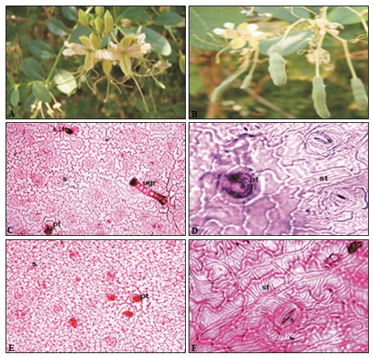
FIG. 1: A) PLANT HABITAT, B) PLANT HABITAT WITH FRUIT, C) ADAXIAL SURFACE, D) ADAXIAL SURFACE (ENLARGED), E) ABAXIAL SURFACE, F) ABAXIAL SURFACE (ENLARGED)
Petiole: Petiole cylindrical. The trace enters the petiole base as ‘C’ shaped arc whose arms are very close. Higher above, only in the base, these arms curve and meet each other. Later the ends of the arm get detached from the main vasculature get fused and as a result, a vascular trace enclosed within arc is produced. Ends of arc remain separate by sclerenchyma. Higher above arms of vasculature meet to form a ring around the central enclosed vascular bundle. At the same time, the central bundle develops fibrous sheath. Main vasculature surrounded by pericyclic sclerenchyma, cortex parenchymatous. Again at the base of lamina, the medullary bundle divides into two.
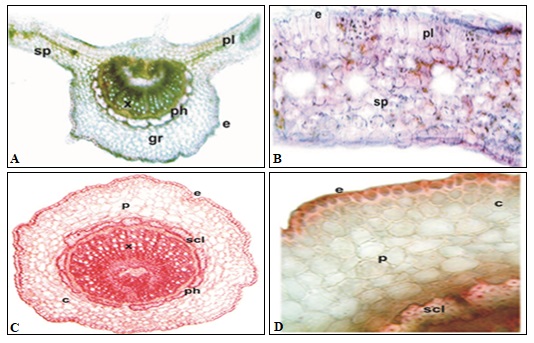
FIG. 2: A) T.S. OF LEAF MIDVEIN, B) T.S. OF LEAF LAMINA, C) T.S. OF THE PETIOLE, D) T.S. OF PETIOLE ENLARGED
The traces are pushed on the upper side and join the main cylinder which opens to form a ‘C’ shaped arc with incurved arms Fig. 2C-D.
DISCUSSION: Little anatomical data is available for comparison. Findings of the present investigation are discussed in the light of data given by Metcalfe and Chalke (1972) 7. Peltate glandular trichomes found in Cadaba fruticosa are reported in species of Cadaba by earlier authors also. Emergences resembling hairs reported earlier in Cadaba species are absent from present species. Trichomes with the bulbous base are reported for the first time. Capparaceae are with anomocytic stomata, however, here anisocytic stomata are present. Both centric and dorsiventral mesophyll reported in the family.
In the present species, palisade is either distinct, single layered or 2-3 layered or may be homogeneous 8. Such a varied type of mesophyll appears to be the characteristic feature of Cadaba fruticosa. The transition in the vascular architecture of petiole is also an interesting feature. Petiole structure varies in Capparidaceae. Petiole architecture of C. fruticosa appears to be a combination of Cadaba linearise Jacq. and Stereophoma elipticum (DC) Spreng.
CONCLUSION: This study will help for the identification of Cadaba fruticosa (L) Druce in fresh and powder form; it can also be useful for standardization purpose.
ACKNOWLEDGEMENT: The authors wish to thank the Head Department of Botany, Osmania University for providing the laboratory facility and constant encouragement. Authors also thankful To UGC, New Delhi for Financial Support in the form of BSR -RFSMS Fellowship.
CONFLICT OF INTEREST: Nil
REFERENCES:
- Tiruchirapalli: The Rapinat Herbarium, St. Josephs College, Tiruchirapalli 1981: 36-38.
- Bhogaonkar PY and Chavhan VN: Traditional Banjara Herbal Medicine of Vidarbha, M.S., India Lambert Germany. Academic. Lap Publishing, 2013.
- Chopra RN, Nayar SL and Chopra IC: Glossary of Indian Medicinal Plants. National Institute of Science Communication, New Delhi, 1996.
- Jain SK: Dictionary of Indian folk medicine and ethnobotany. Deep publication, New Delhi-110015, 1991.
- Reddy KN, Trimurthulu G and Sudhkar Reddy C: Plants used by the ethnic people of Krishna district, Andhra Pradesh. Indian Journal of Traditional Knowledge 2010, 9(2): 313-317.
- Johanson DA: Plant Microtechnique. Tata Mc-Grawhill Publishing Company, Ltd. New Delhi, 1940.
- Metcalfe and Chalke” Anatomy of the Dicotyledones. Oxford University Press, Ely House, London, Vol. I and II, 1972.
- Evans WC: Trease and Evans Pharmacognosy. W. B. Saunders Company Limite Singapore, Edition 14th, 1997.
How to cite this article:
Prabhakar G, Kamalakar P and Shailaja K: Anatomical studies on leaf of Cadaba fruticosa (L.) Druce. Int J Pharmacognosy 2015; 2(11): 546-49. doi link: http://dx.doi.org/10.13040/IJPSR.0975-8232.IJP.2(11).546-49.
This Journal licensed under a Creative Commons Attribution-Non-commercial-Share Alike 3.0 Unported License.
Article Information
5
546-549
555
2037
English
IJP
G. Prabhakar, P. Kamalakar * and K. Shailaja
Department of Botany, Osmania University, Hyderabad, Telangana, India.
kamalakarpalkurthy@gmail.com
26 September 2015
19 November 2015
28 November 2015
10.13040/IJPSR.0975-8232.IJP.2(11).546-49
30 November, 2015