ANATOMICAL AND THIN LAYER CHROMATOGRAPHIC IDENTIFICATION OF ROOT, ROOT-BARK AND LEAF OF PREMNA SERRATIFOLIA L.
HTML Full TextANATOMICAL AND THIN LAYER CHROMATOGRAPHIC IDENTIFICATION OF ROOT, ROOT-BARK, AND LEAF OF PREMNA SERRATIFOLIA L.
K. Babu *, M. Priya Dharishini and Anoop Austin
R & D Center, Cholayil Private Limited, Ambattur, Chennai - 600098, Tamil Nadu, India.
ABSTRACT: Anatomical characters and thin layer chromatographic fingerprint of root, root-bark and leaf of Agnimanthaḥ (Premna serratifolia L.), an important drug used in Ayurvedic system of medicine has been studied in detail to identify the original drug from the substitutes/adulterants.
| Keywords: |
Agnimanthaḥ, Premna serratifolia, Lamiaceae, Identification, Substitute, Adulterant
INTRODUCTION: Agnimanthaḥ is a renowned drug in Ayurveda, constitute one of the drugs in Daśamūla and also forms an ingredient of many important Ayurvedic preparations like Daśamūlāriṣṭaṃ, Dhānvantaraṃ Kaṣāyaṃ, Agastyarasāyanaṃ, Sukumāraghṛtaṃ, etc. The officinal part is root, root-bark and leaves. The roots possess properties like astringent, bitter, acrid, sweet, thermogenic, anodyne, anti-inflammatory, alexiteric, cardiotonic, expectorant, depurative, digestive, carminative, stomachic, laxative, febrifuge, and tonic. They are useful in hepatopathy, cough, asthma, skin diseases, diabetes, and general debility. The leaves are stomachic, carminative and galactagogue and useful in dyspepsia, flatulence, colic, cough, fever, Rheumatologic, hemorrhoids, hepatoprotective and antitumor 1 - 3.
The bark possesses antioxidant, antidiabetic 4 - 5, antimicrobial 6, cardiac stimulant 7 and cardio-protective activity 8.
In all the classical treatises, two types are mentioned viz. agnimanthaḥ and taṛkārī, these are equated with Clerodendrum phlomidis L.f. and Premna serratifolia L. (Syn: Premna corymbosa Rottl. & Wild.; P. obtusifolia R. Br.) respectively, both belonging to the family Lamiaceae 9. The Ayurvedic pharmacopeia of India also accepted Clerodendrum phlomidis as agnimanthaḥ 10. But in the Ayurvedic Formulary of India, contradict stating that Premna serratifolia as the real source of the drug and Clerodendrum phlomidis and Premna mucuronata as the substitute 11. Chunekar in his commentary on Bhāvapṛakāśa nighaṇṭu opines that these two can be used as substitutes for each other, as they have similar properties 9. But most of the authors equate the drug with Premna serratifolia 1 - 2. Vasudeva Nair also mentioned Premna serratifolia is used as agnimanthaḥ in Kerala and Premna latifolia Roxb. and Clerodendrum phlomidis can be used as substitute according to the availability 12.
Though Premna serratifolia is the accepted source of agnimanthaḥ throughout Kerala, which is the main center where Ayurveda has survived through centuries, however, commercial drug invariably mixed with Premna species. Hence, it necessitates standardizing the original drug from adulterants.
Recently, Susan et al., studied the powdery microscopy and phytochemistry of Premna serratifolia root 13, Hurshitha Kumari et al., studied the comparative pharmacognostic evaluation of leaves of four species viz. Premna obtusifolia R. Br., P. latifolia Roxb., Clerodendrum phlomidis L.f., C. inerme (L.) Gaertn. 14 and Gokani et al., studied the comparative pharmacognostic features of Clerodendrum phlomidis and Premna integrifolia roots 15.
However, there is a lack of detailed anatomy of leaf, root-bark and root. Hence, the present investigation was undertaken to provide the complete anatomical diagnostic characters of leaf, root-bark and root of the drug agnimanthaḥ, Premna serratifolia.
MATERIALS AND METHODS:
Anatomical Studies: Fresh root, root-bark, and leaves were collected from the authentic plant (12 years old) grown in R & D center campus, Cholayill Pvt. Ltd., Ambattur, Chennai. The samples were cut into small pieces and fixed immediately in Formalin-Acetic-Alcohol for 24 h. After fixation they were washed thoroughly in distilled water, dehydrated, embedded in paraffin wax after infiltration and sectioned using rotary microtome to the thickness of 8 - 12 µm.
Sections were stained with toluidine blue and photographed. For the study of individual wood elements, wood tissues were macerated employing Jeffrey’s fluid. For leaf clearing, small pieces were treated in 5% NaOH at 60 °C for 3 h, washed with distilled water thoroughly and stained with safranin 16.
Thin Layer Chromatographic (TLC) Analysis: For the TLC analysis, root, root-bark and leaves samples were shade dried for a week and powdered. 2 g of each powdered sample was refluxed with 10 ml of ethyl acetate in a water-bath at 60 °C for 30 min, consecutively 3 times, and then concentrated and dried. The final extract was re-dissolved in ethyl acetate and used for the TLC analysis. Precoated Silica Gel F254 (Merck) plate was used for the stationary phase and chloroform: methanol (9:1) was used as mobile phase. After development, the plate was observed under UV-254 nm and spots recorded. Then the plate was dipped in 0.5% Anisaldehyde sulphuric acid and heated at 105 °C in a hot air oven for 5 min to develop the color and the spots recorded.
RESULTS:
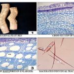
Macroscopic Characters of the Root (Fig. 1: 1): Root about 1 - 1.5 cm thickness shows strong, hard, woody, brown color when fresh, dark brown when dry with clear longitudinal furrows and rootlet scars; aromatic, camphoraceous and characteristic odor.
Microscopic Characters of the Root (Fig. 1: 2 - 4): A transverse section of root about 6 - 7 mm thickness shows distinct periderm, compressed cortex, and tracheary elements. Xylem region shows quite distinct growth rings; diffuse porous, growth rings demarcated by thick-walled, narrow lumened fibers. Vessels are circular or oval, mostly wide, thin-walled, mostly solitary, less frequently in tangential multiples. Individual vessel element cylindrical, short or long, with simple horizontal perforation plate, intervascular pits simple, with or without the tail. Xylem rays distinct, bi- or tri-seriate, rarely tetra-seriate. Fibres libriform, long or short, septate, narrow lumen. Tracheids short, narrow with simple pits.
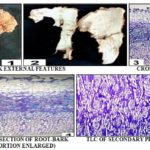
Macroscopic Characters of the Root-Bark (Fig. 2: 1 - 2): The bark has a thickness of about 8 to 10 mm, brown color outer side, pale white inner side, rough surface with peelable fakes and longitudinal furrows, prominent rootlet scars with a characteristic odor.
Microscopic Characters of the Root-Bark (Fig. 2: 3 - 5): Bark differentiated into outer region periderm, middle region secondary cortex and inner region secondary phloem. Periderm is broad and well developed. Phellem and phellogen distinct, phellem cells are homogeneous, thin-walled, large and loosely arranged, somewhat angular in shape. Phelloderm and secondary cortex cells are compressed and distinctly exhibit dark zone. Secondary phloem and cambium present innermost regions. Secondary phloem region broad with bi- or tri-seriate phloem rays. Phloem rays homo or heterocellular. Isolated or group of brachysclereids present in the cortex and secondary phloem region. No calcium oxalate crystals found.
FIG. 1: MACROSCOPIC AND MICROSCOPIC FEATURES OF ROOT OF PREMNA SERRATIFOLIA
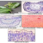
Macroscopic Characters of the Leaf (Fig. 3: 1): Aromatic, thick, shiny, dark green ventral, light green dorsal; petiole 1.9 - 3.5 cm, puberulent; lamina oblong to broadly ovate, 10 - 13 × 5.7 - 7.5 cm, glabrous or pubescent only along veins, base broadly cuneate, rounded or truncate, margin entire, slightly undulate, or crenate, apex acute to rarely acuminate or obtuse.
Microscopic Characters of the Leaf (Fig. 3: 2- 5):
Petiole (Fig. 3: 2): The petiole in transverse sectional view cubical in shape with the distinct concave adaxial surface. The epidermis is a single layer, cubical shaped cells with trichomes. Next to the epidermis 4 - 5 layers of collenchymatous cells present. A large dorsal vascular bundle and two small ventral vascular bundles are present.
Thus, the arrangement is expressed as 1 + 2. The dorsal vascular bundle consists of a central main bowl-shaped xylem and phloem. Patches of sclerenchyma cells covered the phloem. The ventral vascular bundles are somewhat elongate of circular. The center region of ground tissue is made up of large parenchymatous cells. Calcium oxalate crystals are no evident.
Lamina (Fig. 3: 3 - 5): The midrib has convex in the adaxial surface, a single layer epidermis having cuboid shape cells with a thick cuticle. Three to four layers of collenchyma cells followed by epidermis. A bowl-shaped large vascular bundle in the dorsal side and small circular shaped vascular bundle in ventral side consisting xylem and phloem. Patches of sclerenchyma surrounded the phloem region. The center ground tissue is made up of parenchymatous cells.
In lamina, the upper epidermis has large, cuboid or rectangle shaped cells and the lower epidermis has comparatively small cuboid-shaped cells. Both epidermises has thick cuticle. Next to the upper epidermis, the narrowly cylindrical single layer of palisade parenchyma and spherical spongy parenchyma cells forming a network. Vascular bundles are of various sizes. The stomata diacytic type and hypostomatic. Peltate trichomes gland present in the lower epidermis.
FIG. 2: MACROSCOPIC AND MICROSCOPIC FEATURES OF ROOT- BARK OF PREMNA SERRATIFOLIA
FIG. 3: MACROSCOPIC AND MICROSCOPIC FEATURES OF LEAF OF PREMNA SERRATIFOLIA
C – Cortex; Ca – Cambium; CVB – Central vascular bundle; E – Epidermis; EC – Epidermal cell; Fi – Fibre; GR – Growth ring; Gt – Glandular trichome; GT – Ground tissue; LE – Lower epidermis; LVB – Lateral vascular bundle; P – Periderm; Phl – Phloem; PP – Palisade parenchyma; PR – Phloem rays; Sc – Sclerenchyma; Scl – Sclereids; SP – Spongy parenchyma; Sph – Secondary phloem; ST – Sieve tube; St – Stomata; Tr – Tracheid; UE – Upper epidermis; VE – Vessel element; XE – Xylem elements; XP – Xylem parenchyma; XR – Xylem rays.
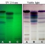
Thin Layer Chromatographic (TLC) Analysis (Table 1; Fig. 4): The Rf - values, and color of the spot shown in Table 1 and Fig. 4 can largely distinguish the parts of the plant.
FIG. 4: TLC FINGERPRINT OF LEAF, ROOT-BARK, AND ROOT OF PREMNA SERRATIFOLIA
A – Leaf, B – Root-bark, C – Root
TABLE 1: THE TLC PROFILE AND Rf - VALUES OF LEAF, ROOT-BARK, AND ROOT AS FOLLOWS
| Leaf | Root-bark | Root | |||
| Rf - Values UV-254 nm | Rf - Values
Visible light (after spray) |
Rf - Values UV-254 nm | Rf - Values
Visible light (after spray) |
Rf - Values UV-254 nm | Rf - Values
Visible light (after spray) |
| 0.16 (Black)
0.46 (Black) 0.51 (Black) 0.67 (Black) 0.84 (Black) |
0.03 (Brown)
0.21 (Brown) 0.25 (Purple) 0.26 (Light brown) 0.34 (Pink) 0.37 (Purple) 0.40 (Light blue) 0.53 (Sky blue) 0.64 (Orange) 0.67 (Violet) 0.73 (Violet) 0.85 (Dark violet) |
0.38 (Black)
0.65 (Black) 0.77 (Black) |
0.25 (Purple)
0.37 (Purple) 0.40 (Light blue) 0.46 (Pink) 0.52 (Sky blue) 0.57 (Blue) 0.60 (Sky blue) 0.63 (Greenish blue) 0.65 (Brown) 0.73 (Violet) 0.79 (Brown) 0.81 (Blue) 0.85 (Violet) |
0.77(Black) | 0.21 (Brown)
0.25 (Purple) 0.37 (Purple) 0.40 (Light blue) 0.53 (Sky blue) 0.64 (Violet) 0.73 (Violet) 0.85 (Violet) |
DISCUSSION AND CONCLUSION: Ayurveda - the ancient indigenous system of medicine is being discovered in our own country and day by day increasing its popularity. The major disadvantage of the Ayurvedic system is the difficulty in identifying the genuine drug prescribed by the founders. Their description of medicinal plants is more poetic than scientific and lacks accuracy because the language they have used is not technical and they did not follow a systematic format for describing the plants. So, this leads to the erroneous identification of more than one plant as the same raw drug by different authors.
Today several plants are used as substitutes for the genuine ones, and the use of such plants is increasing day by day 12. Agnimanthaḥ is such a controversial plant and various plants viz., P. obtusifolia, P. latifolia, P. mucuronata, C. phlomidis, C. inerme are substituted or adulterated to the genuine drug P. serratifolia. Hence, the present investigation was undertaken to provide detailed anatomical features and TLC profile of root, root-bark and leaf of P. serratifolia for the identification purpose. The following anatomical features aid largely to identify the drug in fragmentary form.
Root: Growth rings distinct, diffuse porous, pores circular or oval, wide, solitary, rarely tangential multiples; vessel element cylindrical, short or long, with simple horizontal perforation plate, simple pits, with or without tail; xylem rays bi- or tri-seriate; fibres libriform, septate; tracheids short, narrow with simple holes.
Bark: Distinct three regions - outer periderm, middle compressed cortex (dark zone) and inner secondary phloem; phloem rays bi- or tri-seriate, homo or heterocellular; the presence of brachysclereids in the cortex and secondary phloem region.
Leaf: Petiole - Cubical in shape with the distinct concave adaxial surface; epidermis with trichomes; a large dorsal and two small ventral vascular bundles, the arrangement is 1 + 2; patches of sclerenchyma cells covered the phloem. Lamina - midrib convex in adaxial surface; epidermis cuboid shaped cells with thick cuticle; bowl-shaped large vascular bundle in dorsal side and small circular shaped vascular bundle in ventral side; patches of sclerenchyma surrounded the phloem; upper epidermis large, cuboid or rectangle shaped cells and lower epidermis comparatively small; presence of single layer palisade parenchyma and spherical spongy parenchyma; stomata diacytic type and hypostomatic; presence of peltate trichomes gland in lower epidermis.
ACKNOWLEDGEMENT: The authors are thankful to Mr. Pradeep Cholayil, Chairman and Managing Director and Mrs. Jayadevi Pradeep Cholayil, Directress - Cholayil Private Limited for their support and encouragement in carrying out the study.
CONFLICT OF INTEREST: Nil
REFERENCES:
- Sivarajan VV and Balachandran I: Ayurvedic drugs and their sources. Oxford & IBH Publishing Co. Pvt. Ltd., New Delhi 1994.
- Warrier PK: Indian Medicinal Plants. Orient Longman Pvt. Ltd., Kottakkal, Kerala, India 1995.
- Vadivu R, Suresh AJ, Girinath K, Kannan BP, Vimala R, and Kumar SNM: Evaluation of the hepatoprotective and in-vitro cytotoxic activity of leaves of Premna serratifolia Journal of Scientific Research 2009; 1(1): 145-52.
- Rekha R, Basha SN and Ruby S: Evaluation of in-vitro antioxidant activity of stem-bark and stem-wood of Premna serratifolia (Verbenaceae). Research Journal of Pharmacognosy and Phytochemistry 2009; 1(1): 11-14.
- Majumder R, Akter S, Naim Z, Al-Amin and Alam B: Antioxidant and anti-diabetic activities of the methanolic extract of Premna integrifolia Advances in Biological Research 2014; 8(1): 29-36.
- Rekha R: Antimicrobial activity of different bark and wood of Premna serratifolia International Journal of Pharma and Bio Sciences 2010; 1(1): 1-9.
- Rekha R, Suseela L, Sundaram MR and Basha SN: Cardiac stimulant activity of bark and wood of Premna serratifolia Bangladesh Journal of Pharmacology 2008; 3: 107-13.
- Rekha R and Basha SN: Cardioprotective effect of ethanol extract of stem-bark and stem-wood of Premna serratifolia (Verbenaceae). Research Journal of Pharmacy and Technology 2008; 1(4): 487-91.
- Chunekar KC: Bhāvapṛakāśa nighaṇṭu of Sri Bhavamisra - Commentary. Varansasi, India 1982.
- Anonymous: The Ayurvedic Pharmacopoeia of India. Govt. of India, Ministry of health and family welfare, Dept. of ISM & H, New Delhi 2001; 3.
- Anonymous: The Ayurvedic Formulary of India. Part – I. Govt. of India, Ministry of health and family welfare, Dept. of ISM & H, New Delhi, 2nd Ed, 2003.
- Nair VR: Controversial drug plants. University Press (India) Pvt. Ltd. Hyderabad, India 2004.
- Susan RS, Harini A, Kumar SKN and Hegde PL: Pharmacognostic and phytochemical analysis of Agnimantha (Premna corymbosa) root. Journal of Ayurvedic and Herbal Medicine 2016; 2(6): 204-08.
- Kumari H, Shrikanth P, Pushpan R, Harisha CR and Nishteswar K: Comparative pharmacognostical evaluation of leaves of regionally accepted source plants of agnimantha. Journal of Research and Education in Indian Medicine 2013; 19(1-2): 1-8.
- Gokani RH, Kapadia NS and Shah MB: Comparative pharmacognostic study of Clerodendrum phlomidis and Premna integrifolia. Journal of Natural Remedies 2008; 8(2): 222-31.
- Johansen DA: Plant Microtechnique. McGraw Hill Book Company, New York 1940.
How to cite this article:
Babu K, Dharishini MP and Austin A: Anatomical and thin layer chromatographic identification of root, root-bark and leaf of Premna serratifolia L. Int J Pharmacognosy 2018; 5(5): 302-07. doi link: http://dx.doi.org/10.13040/IJPSR.0975-8232.IJP.5(5).302-07.
This Journal licensed under a Creative Commons Attribution-Non-commercial-Share Alike 3.0 Unported License.
Article Information
8
302-307
990
1825
English
IJP
K. Babu *, M. P. Dharishini and A. Austin
R & D Center, Cholayil Private Limited, Ambattur, Chennai, Tamil Nadu, India.
babuk@cholayil.com
04 January 2018
02 February 2018
13 February 2018
10.13040/IJPSR.0975-8232.IJP.5(5).302-07
01 May 2018


