


A PRELIMINARY PHARMACOGNOSTICAL REPORT ON THE LEAF OF DREGEA VOLUBILIS (L.F.) BENTH. EX HOOK. F.
HTML Full TextA PRELIMINARY PHARMACOGNOSTICAL REPORT ON THE LEAF OF DREGEA VOLUBILIS (L.F.) BENTH. EX HOOK. F.
P. Sudhakar, D. Kavitha and P. Ramachandra Reddy *
Department of Botany, Plant Anatomy and Taxonomy Laboratory, Osmania University, Hyderabad - 500007, Telangana, India.
ABSTRACT: Dregea volubilis (L.f.) Benth. Ex Hook. f. is a large twinning shrub belonging to the family Asclepiadaceae. The leaf is amphistomatic with tetracytic, anomocytic, paracytic stomata. The sides of the leaf in the surface are straight to curved, surface striated on either side. The mesophyll is interspersed with sphaerocrystalliferous idioblasts. Midvein consists of a single arcuate vascular bundle, bicollateral, conjoint, endarch. In T. S. petiole is oval, adaxially grooved at center and with two lateral adaxial ridges, consisting of a large arc shaped vascular bundle at the centre with a 2 small spherical vascular bundles in adaxial ridges.
| Keywords: |
Dregea volubilis, Asclepiadaceae, Sphaero crystals, Pharmacognosy
INTRODUCTION: Dregea volubilis (L.f.) Benth. Ex Hook. F. commonly known as ‘Dudipala’ in Telugu, ‘Nakchhikni’ in Hindi, ‘Madhumalati’ in Sanskrit belongs to the family Asclepiadaceae. It is a large twinning shrub, often with lenticels and sometimes with small black dots. Leaves 6.3-15 by 4.5-11.5 cm, broadly ovate, acuminate, glabrous or more or less softly pubescent, reticulately veined and with a few small glands just above the petiole, base rounded or cordate; petioles 1.3-3.2 cm, long. Flowers numerous, green or yellowish green, in lateral drooping umbellate cymes; pedicels 6-25 mm, long; calyx divided nearly to the base; segments 2.5 mm, long, ovate-oblong, obtuse or subacute, ciliolate. Corolla 6 mm, long, deeply divided, glabrous outside; lobes 5 mm; corona-lobes large, fleshy, the upper free portion rounded on the outer edge.
Staminal column arising from the base of the corolla; anther-tips membranous, broadly ovate-oblong, obtuse; pollen masses oblong, attached to the pollen-carriers by very short caudicles. Style-apex dome-shaped. Follicles 7.5-10 cm, long, slightly tapering to a very blunt point, rugosely striate, glabrous. Seeds broadly ovate, flattened, strongly margined, pale yellowish brown; coma 4.5 cm, long, copious 1. The present describes micro and macroscopic characters and powder microscopy of the leaf of Dregea volubilis. Hence, the information can be of immense use for pharmacognosists, drug manufacturers and medicinal practitioners in the identification of the authentic drug.
MATERIALS AND METHODS: Dregea volubilis plant material was collected from various locations of Warangal district, Telangana, India. The collected material was taxonomically identified and deposited in Herbarium Hyderabad tense, Department of Botany, Osmania University, Hyderabad. The leaves are boiled, fixed in F. A. A. (Formaldehyde -Acetic acid- Alcohol), dehydrated through xylene - alcohol series embedded in paraffin wax. The sections were cut at 10-12 μm on Optica 1090A rotary microtome, stained with crystal violet and basic fuchsin combination and mounted in Canada balsam 2. Epidermal peels were obtained by gently scraping and peeling by razor blade, were stained with saffranine and mounted in glycerine. The powder microscopy characters were studied by boiling the drug in distilled water, stained in saffranine and mounted with glycerine. The photomicrography was done on Olympus BX-53 trinocular microscope attached with a digital Sony camera.
OBSERVATIONS AND RESULTS:
Microscopy: In surface view epidermal cells mostly 5-7 sided, rarely 4 sided, mostly polygonal isodiametric, few anisodiametric, sides thick, straight to curved, surface striated, contents dense and few with calcium oxalate crystals. Stomata mostly tetracytic, few anomocytic, rarely paracytic, subsidiaries 2-5, indistinct, monocyclic, mostly of type, surface striated, guard cells reniform to linear, densely cytoplasmic. Uniseriate macro form cylindrical hair distributed all over on either side.
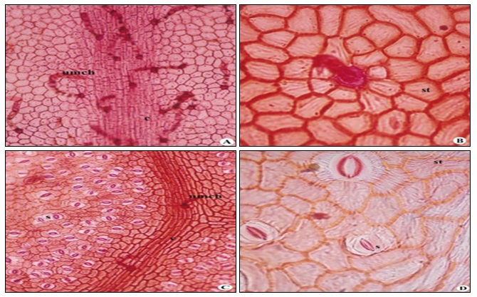
FIG. 1: A) LEAF ADAXIAL SURFACE WITH UNISERIATE CYLINDRICAL HAIR X 139; B) LEAF ADAXIAL SURFACE WITH X 398; C) LEAF ABAXIAL SURFACE WITH COASTAL CELLS X 105; D) LEAF ABAXIAL SURFACE WITH STRIATIONS X 401. umch-uniseriatemacroform cylindrical hair; c-costal cells; st-striations; s-stomata
Epidermal cell frequency 2060 per sq mm, stomatal frequency 78 per sq mm, stomatal index 2.92 (Adaxial surface) Fig. 1 A, B and E. C. F. 2346 per sq mm, stomatal frequency 78 per sq mm, S. I. 2.92 Fig. 1 C, D.
Leaf Anatomy: In T. S. leaf is faintly ribbed adaxially and ribbed conspicuously on the adaxial side at midvein; secondary veins slightly ribbed, lamina wings 151-270 (216) µm and midvein 583- 680 (629) µm in thickness; margins rounded. The epidermis is one layered, cells barrel-shaped, oval to circular and few tabular, contents scanty, cuticle thick, sculptured Fig. 2A, B and C. Mesophyll differentiated into palisade and spongy tissues. Palisade 1- layered, adaxial, throughout, interrupted at midvein and secondary veins, cells columnar, cylindrical, perpendicular to the epidermis; cells 27-68 (43) µm long and 8-19 (13) µm wide; intercellular spaces narrow, contents slightly dense with chloroplasts, cells occasionally interspersed with sphaero crystalliferous idioblasts. Spongy tissue 6-8 celled thick, abaxial, cells circular, oval to oblong and armed, loosely arranged with large intercellular spaces, elongated cells 22 - 38 (30) µm long and 8 - 19 (14) µm wide and isodiametric cells 11 - 27 (19) µm in diameter, contents scanty in few with chloroplasts, cells interspersed with sphaerocrystalliferous idioblasts Fig. 2B.
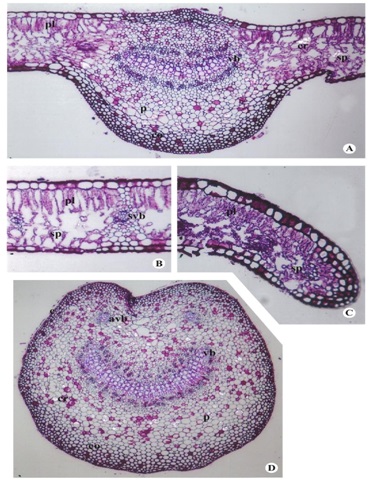
FIG. 2: A) T. S. OF LEAF MIDVEIN X 116; B) T. S. OF LEAF LAMINA X 182; C) T. S. OF LEAF MARGIN X 175; D) T. S. OF PETIOLE X 127. e- epidermis; pl – palisade; sp-spongy tissue; co-collenchyma; p-parenchyma; p-parenchyma; cr-crystal; vb-vascular bundle; svb- secondary vascular bundle; avb- adaxial vascular bundle.
The ground tissue of midvein heterogenous, differentiated into collenchyma and parenchyma tissues. Collenchyma 3 to 4 layered adaxially and 4-6 layered abaxially; cells polygonal, oval to spherical, lamellar; 5-16 (11) µm in diameter on adaxial and cells on abaxial larger, 5-19 (13) µm in diameter, contents scanty, walls thick few tanniniferous idioblasts occur on abaxial and adaxial side.
Parenchyma 10-12 celled in thickness on either sides; cells smaller, polygonal, circular and oval to spherical, cells about 14-27 (19) µm in diameter adaxially, abaxially larger 16-41(28) µm in diameter, walls thin, with small intercellular spaces, contents scanty in few with tanniniferous idioblasts about 11-25 (17) µm in diameter, cells interspersed with sphaerocrystalliferous idioblasts, more frequently on abaxial side. Vascular tissue of midvein consists of the single arcuate vascular bundle 324-410 (364) µm tangentially long 108-132 (173) µm radially wide, bicollateral, conjoint, endarch. Xylem at the center in radial rows, phloem towards abaxially and adaxially; apericyclic, 2-3 celled vascular cambium is present. Tracheary elements 30-35 number at midvein; arranged in radial rows, polygonal, lignified 8-25 (18 ) µm in diameter, walls thick; secondary vascular bundle about 27-41 (35) µm in diameter, arc-shaped, bundle sheath extending to upper epidermis to lower epidermis in minor bundles are present.
Secondary wall thickenings of tracheary elements consist helical thickenings. Xylem parenchyma arranged in between tracheary elements, cells polygonal, slightly thick walled, contents scanty. Phloem is present on sides of midvein bundle consists of phloem parenchyma, sieve tubes, and companion cells; phloem parenchyma is compactly arranged, cells small, polygonal, thin-walled, contents scanty and without intercellular spaces; sieve cells accompanied with companion cells Fig. 2A and B.
Petiole Anatomy: In T. S. petiole is oval shaped, adaxially grooved at the centre and with two lateral adaxial ridges, 1500-1698 (1635) µm laterally long and vertically 1387-1613 (1488) µm wide. Epidermis one layered, cells mostly oval to circular, few barrel-shaped, walls thick, contents dense with few tanniniferous idioblasts and few scanty; cuticle thick over the surface, stomata, and trichomes absent. Ground tissue heterogenous consists of collenchyma and parenchyma tissues. Collenchyma hypodermal, 3-6 layered, throughout, lamellar, cells polygonal, oval to circular, few oblong 14-36 (26) µm in diameter, walls thick, without intercellular spaces; contents scanty in few dense with tannins. Parenchyma 20-23 layered adaxially, 14-17 layered abaxially, beneath the collenchyma; often interspersed with sphaero-crystalliferous idioblasts, 14-50 µm in diameter, contents scanty, with few tanniniferous idioblasts, 14-36 (24) µm in diameter.
Vascular tissue consisting of a large arc-shaped vascular bundle at centre with a 2 small spherical vascular bundles in adaxial ridges; large vascular bundle laterally 821-990 (871) µm long and vertically 226-453 (311) µm wide, conjoint, bicollateral, apericyclic; xylem consists of numerous tracheary elements arranged in radial rows, few laterally aligned; adaxial bundles small, spherical, consisting of few tracheary elements surrounded by phloem; tracheary elements polygonal, oval to circular, 16-36 (25) µm in diameter, walls thick; secondary wall thickenings of tracheary elements in L.S. consist mostly helical, annular and scalariform and few bordered pitted; helices double and single, annular rings free.
Xylem parenchyma is present in between tracheary elements, a 2-3 layered cambium is present in between xylem and phloem. Phloem consists of sieve tubes with companion cells, phloem fibers, and phloem parenchyma; phloem parenchyma cells compactly arranged, cells polygonal, walls thin, intercellular spaces absent, contents scanty, occasionally interspersed with sphaero-crystalliferous cells Fig. 2D.
Powder Microscopy: Powder consists of pieces of the abaxial epidermis with straight to curved sides and tetracytic and anomocytic stomata; pieces of coastal cells with polygonal to linear cells in parallel rows, dense with crystals of calcium oxalates, many; uniseriate cylindrical hair long, either whole or broken.
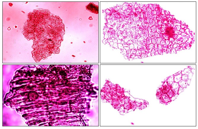
FIG. 3: POWDER MICROSCOPY
DISCUSSION AND CONCLUSION: The anticlinal walls of epidermal cells are reported straight on the adaxial while straight to slightly wavy towards abaxial 3, while presently the sides are thick, straight to curved on either surface. The surface of the epidermal cells has been reported as striated 4 which is also confirmed presently. Further, the epidermal cells are found dense with contents also with calcium oxalate crystals in few. The sides are thick and straight to curved, and the surface is striated. The foliar stomata are reported as hypostomatic 3, 5, 6, 7. Presently, in contrast, the stomata are found to occur on either surface, though few occur near the base, sides of midvein and apex of a leaf on the adaxial surface while dispersed all over the surface except on veins towards abaxial. Further, the stomata are reported paracytic and tetracytic 3; mixed anomocytic and paracytic 4, 6; rubiaceous 7.
Presently they are mostly tetracytic, few anomocytic rarely paracytic with subsidiaries 2-5 in number, indistinct, mostly f-type on either side. Trichomes have been reported as absent 3, while with non glandular uniseriate hairs 4; unicellular and uniseriate hairs 5; uniseriate multicellular and glandular trichomes 6; uniseriate and multicellular hairs 7. But presently they are uniseriate macro form cylindrical hair on either side, distributed commonly all over more on veins in the species studied. The lamina is described as dorsiventral 3, 5, 6, 7 which is presently confirmed. Further, the midrib in T.S. has been reported as ridged adaxially 3. Presently the leaf is faintly ridged adaxially and conspicuously ribbed abaxially at midvein.
The epidermis is reported as 1- layered earlier 3, 5, 6 and is presently confirmed. The shape of the epidermal cells of either side of lamina has been described as cubical to rectangular while those over the midvein as oval to oblong 5, 6. While presently, they are barrel-shaped, oval to circular and tabular over lamina wings on either side but smaller towards on abaxial. Mesophyll has been reported as 1- layered palisade tissue on the upper side and few rows of spongy cells on lower side 3; palisade as row of stalks and 3-5 layered spongy parenchyma 4; palisade as compact, 2-layered followed by spongy parenchyma with oval to oblong, thin-walled, compactly arranged cells 5; 1-2 layered palisade parenchyma followed by rest of spongy parenchyma 6. Presently the palisade is observed as 1-layered, interrupted at midvein and secondary veins. The mesophyll has been reported to possess oil cells and calcium oxalate crystals 3; prismatic rosettes and cluster crystals 6. It is also confirmed in the present study as sphaero-crystalliferous idioblasts are found interspersed in palisade and spongy tissues.
Ground tissue is reported as differentiated into collenchyma and parenchyma 3, 4, 5, 6 which is also presently confirmed. Further, the collenchyma was reported as 1- layered on either side 3 zones of collenchyma adjoining an upper and lower epidermis 5, 2-4 layers beneath the upper epidermis and 6-7 layered below the lower epidermis. Presently the hypodermal collenchyma is 3-6 layered, often interspersed with tanniniferous idioblasts. The parenchymatous ground tissue is reported surrounding the central vascular bundle 3, 4, 6. Parenchyma as lying next to collenchymas 5; Oil cells and calcium oxalate crystals are reported in ground parenchyma 3; stellate crystals in abundance in cortex 5; cluster crystals in midrib 6. Presently the ground parenchyma is found interspersed with sphaerocrystalliferous idioblasts and some tanniniferous idioblasts but without oil cells, as reported earlier 3.
The vascular tissue is reported to possess a crescent-shaped bundle at the center of the midrib 3, 4, 7. Central endarch, bicollateral vascular bundle 5, 6; presently the vascular tissue at midvein is a single arcuate bundle, bicollateral, conjoint, endarch; the xylem consisting of 30-35 tracheary elements in number, arranged in radial rows in contrast to the earlier report of 2-6 vessels arranged parallel to each other 5. In T.S. petiole is grooved adaxially 3; gradual development of a groove on ventral side 4; circular in shape 5. Presently petiole is oval shaped, adaxially grooved at the centre with lateral ridges. The epidermis has been reported as single layered, covered by a thick cuticle 3; single layered epidermis 4; covered by thin cuticle 5. Presently epidermis is 1-layered, covered with thick cuticle also confirms the earlier observations 3, 4. Further, epidermal cells are mostly oval to circular, few barrel shaped, contents dense with tanniniferous idiobalsts in few. Hypodermis is collenchymatous 3; below the epidermis colleen-chyma is 3-4 layered with circular to oval cells, angularly thickened cells 5; cortex composed with collenchyma and parenchyma 4. Presently ground tissue is heterogenous and consists of collenchyma and parenchyma tissues as reported earlier 3, 4, 5.
The cortex consists of thin-walled, circular to oval parenchymatous cells with having distinct intercellular spaces, some of these cells contain chloroplasts 5; ground tissue is parenchymatous, surrounding the vascular bundle 3. Present studies while confirming earlier observations 3, 4, 5 shows that parenchyma is 20-23 layered adaxially and 14-17 layered on abaxial. Cells polygonal, oval to circular, walls thin with small intercellular spaces as observed earlier 5; the calcium oxalate crystals are present in the ground tissue 3; rosette and cluster crystals were in ground tissue 6 is confirmed in the present studies. Vascular tissue is reported as a crescent-shaped, bicollateral, endarch bundle at the centre of the petiole 3; centric vascular strand 5; large crescent shape bundle at the centre while 2 smaller circular bundles close to the adaxial ridges 4. Presently, vascular tissue consists of a large arc-shaped vascular bundle at centre with a 2 small spherical vascular bundles in adaxial ridges which is conjoint, bicollateral, apericyclic and confirms with earlier observations 3, 4, 5. Xylem consists of numerous tracheary elements arranged in radial rows. A 2-3 layered cambium is present.
ACKNOWLEDGEMENT: The authors are thankful to Dr. P. Padma Rao, A.R.O. (Pharmacognosy), Drug Standardisation Unit (CCRH), Hyderabad for valuable suggestions and encouragement, Shri. Y. Sairam, Vice President, Anchrom enterprises Pvt. Ltd., Mumbai, for HPTLC facility, Dr. P. Uday Kumar, Deputy Director, Pathology Division, National Institute of Nutrition, Hyderabad for the microtome facility and thankful to the Head, Department of Botany, Osmania University, Hyderabad for providing facilities.
CONFLICT OF INTEREST: Nil
REFERENCES:
- Kirtikar KR and Basu BD: Indian medicinal plants, Bishen Singh Mahendra Pal Singh, Dehradun, 2012.
- Johansen DA: Plant micro technique. Mc. Graw Hill, Book Co., New York, 1940.
- Deepalaxmi T and Roseline A: Pharmacognostic studies of some medicinally valuable greens. J Swamy Bot Cl 2003; 20: 85-94.
- Gupta RC: Pharmacognostic studies on Jivanti, part IV- Dregea volubilis (Linn.f.) Benth. Bull Bot Surv India 1985; 27(1-4): 41-57.
- Monithomas PTA, Hespibah, Prasad NBR and Sanjeev Kumar P: Pharmacognostical and clinical studies on Wattakaka volubilis (Linn.F.). Ancient Science of Life 1996; 15(4): 277-281.
- Kartika KS, Sanjaya KS, HariVenkatesh KR and Jyothi T: A Pharmacognostic evaluation on moorvadheda-Dregea volubilis (L.f.) Benth. Ex Hook. F. International Research Journal of Pharmacy 2012; 3(9): 127-130.
- Chavre B: Pharmacognostic and phytochemical investigation of leaves of Wattakaka volubilis (L.F.) Stap. Journal of Basic Sciences 2015, 1: 1-5.
How to cite this article:
Sudhakar P, Kavitha D and Reddy PR: A preliminary pharmacognostical report on the leaf of Dregea volubilis (L.F.) Benth. Ex Hook. F. Int J Pharmacognosy 2018; 5(2): 97-02. doi link: http://dx.doi.org/10.13040/IJPSR.0975-8232.IJP.5(2).97-02.
This Journal licensed under a Creative Commons Attribution-Non-commercial-Share Alike 3.0 Unported License.
Article Information
4
97-102
658
1883
English
IJP
P. Sudhakar, D. Kavitha and P. R. Reddy*
Department of Botany, Plant Anatomy and Taxonomy Laboratory, Osmania University, Hyderabad, Telangana, India.
prreddybotany7@gmail.com
10 October 2017
14 November 2017
18 November 2017
10.13040/IJPSR.0975-8232.IJP.5(2).97-02
01 February 2018