PHYTOCHEMICAL SCREENING OF GUM EXTRACTED FROM CURCUMA AMADA
HTML Full TextPHYTOCHEMICAL SCREENING OF GUM EXTRACTED FROM CURCUMA AMADA
Nishant Thakur *, Payal Mittal, Rajwinder Kaur and Manish Goswami
UIBPS, Chandigarh University, Gharuan, Mohali - 140413, Punjab, India.
ABSTRACT: The aim of the present study is the phytochemical screening of gum of Curcuma amada (Mango ginger). Natural gums are economic, easily available and found useful as a tablet binder. The phytochemical properties of a gum obtained from the rhizomes of Curcuma amada were characterized by various test including a test for steroids, tannins, flavonoids, amino acids, carbohydrates, and glycosides. All of the test except for carbohydrates were negative confirms the presence of gum.
| Keywords: |
Curcuma amada gum, Tablet binder, Carbohydrates, Natural gum, Mucilages
INTRODUCTION: As the man learned to grow and got civilized, he started plasticizing much herbal medicine by trial or error. The human beings have always turned to plants for food, shelter, clothing, weapons, and healing. In the past 1, when pain, injury, and disease struck to traditional people or ancient civilization, they depended greatly on local flora and fauna for their survival. Even though synthetic medicine or polymer has taken a front seat during 1920-1930 but 25 to 30 percent of modern drugs or polymer are derived from some parts of higher plants, e.g., Cinchona, Quinine, Turmeric, etc. Plants have provided humanity a large variety of potent drugs to alleviate suffering from diseases. In spite of spectacular advances in synthetic drugs in recent years, some of medicinally drugs and pharmaceutical used additive of plant origin have still retained their importance.
Mainly 2 plant and animal species utilized for traditional medicines are sourced from the wild, a situation highlighted by the results of the surveys conducted for this study; increasing demand for traditional medicine has important implications for the conservation of the many species of flora and fauna upon which traditional remedies are largely based. While some medicinal species are used whole, in other particular parts are used for their different medicinal properties.
The majority are traded and used in their raw and dried forms. A wide range of medicinal plant is used for extract the raw drugs and possess medicinal properties. Different parts are including roots, stem, fruit, bark, flower, rhizomes and leave, bark, gum or mucilage, etc. substances extracted for medicinal purposes. These extracted substances are often used in large quantities in the pharmaceutical industry. Gums are used for the formulation of ocular jells, topical formulation and cosmetics products.
Gums 3 are considered to be pathological products formed following the injury to the plant or owing to unfavorable conditions, such as drought, by a breakdown of cell walls while, mucilage is generally normal products of metabolism, formed within the cell and are produced without injury to the plant. Gums readily dissolve in water, whereas, mucilage form slimy masses.
Gums are pathological products, whereas mucilages are physiological products. Natural gums are often preferred to synthetic materials due to their nontoxicity, low cost, and free availability.
Advantages 4 of Natural Gums and Mucilage’s Polymer in Pharmaceutical Sciences: The following are a number of the advantages of natural plant-based materials.
- All living organisms produce Biodegradable- Naturally available biodegradable polymers. They represent truly renewable source, and they have no adverse impact on humans or environmental health (g., skin and eye irritation).
- Biocompatible and non-toxic-chemically, nearly all of these plant materials are carbohydrates composed of repeating sugar (monosaccharide) units. Hence, they are non- toxic.
- Low cost-It is always cheaper to use natural sources. The production cost is also much lower compared with that for a synthetic material. India and many developing countries are dependent on agriculture.
- Environmental-Friendly processing-Gums and mucilage are from different sources are easily collected in different seasons in large quantities due to the simple production processes involved.
- Local availability- In developing countries, governments promote the production of plant-like guar gum and tragacanth because of the wide applications in a variety of industries. Better patient tolerance as well as public acceptance. There is less chance of side and adverse effects with natural materials compared with synthetic one. For example PMMA and povidone.
- Edible sources-Most gums and mucilage’s are obtained from edible sources.
Taxonomy of Mango ginger (Curcuma amada Roxb.):
Taxonomy and Botony of Curcuma amada:
Kingdom: Plantae
Superdivision: Spermatophyta
Division: Magnoliophyta
Class: Monocotyledonae
Order: Zingiberales
Family: Zingiberaceae
Genus: Curcuma
Species: C. amada Roxb.
Curcuma amada is a unique spice having morphological resemblance with ginger but imparts a raw mango (Mangifera indica) flavor. The word probably derives from the Arabic word ‘kurkum,’ which means yellow color. Curcuma amada Roxb. is commonly known as mango ginger. It is a perennial, rhizomatous, aromatic herb belonging to the family Zingiberaceae.
Occurrence and Distribution: The geographical distribution of this genus ranges from India to Thailand, Indo-China, Malaysia, Indonesia, and northern Australia. C. amada is found the wild in parts of West Bengal and is cultivated in Gujarat, Uttar Pradesh, Kerala, Karnataka, Tamil Nadu, and the north-eastern states. They originated in the Indo-Malayan region and distributed widely in the tropics from Asia to Africa and Australia.
Functional Attributes of Curcuma amada Starch: It appears that starch from mango ginger has potential functional properties. The starch from an unconventional source like mango ginger. It has distinct structural and biochemical features of its own. Mango ginger contains 1.3% ash, 9.8% moisture and 45% starch with 43% amylase.
Morphological and Anatomy Profile of Curcuma Amada Rhizomes:
Rhizomes: Profusely branched rhizomes more bulbous.
Colour of Rhizomes: Creamy white
Starch grains: Mostly rod-shaped. Varies from 5-20 in each cell.
MATERIALS AND METHODS: The present investigation is an effort to study the phyto-chemical constituents present in the gum of Curcuma amada which can later be used as a tablet binder. Curcuma amada, family Zingiberaceae rhizome was authenticated by DR. K. Madhava Chetty in the department of Botany from Sri Venkateshwara University, Tirupati - 517502, (Andhra Pradesh) India.
All the chemicals used were of analytical grade and procured from yarrow chem. Mumbai, India. The rhizomes Curcuma amada were dried and powdered. The powder of rhizomes was soaked demineralized water for one day. Boiled for 30 min and put to cool at room temperature for 1 h for complete release of gum into the water.
The material was squeezed within eightfold muslin cloth. The filtrate was precipitated from solution using absolute acetone. The precipitates were separated and dried on hot air oven. The dried gum was stored in desecrator to prevent from moisture.
Phytochemical Investigation: 5, 6
1. Tests for Steroids:
Salkowaski Reaction: Few milligrams of gum was taken in the test tube. 2 ml of chloroform and 2 ml of conc. sulphuric acid was added from the side of the test-tube. The test-tube was shaken for a few minutes. The development of red color in the chloroform layer indicated the presence of sterols.
Liebermann’s Test: Few milligrams of gum was taken in the test tube. Few ml of acetic anhydride was added and gently heated. The contents of the test-tube were cooled. Few drops of concentrated sulphuric acid were added from the side of the test-tube. A blue color gave evidence of the presence of sterols.
Liebermann-Burchard’s Reaction: Few milli- grams of gum was dissolved in chloroform, and few drops of acetic anhydride were added to it, followed by concentrated sulphuric acid from the side of the tube. A transient color development from red to blue and finally green indicated the presence of sterols.
2. Tests for Alkaloids: Few milligrams of gum was taken separately in 5 ml of 1.5% v/v hydrochloric acid and filtered. These filtrates were then used for testing alkaloids with following reagents.
Mayer’s Test: Filtrates were treated with Mayer’s reagent (Potassium mercuric iodide). Formation of a yellow colored precipitate indicates the presence of alkaloids.
Wagner’s Test: Filtrates were treated with Wagner’s reagent (Iodine in potassium iodide). Formation of brown/reddish precipitate indicates the presence of alkaloids.
Dragendroff’s Test: Filtrates were treated with Dragendroff’s reagent (Solution of potassium bismuth iodide). Formation of red precipitate indicates the presence of alkaloids.
Hager’s Test: Filtrates were treated with Hager’s reagent (saturated picric acid solution). Presence of alkaloids confirmed by the formation of a yellow colored precipitate.
3. Tests for Tannins: The test residue of each extract was taken separately in water, warmed and filtered. Tests were carried out with the filtrate using the following reagents.
Ferric Chloride Reagent: A 5% w/v solution of ferric chloride in 90% alcohol was prepared. Few drops of this solution were added to a little of the above filtrate. If dark green or deep blue color is obtained, tannins are present.
Lead Acetate Test: A 10% w/v solution of basic lead acetate in distilled water was added to the test filtrate. If the precipitate is obtained, tannins are present.
Potassium Dichromate Test: If on the addition of a solution of potassium dichromate in a test filtrate, dark color is developed, tannins are present.
Gelatin Solution Test: 1% w/v solution of gelatin in water, containing 10% sodium chloride was prepared. A little of this solution was added to the filtrate. If the white precipitate is obtained, tannins are presents.
Bromine Water Test: Bromine solution was added to the test filtrate. If decolorization of bromine water occurs, tannins are present.
4. Tests for Flavonoids:
Shinoda Test: Few milligrams of gum was taken in the test tube and dissolved in 5 ml ethanol (95% v/v) and reacted with few drops of concentrated hydrochloric acid and 0.5 g of magnesium metal. The pink, crimson or magenta color is developed within a minute or two if flavonoids are present.
Alkaline Reagent Test: Extracts were treated with a few drops of sodium hydroxide solution. Formation of intense yellow color, which becomes colorless on the addition of dilute acid, indicates the presence of flavonoids.
Lead Acetate Test: Extracts were treated with a few drops of lead acetate solution. Formation of yellow color precipitate indicates the presence of flavonoids.
5. Tests for Amino Acids:
Ninhydrin Test: To the extract, 0.25% w/v ninhydrin reagent was added and boiled for a few minutes. Formation of a blue color indicates the presence of amino acid.
Xanthoproteic Test: The extracts were treated with a few drops of conc. Nitric acid. Formation of yellow color indicates the presence of proteins.
6. Tests for Sugars:
Molisch’s Test: The Molisch’s reagent was prepared by dissolving 10g of a-naphthol in 100 ml of 95% alcohol. A few mg of the test residue was placed in a test-tube containing 0.5 ml of water, and it was mixed with 2 drops of Molisch’s reagent. To this solution, was added 1 ml of concentrated sulphuric acid from the side of the inclined test-tube, so that the acid formed a layer beneath the aqueous solution without mixing with it. If a red-brown ring appears at the common surface of the liquids, sugars are present.
Barfoed’s Test: This reagent was prepared by dissolving 13.3 gm of crystalline neutral copper acetate in 200 ml of 1% acetic acid solution. The test residue dissolved in water and heated with a little of the reagent. If a red precipitate of cuprous oxide is formed within two minutes, mono-saccharides are present.
Fehling’s Solution Test: Fehling solutions A and Fehling solution B both were mixed in equal volumes immediately before use. A little of the test residue was dissolved in water, and a few ml of the Fehling’s solution was added to it. This mixture was then warmed. If a red precipitate of cuprous oxide is obtained, reducing sugars are present.
7. Test for Glycosides: Extracts were hydrolyzed with dil. HCl, and then subjected to test for glycosides.
Modified Borntrager’s Test: Gum was treated with Ferric Chloride solution and immersed in boiling water for about 5 minutes. The mixture was cooled and extracted with equal volumes of benzene. The benzene layer was separated and treated with ammonia solution. Formation of rose-pink color in the ammonical layer indicates the presence of anthranol glycosides.
Legal’s Test: Extracts were treated with sodium nitroprusside in pyridine and sodium hydroxide. Formation of pink to blood red color indicates the presence of cardiac glycosides.
Evaluation of Antimicrobial Studies:
Microbiological Studies: The term micro-biological study designates a type of biological assay, specifically, a biological assay performed with microorganisms. e.g., Bacteria, yeasts and molds. This involves the measurement of the relative potency of activity of compounds by determining the amount required to produce a stipulated effect on the suitable organism under standard conditions.
Determination of Minimum Inhibitory Concentration (MIC) using the Micro-Well Dilution Method: Antimicrobial activity was evaluated by measuring the inhibition zone against test microorganisms. The aqueous soluble of Curcuma amada gum was studied for their anti-bacterial activity by determining their MIC. The minimal inhibitory concentration (MIC) was also determined for each microorganism that was sensitive by the disc-diffusion assay. MIC values for extracts against microbial strains were determined based on micro-well dilution method.
(1) Antibacterial Studies: 7
Bacteria: The following gram positive and gram negative strains were used for our study:
1. Staphylococcus aureus
2. Pseudomonas aeruginos
Preparations of Media: Muller Hinton Agar (MH, Hi-media) was used.
Formula (gm/liter)
Beef 2g
Casein acid hydrolysate 17.5g
Starch 1.5 g
Agar 17g
pH 7.4.
About 38 g of MH agar was weighed and dissolved in 1000 ml of distilled water and adjusted to pH 7.3 sterilized by autoclaving at 121°C for 15 min at 15 psi pressure and was used for sensitivity tests.
(2) Anti-Fungal Activity: Aspergillus niger
Preparation of Media: Sabourand dextrose agar (Hi-media) media (SDA) was used for cultivation of fungi and particularly pathogenic fungi associated with skin infections.
Formula (gm/liter)
Peptone 10g
Dextrose 40g
Agar 15g
pH 5.6
65 gm of SDA was dissolved in 1000 ml of distilled water. The medium was sterilized by autoclaving at 121 °C for 15 min at 15 psi pressure.
Cup-Plate Method: The cup-plate agar diffusion method was used to assess the antifungal activity of the prepared extracts. 0.6 ml of standardized bacterial and fungal stock suspensions (108-109) colony forming units and thoroughly mixed with 60 ml of sterile nutrient agar.
20 ml of the prepared nutrient agar were distributed into sterile Petri dishes. The agar was left to set and in each of these plates 4 cups, 6 mm in diameter, was cut using a sterile cork borer no. 4 and the agar discs were removed. Cups were filled with 0.1 ml of extracts; the different concentration of gum (sample) was disc is put in the plate. The plates were incubated in the upright position at 37 °C for 18 h. After incubation, the diameter of inhibition zones was measured and values were recorded.
RESULTS AND DISCUSSION: India is the major producer of the turmeric in Asia and turmeric is the part of its culture from long ago. Rhizomes are soaked in hot water during production of the turmeric and thereof the gum, and mucilaginous part comes in the water. That gum is procured and phytochemically tested to confirm the presence of carbohydrates in it. The tests for steroids, Tannins, flavonoids, and amino acids proved out to negative but positive in case of carbohydrates that confirming the presence of the carbohydrates. Microbiological studies were also carried out using standard test organisms which indicated no zone of inhibition. This study provided the concept of best out of waste because earlier that water containing the gum was drained off. However, it can be utilized to procure the gum of natural origin.
TABLE 1: PHYTOCHEMICAL SCREENING
| Test | Tests name | Results |
| Steroids | Salkowaski reaction
Liebermann’s test |
-ve
-ve |
| Tannin | Ferric chloride reagent
Lead acetate test Potassium dichromate test Gelatin solution test Bromine water test |
-ve
-ve -ve -ve -ve |
| Flavonoids | Shinoda test
Alkaline Reagent Test Lead acetate Test |
-ve
-ve -ve |
| Amino acid | Ninhydrin test
Xanthoproteic Test |
-ve
-ve |
| Carbohydrates | Molisch’s test
Barfoed’s test Fehling’s solution test |
+ve
+ve +ve |
| Glycoside | Borntrager’s Test
Legal test |
-ve
-ve |
+ve present, -ve absent
Phytochemical screening gum showed only carbohydrates were a presence and all other i.e. glycoside, flavanoid, steroid, tannin, amino acid, and tannin were absent.
Antimicrobial and Antifungal activity: The extract of gum from rhizomes of Curcuma amada studied antimicrobial and antifungal activity by cup and plate method.
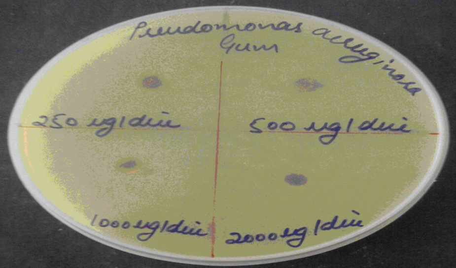
FIG. 1: ANTIMICROBIAL TESTING ON PSEUDOMONAS AERUGINOSA
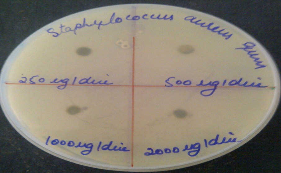
FIG. 2: ANTIMICROBIAL TESTING ON STAPHYLOCOCCUS AUREUS
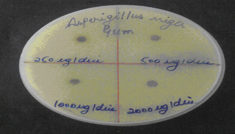
FIG. 3: ANTIFUNGAL ACTIVITY ON ORGANISM ASPERGILLUS NIGER
TABLE 2: ANTIMICROBIAL AND ANTIFUNGAL ACTIVITY
| Organism | Concentration | Disc size | Zone diameter |
| Pseudomonas aeruginosa | 0.250 mg | 6 mm | Nil |
| 0.500 mg | 6 mm | Nil | |
| 1 mg | 6 mm | Nil | |
| 2 mg | 6 mm | Nil | |
| Staphylococcus aureus | 0.250 mg | 6 mm | Nil |
| 0.500 mg | 6 mm | Nil | |
| 1 mg | 6 mm | Nil | |
| 2 mg | 6 mm | Nil | |
| Aspergillus niger | 0.250 mg | 6 mm | Nil |
| 0.500 mg | 6 mm | Nil | |
| 1 mg | 6 mm | Nil | |
| 2 mg | 6 mm | Nil |
No zone inhibition was observed for Pseudomonas aeruginosa, Staphylococcus aureus and Aspergillus niger with different concentration of gum is used as above.
ACKNOWLEDGEMENT: The authors are highly thankful to Akal College of Pharmacy for providing their laboratories and financial assistance in carrying out this study.
CONFLICT OF INTEREST: Nil
REFERENCES:
- Lewis EM: Should we be concerned about herbal remedies. Jo of Ethnopharmacology 2001; 75: 141-164.
- Van NDN and Tap N: An overview of the use of plants and animals in traditional medicine systems in Vietnam. Traffic Southeast Asia 2008.
- Beneke CE, Viljoen AM and Hamman JH: Polymeric plant-derived excipients. Drug Delivery Molecules 2009; 14: 2602-2620.
- Malviya R, Srivastava L and Kulkarni GT: Applications of mucilages in drug delivery. Advances in Biological Research 2011: 1 -7.
- Ali M: Textbook of Pharmacognosy, CBS Publishers and Distributors, New Delhi, 1998: 111-115, 385-387.
- Khandelwal KR: Practical Pharmacognosy Techniques and Experiments, Nirali Prakashan, Pune, 2005.
- Gupta AK: Quality Standards of Indian Medicinal Plants. Published by Indian J Med Res, New Delhi. Vol. I, 2003.
How to cite this article:
Thakur N, Mittal P, Kaur R and Goswami M: phytochemical screening of gum extracted from Curcuma amada. Int J Pharmacognosy 2015; 2(8): 419-25. doi link: http://dx.doi.org/10.13040/IJPSR.0975-8232.IJP.2(8).419-25.
This Journal licensed under a Creative Commons Attribution-Non-commercial-Share Alike 3.0 Unported License.